. 2020 Jun 16;117(24):13529-13540.
doi: 10.1073/pnas.1917969117. Epub 2020 Jun 1.

MAML1/2 promote YAP/TAZ nuclear localization and tumorigenesis

Affiliations

MAML1/2 promote YAP/TAZ nuclear localization and tumorigenesis

Jiyoung Kim et al. Proc Natl Acad Sci U S A. .

Abstract

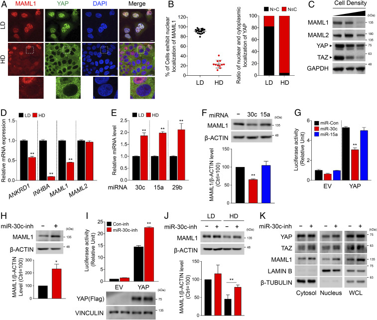
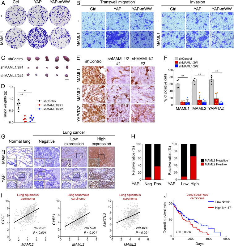
The Hippo pathway plays a pivotal role in tissue homeostasis and tumor suppression. YAP and TAZ are downstream effectors of the Hippo pathway, and their activities are tightly suppressed by phosphorylation-dependent cytoplasmic retention. However, the molecular mechanisms governing YAP/TAZ nuclear localization have not been fully elucidated. Here, we report that Mastermind-like 1 and 2 (MAML1/2) are indispensable for YAP/TAZ nuclear localization and transcriptional activities. Ectopic expression or depletion of MAML1/2 induces nuclear translocation or cytoplasmic retention of YAP/TAZ, respectively. Additionally, mutation of the MAML nuclear localization signal, as well as its YAP/TAZ interacting region, both abolish nuclear localization and transcriptional activity of YAP/TAZ. Importantly, we demonstrate that the level of MAML1 messenger RNA (mRNA) is regulated by microRNA-30c (miR-30c) in a cell-density-dependent manner. In vivo and clinical results suggest that MAML potentiates YAP/TAZ oncogenic function and positively correlates with YAP/TAZ activation in human cancer patients, suggesting pathological relevance in the context of cancer development. Overall, our study not only provides mechanistic insight into the regulation of YAP/TAZ subcellular localization, but it also strongly suggests that the miR30c-MAML-YAP/TAZ axis is a potential therapeutic target for developing novel cancer treatments.

Keywords: Hippo signaling; MAML1/2; TEAD; YAP/TAZ; nuclear localization.


Conflict of interest statement

The authors declare no competing interest.

Figures

Fig. 1.
Fig. 1.
MAML1/2 promote the nuclear localization and transcriptional activities of YAP/TAZ. (A) Ectopically expressed MAML1 and YAP in HEK293 cells colocalized in the nucleus. (Scale bars, 10 μm.) (B) Co-IP experiments showed that endogenous MAML1 interacts with YAP/TAZ. (C) The fluorescence signal generated by proximity ligation assay demonstrates that endogenous MAML1 and YAP are closely located in the nucleus. (Scale bars, 10 μm.) (D) MAML1 and MAML2, but not MAML3, increased YAP reporter activity in HCT116 cells. (E) Knockdown of MAML1/2 in HEK293A reduced nuclear localization of YAP. The areas marked with white rectangles were magnified and shown in E, Right. (Scale bars, 50 μm.) (F) Knockdown of MAML1/2 in HEK293T cells reduced YAP reporter activity. (G) Quantitative real-time PCR showed that knockdown of MAML1/2 in HeLa cells reduced mRNA levels of Hippo and Notch signaling target genes. (H) A wound-healing assay showed that knockdown of MAML1/2 in HeLa cells reduced migration rate, which was induced by the ectopic expression of YAP. In D and FH, the statistical analyses represent average values from a representative experiment performed in triplicate. Error bars represent SDs of triplicate assays. **P < 0.01. n.s., not significant. Student’s t test was used for statistical analysis.
Fig. 2.
Fig. 2.
Nuclear localization of MAML is essential for the retention of YAP/TAZ in the nucleus. (A) Schematic diagram of human MAML1. The red line indicates a putative NLS sequence, and “KKTRR” was mutated to “AATLL” for inducing loss of NLS function. (B) MAML1-mNLS is present in the cytoplasm, and it does not promote nuclear localization of YAP. (Scale bars, 10 μm.) (C) MAML1–mNLS did not increase YAP reporter activity in HCT116 cells when compared to wild-type (WT) MAML1. Ctrl, control. (D, Left and Center) MAML1–mNLS did not rescue retarded HEK293 cell migration induced by MAML1/2 depletion. (D, Right) Western blot shows normal expression of proteins. (E) Live cell imaging suggests that MAML1 retains YAP in the nucleus, which enhances YAP nuclear localization. (Scale bars, 10 μm.) In C, the statistical analysis represents average values from a representative experiment performed in triplicate. Error bars represent SDs of triplicate assays. In D, Center, the statistical analysis represents average values of three different regions from a representative experiment. Error bars represent SDs of values from three different regions. **P < 0.01. n.s., not significant. Student’s t test was used for statistical analysis.
Fig. 3.
Fig. 3.
The WW domains of YAP/TAZ and the PPxY motifs of MAML1/2 are required for the interaction between YAP/TAZ and MAML1/2. (A) YAP-mWW does not interact with MAML1. (B) Nuclear localization of YAP-mWW is not induced by ectopic expression of Flag-MAML1. (Scale bars, 10 μm.) (C) YAP-mWW–mediated reporter activity in HEK293T cells is not further enhanced by MAML1. (D) PPxY motifs in MAML1/2 are highly conserved in various species. (E) MAML1–PPxA shows reduced interaction with YAP. (F) MAML1–PPxA does not enhance YAP reporter activity in HCT116 cells as much as wild-type MAML1. (G) The nuclear localization of YAP was not induced by ectopic expression of MAML1–PPxA. (Scale bars, 10 μm.) (H) MAML1–PPxA did not rescue retarded HEK293 cell migration induced by MAML1/2 depletion. Wound-healing assay and analysis were performed as described in Fig. 2D. In C and F, the statistical analysis represents average values from a representative experiment performed in triplicate. Error bars represent SDs of triplicate assays. **P < 0.01. n.s., not significant. Student’s t test was used for statistical analysis. Ctrl, control; WT, wild type.
Fig. 4.
Fig. 4.
MAML1/2 act as transcriptional coactivators by forming a trimeric complex with YAP/TAZ and TEAD. (A) MAML1/2 KO cell pools exhibited reduced NLS–YAP-mediated reporter activity. (B) NLS–YAP-mediated reporter activity was further enhanced by MAML1 overexpression. (C) MAML1ΔTAD did not induce YAP reporter activity when compared to wild type. (D) Co-IP assay showed that MAML1 interacted with YAP and TEAD1. (E) Depletion of YAP/TAZ reduces interaction between MAML1 and TEAD. (F) ChIP assay showed that MAML1 and YAP co-occupies on the promoters of YAP target genes. FAT3 was used for negative control. (G) ChIP assay showed that TEAD1/3/4 are required for MAML1 to bind the promoters of YAP target genes. HBB was used as a negative control. The statistical analyses represent average values from a representative experiment performed in triplicate. Error bars represent SDs of triplicate assays. *P < 0.05; **P < 0.01. n.s., not significant. Student’s t test was used for statistical analysis. Ctrl, control.
Fig. 5.
Fig. 5.
Cell-density-dependent regulation of MAML1 and MAML2 and the involvement of miR-30c in the regulation of MAML1. (A) MAML1 and YAP colocalize in the nucleus or cytoplasm of HEK293 cells in a cell-density-dependent manner. HD, high cell density; LD, low cell density. (Scale bars, 10 μm.) (B) Quantification of MAML1 and YAP cellular localization shown in A. (C) Protein levels of MAML1/2 are reduced in HEK293 cells grown in high density. (D) qPCR assay showed that mRNA levels of MAML1, but not MAML2, were decreased in HEK293A cells grown in high density. (E) The level of miR-30c is increased in cells grown at high density. miR-15a and miR-29b were used for positive controls. (F, Upper) Ectopic expression of miR-30c mimics reduced MAML1 protein levels. (F, Lower) ImageJ was used to calculate the relative level of MAML1. (G) Ectopic expression of miR-30c mimics in HEK293 cells reduced YAP-mediated reporter activity. (H, Upper) Ectopic expression of an inhibitor of miR-30c in HEK293 cells increased MAML1 protein levels. (H, Lower) The relative level of MAML1 was analyzed by ImageJ. (I, Upper) Ectopic expression of miR-30c inhibitor in HEK293 cells increased YAP-mediated reporter activity. (I, Lower) Western blotting shows an equal amount of YAP expression. (J, Upper) Ectopic expression of a miR-30c inhibitor in HEK293 cells increased MAML1 protein level grown in high cell density. (J, Lower) The relative level of MAML1 was analyzed by ImageJ. (K) Ectopic expression of an miR-30c inhibitor in HEK293 cells increased the MAML1 level as well as nuclear localization of YAP and TAZ. The statistical analyses represents the average values from a representative experiment performed in triplicate. Error bars represent SDs of triplicate assays. *P < 0.05; **P < 0.01. Student’s t test was used for statistical analysis.
Fig. 6.
Fig. 6.
MAML1/2 display oncogenic activities, and MAML2 protein levels are strongly correlated with the expression of YAP/TAZ target genes in lung cancer patients. (A) MAML1 enhances colony-forming activity of YAP. (B) Migration and invasion ability of HCT116 cells were enhanced upon cotransfection with MAML1 and YAP. (Magnification, x200.) (C and D) Knockdown of MAML1/2 in HCT116 cells shows reduced tumor growth in xenograft experiments. (Scale bar, 1,000 μm [C].) (E and F) MAML1/2-depleted cell-driven tumors exhibit significantly reduced YAP/TAZ levels when compared to control cell driven tumors. (Scale bars, 100 μm.) (F) Graph shows percentage of positive MAML1, MAML2, and YAP/TAZ-stained cells in control and MAML1/2-depleted cell-driven tumors displayed in E. (G and H) The tissue microarray analysis of lung cancer patients showed a positive correlation between MAML2 and YAP protein levels. (G) Histochemical analysis was performed with antibodies indicated on the left. G, Insets show magnification of the small rectangular region. (Scale bars, 20 μm.) (H) Graphs showing correlations between the levels of MAML2 and YAP (raw data were presented in SI Appendix, Fig. S6 F and G). Neg., negative; pos., positive. (I) The mRNA expression datasets of lung squamous carcinoma patients from the TCGA database show a positive correlation between MAML2 and YAP target genes. The correlation coefficient (r) was calculated by Pearman’s linear correlation. Two-tailed P value was used for analyzing statistical significance. (J) Kaplan–Meier plot shows that high expression of MAML2 is associated with poor prognosis of lung cancer patients. Lower 33rd percentile (n = 161) and upper 24th percentile (n = 117) were analyzed. **P < 0.01.

Similar articles

Cited by

References

    1. Pan D., The hippo signaling pathway in development and cancer. Dev. Cell 19, 491–505 (2010). - PMC - PubMed
    1. Fu V., Plouffe S. W., Guan K. L., The Hippo pathway in organ development, homeostasis, and regeneration. Curr. Opin. Cell Biol. 49, 99–107 (2017). - PMC - PubMed
    1. Misra J. R., Irvine K. D., The Hippo signaling network and its biological functions. Annu. Rev. Genet. 52, 65–87 (2018). - PMC - PubMed
    1. Piccolo S., Dupont S., Cordenonsi M., The biology of YAP/TAZ: Hippo signaling and beyond. Physiol. Rev. 94, 1287–1312 (2014). - PubMed
    1. Kim W., Jho E. H., The history and regulatory mechanism of the Hippo pathway. BMB Rep. 51, 106–118 (2018). - PMC - PubMed

Publication types

MeSH terms