. 2020 Jun 16;117(24):13457-13467.
doi: 10.1073/pnas.1919507117. Epub 2020 Jun 1.

MYC protein stability is negatively regulated by BRD4

Affiliations

MYC protein stability is negatively regulated by BRD4

Ballachanda N Devaiah et al. Proc Natl Acad Sci U S A. .

Abstract

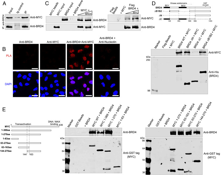
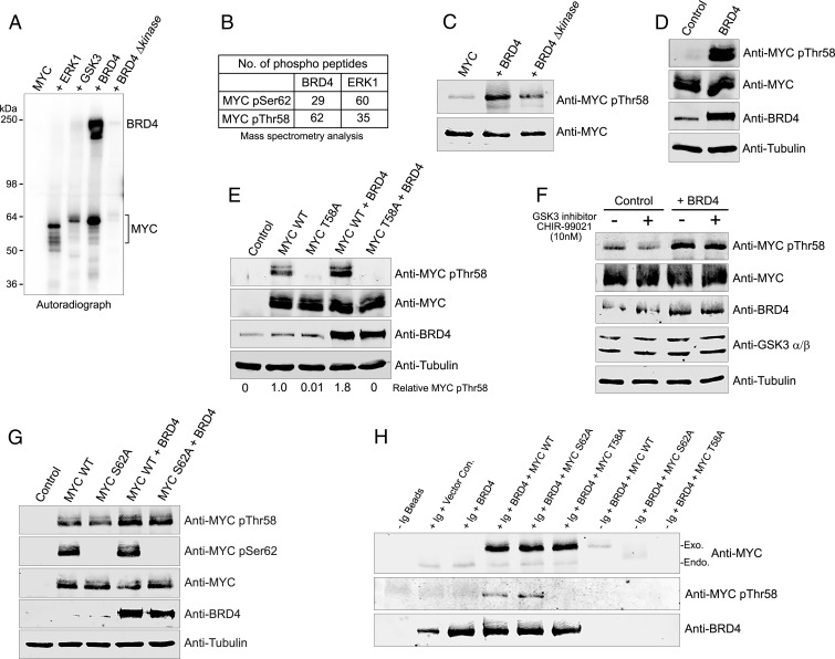
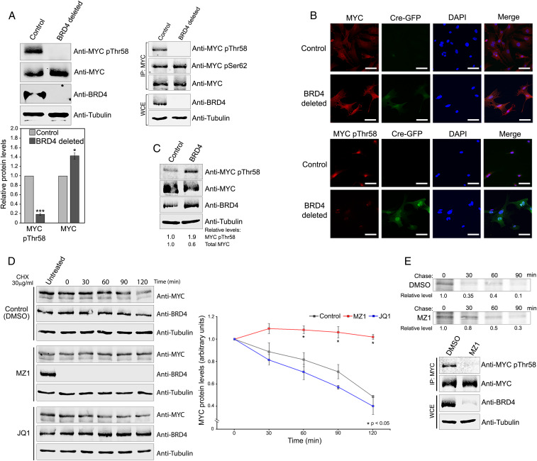
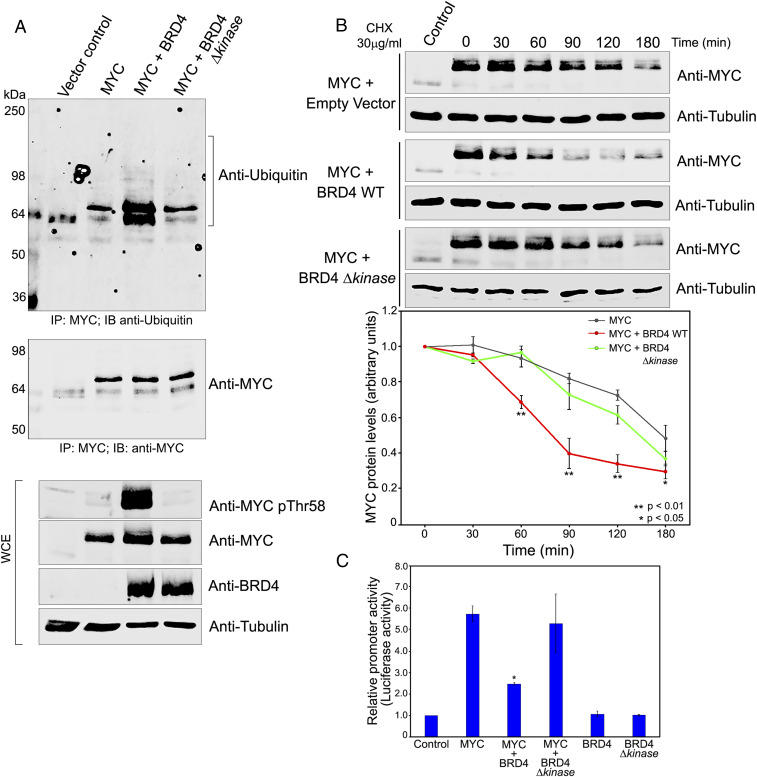
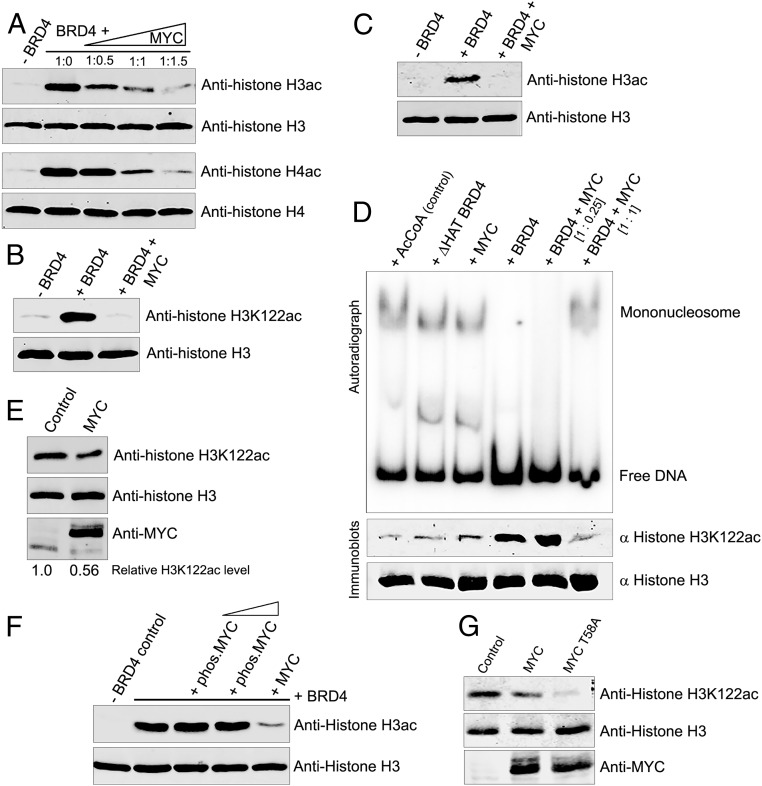
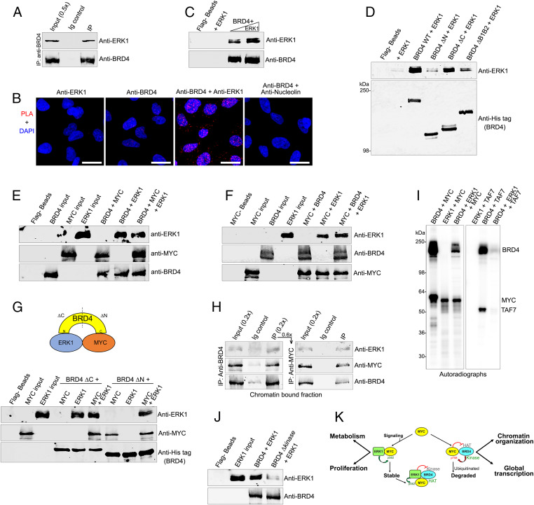
The protooncogene MYC regulates a variety of cellular processes, including proliferation and metabolism. Maintaining MYC at homeostatic levels is critical to normal cell function; overexpression drives many cancers. MYC stability is regulated through phosphorylation: phosphorylation at Thr58 signals degradation while Ser62 phosphorylation leads to its stabilization and functional activation. The bromodomain protein 4 (BRD4) is a transcriptional and epigenetic regulator with intrinsic kinase and histone acetyltransferase (HAT) activities that activates transcription of key protooncogenes, including MYC We report that BRD4 phosphorylates MYC at Thr58, leading to MYC ubiquitination and degradation, thereby regulating MYC target genes. Importantly, BRD4 degradation, but not inhibition, results in increased levels of MYC protein. Conversely, MYC inhibits BRD4's HAT activity, suggesting that MYC regulates its own transcription by limiting BRD4-mediated chromatin remodeling of its locus. The MYC stabilizing kinase, ERK1, regulates MYC levels directly and indirectly by inhibiting BRD4 kinase activity. These findings demonstrate that BRD4 negatively regulates MYC levels, which is counteracted by ERK1 activation.

Keywords: BRD4 histone acetyltransferase; BRD4 kinase; ERK1; MYC phosphorylation; MYC stability.


Conflict of interest statement

The authors declare no competing interest.

Figures

Fig. 1.
Fig. 1.
BRD4 and MYC interact in the nucleus. (A) BRD4 associates with MYC. BRD4 was immunoprecipitated from HeLa nuclear extract using anti-BRD4 and immunoblotted with anti-MYC. (Anti-BRD4 specificity is shown in SI Appendix, Fig. S1A) (B) BRD4 colocalizes with MYC in the nucleus. PLAs with anti-BRD4 and anti-MYC on fixed HeLa cells. PLA with anti-BRD4 and nucleolin as a negative control. PLA, red; DAPI nuclei staining, blue. (Scale bars, 20 µM.) (C) BRD4 binds MYC directly. (Left) rBRD4 (0.5 and 0.75 µg) was pulled down with 0.2 µg rMYC immobilized on anti-MYC-agarose beads. (Right) rMYC (0.2 and 0.4 µg) was pulled down with 0.5 µg rBRD4 immobilized on Flag-beads (Right). Beads with or without prey protein are controls; input was the same for both pull-downs. (D) MYC binds BRD4 C-terminal region. (Top) Map of BRD4 and deletion mutants. (Bottom) Anti-MYC immunoblot of 0.2 µg rMYC recovered by pull-down with 0.5 µg wild-type His-BRD4-Flag or equimolar amounts of His-BRD4-Flag mutants on Flag beads. Anti-His immunoblot shows BRD4 retained on Flag beads; beads with or without rMYC are controls. (E) BRD4 binds MYC transactivation domain. (Left) Map of MYC and deletion mutants. (Middle) Anti-BRD4 immunoblots of 0.5 µg BRD4 recovered by pull-down with 0.25 µg wild-type GST-MYC or equimolar amounts of GST-MYC mutants on GST-Sepharose beads. Anti-GST immunoblots show MYC on beads; beads with or without BRD4 are controls. (Right) As in Middle, using additional MYC deletion mutants to fine map BRD4-binding region on MYC. MYC degradation products (*).
Fig. 2.
Fig. 2.
BRD4 phosphorylates MYC at threonine 58. (A) BRD4 phosphorylates MYC. Autoradiograph of kinase assays with BRD4 (0.3 µg) or equimolar amounts of BRD4 ∆kinase, ERK1, or GSK3 with 0.5 µg rMYC substrate. (B) BRD4 preferentially phosphorylates MYCThr58. rMYC phosphorylated by rBRD4 (0.3 µg) or equimolar amounts of rERK1 was analyzed by mass spectrometry. Table shows number of MYC pSer62 and pThr58 containing phosphopeptides are identified. (C) BRD4 phosphorylates MYCThr58. rMYC (0.5 µg) phosphorylated by BRD4 (0.3 µg) or equimolar amounts of BRD4 ∆kinase in in vitro kinase assays was immunoblotted with anti-MYC pThr58 or anti-MYC. (D) BRD4 mediates endogenous MYCThr58 phosphorylation. MYCpThr58, MYC, BRD4, and tubulin immunoblots of whole cell extracts (WCEs) from HeLa cells transfected with 3 µg BRD4 or vector (control). (E) MYCpThr58 antibody specifically recognizes exogenous MYC phosphorylation by BRD4. MYCpThr58, MYC, BRD4, and tubulin immunoblots of WCEs from HeLa cells transfected with 3 µg vector, MYC WT, or MYC T58A mutant with or without 3 µg BRD4. Relative MYC pThr58 levels, normalized to tubulin, are shown. (F) BRD4 phosphorylation of endogenous MYC pThr58 does not depend on GSK3β. MYC pThr58, MYC, BRD4, GSK3β, and tubulin immunoblots of WCEs from HeLa cells transfected with 3 µg vector or BRD4 and treated with DMSO or GSK3β inhibitor CHIR-99021 overnight. (G) BRD4 phosphorylation of exogenous MYCThr58 is independent of MYCSer62. MYC pThr58, pSer62, MYC, BRD4, and tubulin immunoblots of WCEs from HeLa cells transfected with 3 µg vector, MYC WT, or MYC S62A mutant with or without 3 µg BRD4. (H) BRD4 interacts with MYCWT, MYCT58, and MYCS62 and phosphorylates Thr58. BRD4, MYC, and pT58 immunoblots of WCEs from HeLa cells transfected with 3 µg MYCT58, MYCS62 mutants, or vector, with or without 3 μg BRD4 and immunoprecipitated with anti-BRD4.
Fig. 3.
Fig. 3.
Endogenous BRD4 kinase controls endogenous MYC Thr58 phosphorylation and ubiquitination. (A) BRD4 deletion abrogates endogenous MYC pThr58 and increases total MYC in MEFs. (Top, Left) MYCpThr58, MYC, BRD4, and tubulin immunoblots of WCEs from Brd4-floxed MEFs uninfected (control) or infected (BRD4 deleted) with Cre-GFP-expressing retrovirus. (Right) WCEs immunoprecipitated with anti-Myc antibody and immunoblotted with indicated antibodies. (Bottom) Quantification of MYCpThr58 and total MYC levels in WCEs from three independent experiments performed as above. Error bars represent ±SEM (*P < 0.05; ***P < 0.0001). (B) Endogenous MYC pThr58 and total MYC levels depend on BRD4. Brd4-floxed MEFs uninfected (control) or infected (BRD4 deleted) with a Cre-GFP-expressing retrovirus were stained with DAPI and analyzed by immunofluorescence with anti-MYC and anti-MYCpThr58 antibodies. GFP fluorescence identifies Cre-GFP-infected, BRD4-depleted cells. Representative images are shown. (Scale bars, 20 µM.) (C) BRD4 overexpression increases MYCpThr58 and decreases total endogenous MYC in MEFs. MYC pThr58, MYC, BRD4, and tubulin immunoblots of WCEs from MEFs infected with vector only or with BRD4 expressing retrovirus. Relative MYCpThr58 and total MYC levels, normalized to tubulin, are shown. (D) Degradation of endogenous BRD4, but not its inhibition by JQ1, stabilizes endogenous MYC. (Left) MYC and BRD4 immunoblots of WCEs from HCT116 cells grown in the presence of PROTAC MZ1 (0.1 µM), 500 nM JQ1, or dimethyl sulfoxide (control) overnight, treated with 30 µg/mL cycloheximide (CHX), and harvested at 0, 30, 60, 90, and 120 min after CHX treatment. Tubulin, loading control. (Right) Quantification of total MYC protein levels in WCEs from three independent experiments performed as above. Error bars represent ±SEM (*P < 0.05). (E) Degradation of BRD4 leads to endogenous MYC stabilization. (Top) Autoradiographs of MYC immunoprecipitated from HeLa cells treated with either DMSO or MZ1 (0.1 µM) for 2 h and pulse chased after labeling with [35S]methionine/cysteine for 20 min (pulse) and incubated in unlabeled complete medium for the indicated times (chase). (Bottom) MYC pThr58, MYC, BRD4, and tubulin immunoblots of immunoprecipitated MYC and WCEs from above cells.
Fig. 4.
Fig. 4.
MYC stability and function are regulated by BRD4 kinase. (A) BRD4 phosphorylates exogenous MYC Thr58 resulting in MYC ubiquitination. WCEs from HeLa cells transfected with 3 µg vector or MYC, with or without BRD4 and BRD4 Δkinase were immunoblotted with anti-MYC pThr58, MYC, BRD4, and tubulin antibodies (Bottom). Total MYC was immunoprecipitated from WCEs using MYC-agarose beads and immunoblotted with anti-MYC (Middle) or anti-ubiquitin (Top). (B) Exogenous MYC stability is regulated by BRD4 kinase activity. (Left) MYC immunoblots of WCEs from HeLa cells transfected with 3 µg vector or MYC with or without BRD4 and BRD4 Δkinase, treated with 30 µg/mL CHX 18 h posttransfection and harvested at 0, 30, 60, 90, 120, and 180 min after CHX treatment. Tubulin, loading control. (Right) Quantification of MYC protein levels in WCEs from three independent experiments performed as above. Error bars represent ±SEM (*P < 0.05; **P < 0.01). (C) MYC transactivation of CDK4 promoter is modulated by BRD4 kinase activity. Relative luciferase activity in HeLa cells transfected with 1 µg CDK4 E-box luciferase reporter plasmid alone (control) or cotransfected with 1 µg MYC and BRD4 or BRD4 Δkinase in combination or individually. Error bars represent ±SEM (*P < 0.05, relative to MYC alone).
Fig. 5.
Fig. 5.
MYC inhibits BRD4 histone acetylation and nucleosome disassembly. (A) MYC inhibits BRD4 acetylation of histones H3 and H4. Histone H3ac, H3, H4ac, and H4 immunoblots of 1 µg H3 and H4 subjected to HAT assays with 0.5 µg BRD4 in the presence or absence of MYC, at BRD4:MYC molar ratios of 1:0.5, 1:1, and 1:1.5. (B) MYC inhibits BRD4 acetylation of H3K122. Histone H3K122ac and H3 immunoblots of 1 µg H3 subjected to HAT assays with 0.5 µg BRD4 alone or with 0.2 µg MYC. (C) MYC inhibits BRD4 acetylation of histone H3 in assembled mononucleosomes. H3ac and H3 immunoblots of mononucleosomes (10 pmol), assembled in vitro with 5S rDNA and subjected to a HAT assay with 0.5 µg BRD4 alone or with 0.2 µg MYC (1:1 molar ratio). (D) BRD4-mediated nucleosome disassembly is inhibited by MYC. Mononucleosomes (10 pmol) assembled with radiolabeled 5S rDNA were subjected to HAT assays with or without 0.5 µg BRD4 or BRD4 ΔHAT mutant in the presence or absence of MYC added at 1:0.25 and 1:1 BRD4:MYC molar ratios. Reactions with only AcCoA or MYC were controls. Free radiolabeled DNA was resolved from mononucleosomes by electrophoresis (autoradiogram, Upper). Identical HAT assays were done in parallel with nonradiolabeled mononucleosomes and the extent of H3K122 acetylation was determined by immunoblotting; histone H3 immunoblot served as control (Lower). (E) Exogenous MYC inhibits acetylation of histone H3K122. Histone H3, H3K122ac, and MYC immunoblots of WCEs from HeLa cells transfected with 3 µg MYC or vector (control). Relative H3K122ac levels, normalized to total histone H3, are shown. (F) BRD4 HAT activity is not inhibited by MYC prephosphorylated by BRD4. Histone H3ac and H3 immunoblots of 1 µg histones H3 subjected to HAT assays with 0.5 µg BRD4 in the presence or absence of purified, BRD4 prephosphorylated MYC (0.2 and 0.4 µg), or unphosphorylated MYC (0.2 µg). (G) Exogenous MYC T58A mutant inhibits acetylation of histone H3K122. H3K122ac, histone H3, and MYC immunoblots of WCEs from HeLa cells transfected with 3 µg MYC, MYC T58A mutant, or vector (control).
Fig. 6.
Fig. 6.
ERK1 interacts with BRD4 and inhibits its kinase activity. (A) BRD4 associates with ERK1 in vivo. BRD4 was immunoprecipitated from HeLa nuclear extract using anti-BRD4 and immunoblotted with anti-ERK1. (B) BRD4 interacts with ERK1 in situ. PLAs with anti-BRD4 and anti-ERK1 on fixed HeLa cells. PLA with anti-BRD4 and nucleolin as a negative control. PLA, red; DAPI nuclei staining, blue. (Scale bars, 20 µM.) (C) BRD4 interacts directly with ERK1. Recombinant ERK1 (0.1, and 0.2 µg) was pulled down with 0.4 µg recombinant BRD4 immobilized on Flag beads. Beads with or without ERK1 are controls. (D) ERK1 binds the BRD4 N-terminal region. Anti-ERK1 immunoblot of 0.2 µg ERK1 recovered by pull-down with 0.75 µg wild-type His-BRD4-Flag or equimolar amounts of ΔN, ΔC, and ΔB1B2 His-BRD4-Flag mutants on Flag beads (Fig. 1D). Anti-His immunoblot shows BRD4 on beads; beads with or without ERK1 are controls. (E) MYC and ERK1 do not compete for binding to BRD4. ERK1, MYC, and BRD4 immunoblots of pull-down reactions done by incubating BRD4-Flag (0.75 μg) immobilized on beads with MYC (0.3 µg) alone (1:1 molar ratio), with ERK1 (0.2 µg) alone (1:1 molar ratio), or with equimolar amounts of both. Beads alone and the input proteins are controls. (F) BRD4 and ERK1 do not compete for binding to MYC. ERK1, BRD4, and MYC immunoblots of pull-down reactions with equimolar amounts of MYC (0.3 µg) immobilized on agarose beads with ERK1 (0.2 µg) alone, BRD4 (0.75 µg) alone, or both. Beads alone and the input proteins are controls. (G) BRD4, ERK1, and MYC directly interact in a trimeric complex in vitro. (Top) Diagram of BRD4-ERK1-MYC complex. (Bottom) ERK1, BRD4, and MYC immunoblots of pull-down reactions done by incubating either MYC (0.22 µg), ERK1 (0.15 µg), or both with BRD4 ΔC or ΔN proteins (0.3 µg) immobilized on Flag beads. Beads alone and the input proteins are controls. (H) BRD4, ERK1, and MYC exist in a trimeric complex in cellulo. ERK1, BRD4, and MYC immunoblots of BRD4 immunoprecipitated using anti-BRD4 in a primary IP (Left) from chromatin-bound HeLa nuclear protein fraction, subjected to sequential IP with MYC agarose beads (Right). (I) ERK1 inhibits BRD4 kinase activity. Autoradiograph of kinase assays with BRD4 (0.5 µg) or equimolar amounts of ERK1 using 0.5 µg of MYC or TAF7 as substrates. (J) BRD4 Δkinase does not bind ERK1. Anti-ERK1 immunoblot of 0.2 µg ERK1 recovered by pull-down with 0.75 µg wild-type BRD4 or BRD4 Δkinase (lacking aa 502 to 548) on Flag beads. Anti-BRD4 immunoblot shows BRD4. Beads alone and ERK1 input are controls (K). Model of regulatory interactions of MYC, BRD4, and ERK1. BRD4 binding to MYC maintains homeostatic levels of MYC protein and global transcription. MYC stability is regulated by interactions with BRD4 and ERK1 which phosphorylate MYC at Thr58 and Ser62, leading to its degradation and stabilization, respectively. (Right) BRD4 regulates transcription of MYC gene, resulting in increased levels of MYC protein (not shown) and amplified MYC-targeted transcription. MYC inhibits BRD4 HAT activity, limiting chromatin remodeling and transcription. Conversely, BRD4 kinase deregulates MYC protein stability by phosphorylating Thr58. Paradoxically, this enhances pause release while moderating transcriptional amplification. (Left) Signaling through the MAP kinase pathway leads to nuclear localization of ERK1, which phosphorylates MYC at Ser62 and inhibits BRD4 kinase activity, together resulting in stabilized MYC. Phosphorylated MYC is unable to inhibit BRD4 HAT, enabling renewed chromatin remodeling and transcriptional activation.

References

    1. Eilers M., Eisenman R. N., Myc’s broad reach. Genes Dev. 22, 2755–2766 (2008). - PMC - PubMed
    1. Nie Z. et al. ., c-Myc is a universal amplifier of expressed genes in lymphocytes and embryonic stem cells. Cell 151, 68–79 (2012). - PMC - PubMed
    1. Lin C. Y. et al. ., Transcriptional amplification in tumor cells with elevated c-Myc. Cell 151, 56–67 (2012). - PMC - PubMed
    1. Bretones G., Delgado M. D., León J., Myc and cell cycle control. Biochim. Biophys. Acta 1849, 506–516 (2015). - PubMed
    1. Carroll P. A., Freie B. W., Mathsyaraja H., Eisenman R. N., The MYC transcription factor network: Balancing metabolism, proliferation and oncogenesis. Front. Med. 12, 412–425 (2018). - PMC - PubMed

Publication types

MeSH terms