. 2020 Jun 1;11(6):410.
doi: 10.1038/s41419-020-2553-6.

CAMK2A supported tumor initiating cells of lung adenocarcinoma by upregulating SOX2 through EZH2 phosphorylation

Affiliations

CAMK2A supported tumor initiating cells of lung adenocarcinoma by upregulating SOX2 through EZH2 phosphorylation

Si-Qi Wang et al. Cell Death Dis. .

Abstract

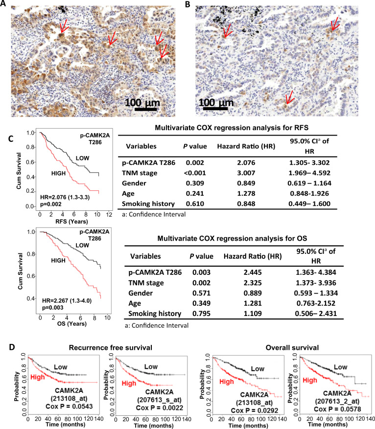
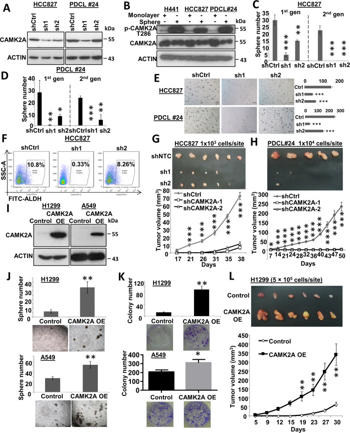
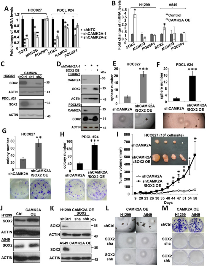
Tumor initiating cells (TIC) of lung cancer are mainly induced by stress-related plasticity. Calcium/Calmodulin dependent protein kinase II alpha (CAMK2A) is a key calcium signaling molecule activated by exogenous and endogenous stimuli with effects on multiple cell functions but little is known about its role on TIC. In human lung adenocarcinomas (AD), CAMK2A was aberrantly activated in a proportion of cases and was an independent risk factor predicting shorter survivals. Functionally, CAMK2A enhanced TIC phenotypes in vitro and in vivo. CAMK2A regulated SOX2 expression by reducing H3K27me3 and EZH2 occupancy at SOX2 regulatory regions, leading to its epigenetic de-repression with functional consequences. Further, CAMK2A caused kinase-dependent phosphorylation of EZH2 at T487 with suppression of EZH2 activity. Together, the data demonstrated the CAMK2A-EZH2-SOX2 axis on TIC regulation. This study provided phenotypic and mechanistic evidence for the TIC supportive role of CAMK2A, implicating a novel predictive and therapeutic target for lung cancer.


Conflict of interest statement

The authors declare that they have no conflict of interest.

Figures

Fig. 1
Fig. 1. Correlation of activated CAMK2A expression in lung AD with patient survivals.
a, b Immunohistochemical (IHC) evaluation of p-CAMK2A T286 expression in lung adenocarcinoma. a High-level expression, showing higher frequency of tumor cells staining positively for cytoplasmic and nuclear signals (arrows). b Low-level expression, showing fewer positively stained tumor nuclei (arrows) in the same unit area as shown in Fig. 1b. c COX regression analysis for recurrence-free survival (RFS) and overall survival (OS) stratified by p-CAMK2A T286 levels. Survival curves were generated by SPSS 18.0. d COX regression analysis of CAMK2A effects on RFS and OS using public expression array data (www.kmplot.com/lung).
Fig. 2
Fig. 2. Effects of CAMK2A on TIC properties in lung AD cells.
a Western blot of shRNA-mediated CAMK2A KD in HCC827 and PDCL#24 cells. b Activated CAMK2A (p-T286) relative to total CAMK2A expression was increased in spheres compared with cell monolayers of H441, HCC827 and PDCL#24. c, d Sphere formation of both cell lines with CAMK2A KD were inhibited for two generations. e Colony formation of CAMK2A KD cells were decreased in both cell lines. f Flow cytometry of HCC827 with CAMK2A KD showed reduction of ALDH-expressing cells. g, h Tumor sizes and growth curves of xenografts with CAMK2A KD from HCC827 (g) and PDCL#24 (h) were suppressed compared with controls (shNTC). i Western blot of CAMK2A OE in H1299 and A549 cells. j, k Sphere formation (j) and colony formation (k) of H1299 and A549 with CAMK2A OE were increased. l Tumor growth of H1299-derived xenografts with CAMK2A OE was facilitated. *p < 0.05; **p < 0.01; ***p < 0.001 compared with control. Data represented mean ± SD.
Fig. 3
Fig. 3. Effects of CAMK2A on responses of lung AD cells to target therapy and cytotoxic drugs.
a, b Cell viability of HCC827 with CAMK2A KD showed increased sensitivity to gefitinib (a) and cisplatin (b). c, d Cell viability of A549 (c) and H1299 (d) with CAMK2A OE showed increased resistance to cisplatin. e, f Western blot showed that short term treatment of gefitinib and cisplatin increased p-CAMK2A level in HCC827 (e) and H1299 cells (f). g, h Western blot of gefitinib-resistant HCC827 (GR) (g) and cisplatin-resistant A549 (CR) cells (h) showed increased p-CAMK2A T286 relative to total CAMK2A compared with parental control cells. i, j Cell viability of HCC827 GR cells (i) and A549 CR cells (j) under CAMK2A inhibition by KN93 showed sensitization to gefitinib and cisplatin, respectively. k Flow cytometry analysis of cisplatin treated HCC827 and PDCL#24 cells with CAMK2A KD showed increased apoptosis. *p < 0.05; **p < 0.01, compared with control, mean ± SD was presented.
Fig. 4
Fig. 4. SOX2-mediated effects of CAMK2A TIC in lung AD.
a, b Quantitative PCR of SOX2, NANOG and POU5F1 transcripts in HCC827 and PDCL#24 cells with CAMK2A KD (a), and in H1299 and A549 cells with CAMK2A OE (b). c Western blot showing SOX2 decrease in HCC827 and PDCL#24 cells with CAMK2A KD. d Western blot showing SOX2 changes in HCC827 and PDCL#24 cells with CAMK2A KD and SOX2 OE. eh Overexpression of SOX2 increased sphere formation (e, f) and colony formation (g, h) in CAMK2A-downregulated HCC827 and PDCL#24 cells. i SOX2 overexpression enhanced tumorigenicity in HCC827 shCAMK2A-1-derived xenografts. n = 4. j Expression of SOX2 in CAMK2A-overexpressed H1299 and A549 cells by western blot. k Downregulation of SOX2 in CAMK2A-overexpressed H1299 and A549 cells was confirmed by western blot. l, m Downregulation of SOX2 abrogated CAMK2A’s tumor initiating properties in sphere formation (l) and colony formation (m) in H1299 and A549 CAMK2A-overexpressed cells. *p < 0.05; **p < 0.01; ***p < 0.001, compared with control, mean ± SD was presented.
Fig. 5
Fig. 5. CAMK2A enhanced SOX2 expression through decreasing EZH2 mediated H3K27me3 in the regulatory region of SOX2.
a, b Expression of EZH2 and H3K27me3 in CAMK2A-downregulated HCC827 and PDCL #24 cells (a) as well as CAMK2A-overexpressed H1299 and A549 cells (b) by western blot. c, d Chromatin immunoprecipitation (ChIP) assay coupled with qPCR (ChIP-qPCR) analysis revealed the relative enrichment of H3K27me3 (c) and EZH2 (d) on SOX2 regulatory region in CAMK2A-downregulated HCC827 cells. e, f ChIP-qPCR analysis revealed the relative decrease of H3K27me3 (e) and EZH2 (f) on SOX2 regulatory region in CAMK2A-overexpressed H1299 cells. gj Downregulation of EZH2 rescued the inhibitory effects of CAMK2A knockdown on SOX2 expression (g), sphere formation (h), ALDH+ population (i) and cisplatin sensitivity (j) in HCC827 cells. *p < 0.05; **p < 0.01; ***p < 0.001, compared with control, ##p < 0.01 compared with shCAMK2A, mean ± SD was presented.
Fig. 6
Fig. 6. CAMK2A phosphorylated EZH2 at T487.
a Co-immunoprecipitation (CoIP) in HEK293T cells with dual overexpression of CAMK2A and EZH2, either using CAMK2A or EZH2 as bait. b CoIP of EZH2 in HCC827 cells using CAMK2A as bait. c CoIP of EZH2 in H1299 CAMK2A-OE cells. d CoIP of EZH2 in A549 CAMK2A-OE cells with or without EZH2 knock-down using CAMK2A anti-body. e CoIP of EZH2 in HEK293T cells with overexpression of EZH2, CAMK2A WT or CAMK2A T286A using CAMK2A as bait. f Expression of total EZH2 and p-EZH2 T487 in monolayer and spheres from PDCL#24, HCC827, and A549 cells. g Expression of total EZH2 and p-EZH2 T487 in HCC827, H1299 and A549 cells with CAMK2A knock-down or overexpression by immunoblot. h Expression of p-EZH2 T487 in HCC827 cells treated with KN92/KN93 by immunoblot. i Expression of p-EZH2 T487 in HCC827 GR cells by immunoblot. j Immunoprecipitated p-EZH2 T487 level by EZH2 in HCC827 cells with or without CAMK2A knock-down. k CoIP of EZH2 in HEK293T cells with overexpression of CAMK2A, EZH2 WT or EZH2 T487A using CAMK2A as bait. j Correlation analysis between p-CAMK2A T286 and p-EZH2 T487 expression in 169 primary resected lung AD by immunohistochemistry. k Schematic representation depicting supportive effect of CAMK2A on TIC maintenance through EZH2/H3K27me3/SOX2 pathway.

References

    1. Kreso A, Dick JE. Evolution of the cancer stem cell model. Cell Stem Cell. 2014;14:275–291. doi: 10.1016/j.stem.2014.02.006. - DOI - PubMed
    1. Saygin C, Matei D, Majeti R, Reizes O, Lathia JD. Targeting cancer stemness in the clinic: from hype to hope. Cell Stem Cell. 2019;24:25–40. doi: 10.1016/j.stem.2018.11.017. - DOI - PubMed
    1. Roderick HL, Cook SJ. Ca2+ signalling checkpoints in cancer: remodelling Ca2+ for cancer cell proliferation and survival. Nat. Rev. Cancer. 2008;8:361–375. doi: 10.1038/nrc2374. - DOI - PubMed
    1. Stewart TA, Yapa KT, Monteith GR. Altered calcium signaling in cancer cells. Biochimica et. Biophysica Acta. 2014 doi: 10.1016/j.bbamem.2014.08.016. - DOI - PubMed
    1. Hempel N, Trebak M. Crosstalk between calcium and reactive oxygen species signaling in cancer. Cell Calcium. 2017;63:70–96. doi: 10.1016/j.ceca.2017.01.007. - DOI - PMC - PubMed

Publication types

MeSH terms

Substances