. 2020 Jun 1;11(6):408.
doi: 10.1038/s41419-020-2621-y.

Exosomal miR-130b-3p targets SIK1 to inhibit medulloblastoma tumorigenesis

Affiliations

Exosomal miR-130b-3p targets SIK1 to inhibit medulloblastoma tumorigenesis

Saihua Huang et al. Cell Death Dis. .

Abstract

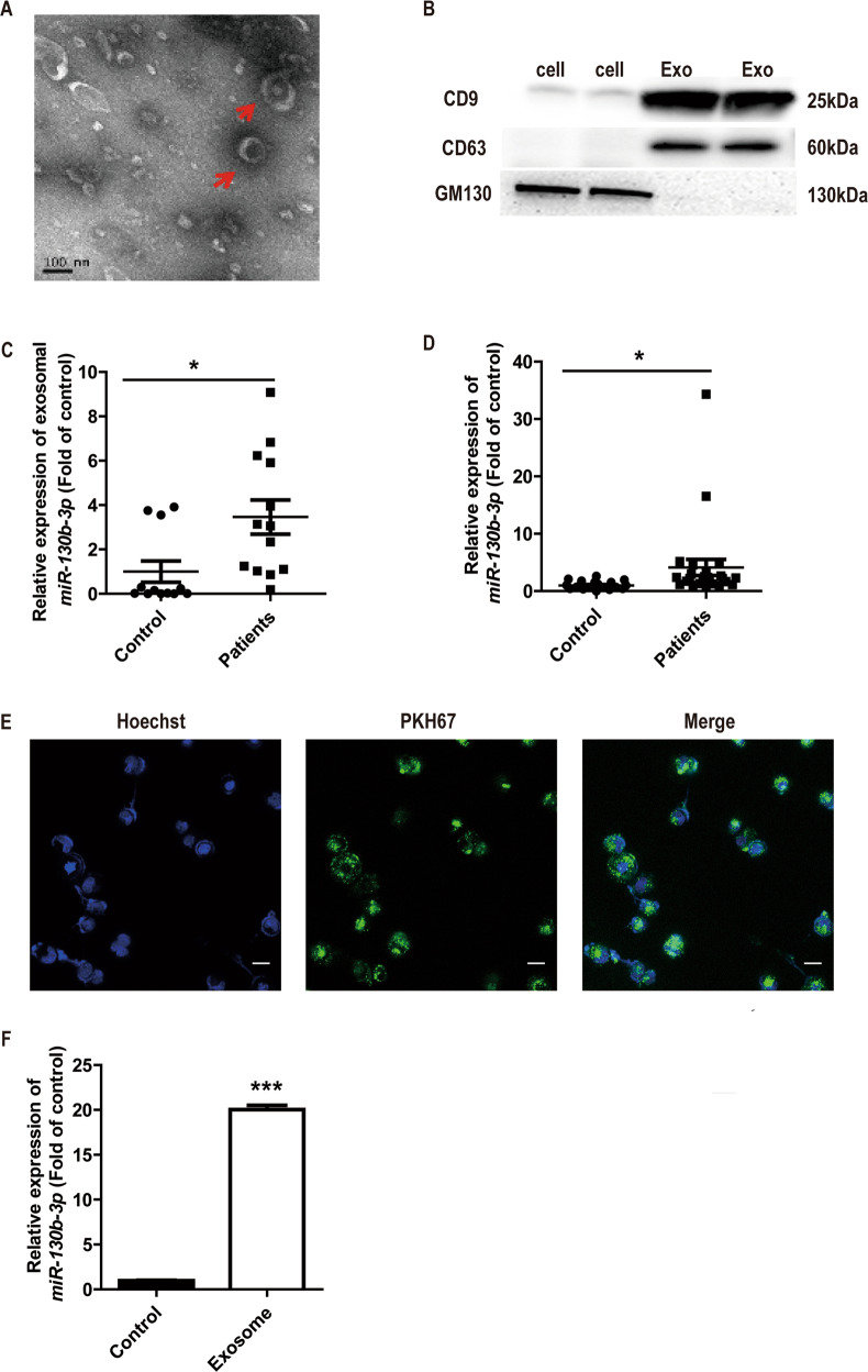
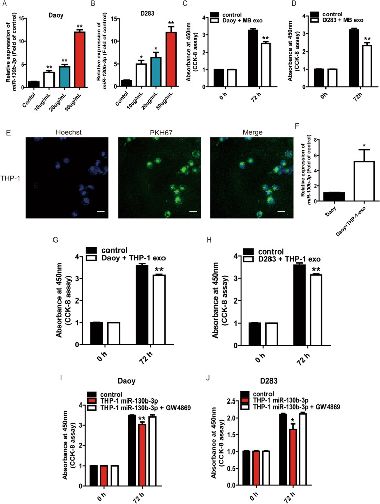
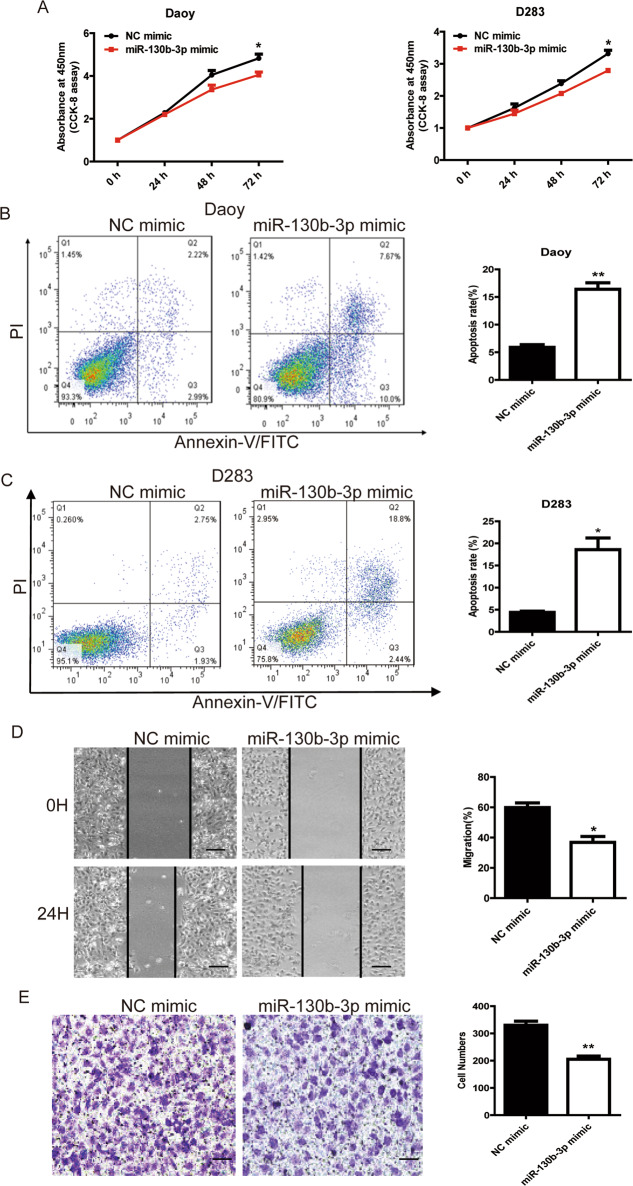
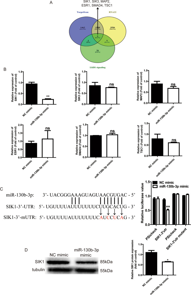
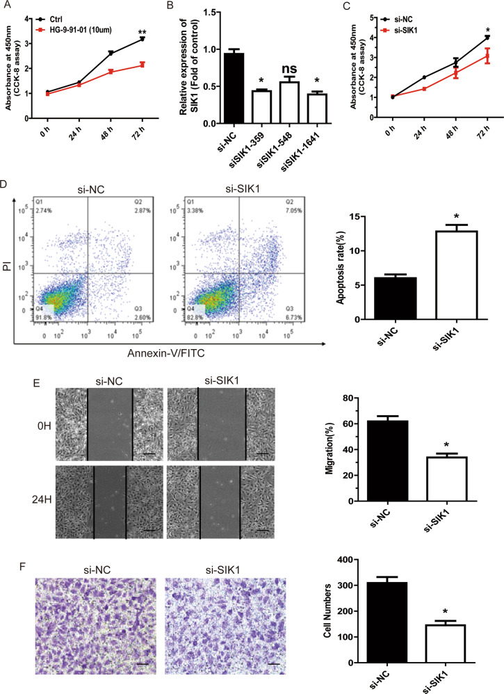
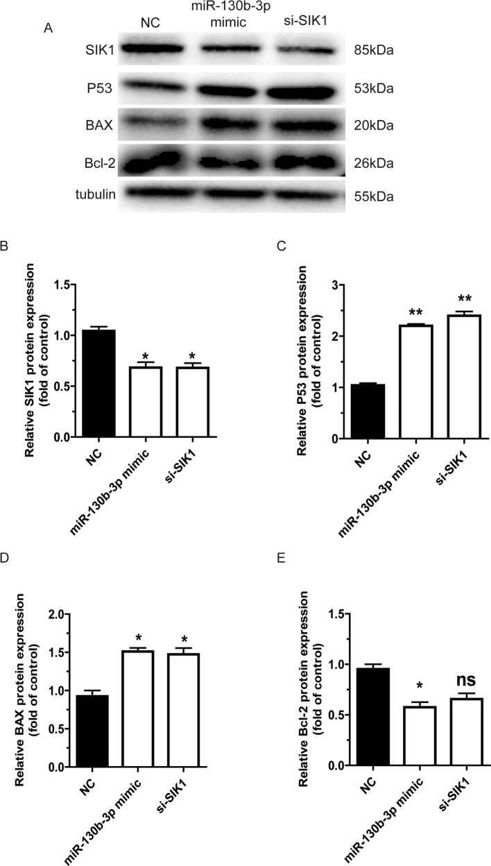
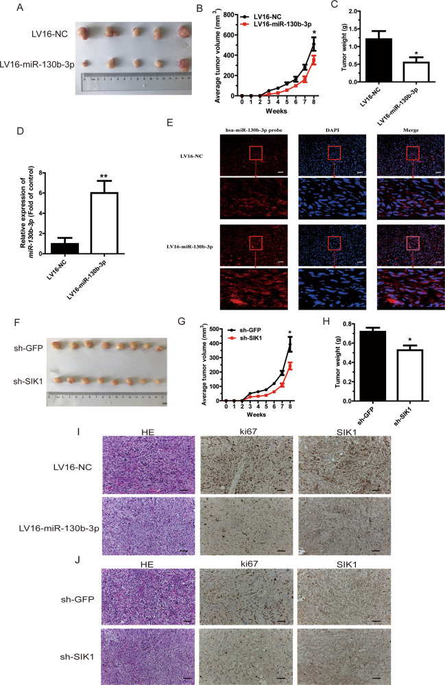
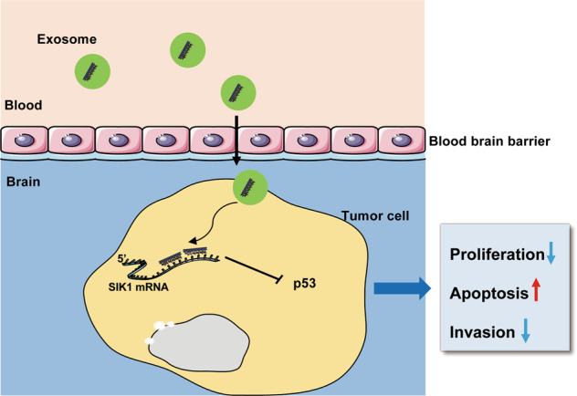
Exosomes are an important carrier for cell communication. miRNAs in exosomes are potential biomarkers and therapeutic targets in different types of cancer. However, the role of exosomal miRNAs in medulloblastoma (MB) patients is largely unknown. In this study, we reported that there was a higher level of miR-130b-3p in exosomes derived from MB patient plasma compared with exosomes from healthy control plasma. Exosomes from MB patient plasma could transfer miR-130b-3p to an MB cell line and played suppressor roles for cell proliferation. miR-130b-3p suppressed MB tumorigenesis by targeting a previously unknown target, serine/threonine-protein kinase 1 (SIK1), through the p53 signaling pathways. In addition, we found an unreported role of SIK1 in promoting MB tumor growth and an SIK1 inhibitor could inhibit MB cell proliferation. This research provides new insight into the molecular mechanism of MB and may provide a new therapeutic strategy for MB treatment.


Conflict of interest statement

The authors declare that they have no conflict of interest.

Figures

Fig. 1
Fig. 1. miR-130b-3p can be transferred to tumor cells through exosomes.
a The exosomes was assessed using transmission electron microscopy. Scale bar, 100 nm. b Western blot analysis of the exosomal marker, CD9 and CD63; Golgi marker, GM130. c Exosomal miR-130b-3p expression in healthy and MB patient plasma detected by RT-qPCR. d The expression of miR-130b-3p in healthy and MB patient PBMCs detected by RT-qPCR. e Exosomes were isolated from MB patient plasma, dyed with PKH67 (green) and cocultured with Daoy cells for 12 h, then dyed with Hoechst33258 (blue) and viewed with confocal microscopy (original magnifications ×200). f The expression of miR-130b-3p in Daoy cells cocultured with the exosomes isolated from MB patient plasma, detected by RT-qPCR. Data represent means ± SEMs from three independent experiments. *p < 0.05, ***p < 0.001.
Fig. 2
Fig. 2. Exosomal miR-130b-3p inhibit MB cell proliferation in vitro.
a, b RT-qPCR assay was used to detect the expression of miR-130b-3p in Daoy and D283 cocultured with the different concentrations of exosomes (10, 20, 50 μg/mL) collected from MB patient plasma. c, d The proliferative ability of Daoy and D283 treated with the different concentrations of exosomes derived from MB patient plasma by CCK-8. e Exosomes were isolated from culture supernatant from THP-1, dyed with PKH67 (green) and cocultured with Daoy, then dyed with Hoechst33258 (blue) and viewed with confocal microscopy (original magnifications ×200). f The expression of miR-130b-3p in Daoy cocultured with the exosomes collected from THP-1 culture supernatant, detected by RT-qPCR. The proliferative ability of Daoy (g) and D283 (h) treated with the exosomes derived from supernatant of THP-1 transfected with miR-130b-3p mimic or NC was tested by CCK-8. Cell proliferation of Daoy (i) and D283 (j) treated with supernatant of THP-1 transfected with miR-130b-3p mimic or NC and GW4869 was assessed by CCK-8. Data are presented as mean ± SEM. *p < 0.05, **p < 0.01.
Fig. 3
Fig. 3. miR-130b-3p functions as a tumor suppressor of MB cells in vitro.
a The proliferative ability of MB cells transfected with miR-130b-3p mimic or NC was tested through CCK-8. b, c Cell apoptosis was detected through flow cytometry in MB cells transfected with miR-130b-3p mimic or negative control. d Cell migration was detected through a wound-healing assay in MB cells transfected with miR-130b-3p or NC (original magnifications ×200). e Cell invasion analysis was performed through transwell invasion assays in MB cells transfected with miR-130b-3p or NC (original magnifications ×200). Data represent means ± SEMs from three independent experiments. *p < 0.05, **p < 0.01.
Fig. 4
Fig. 4. SIK1 is a target of miR-130b-3p.
a Venn diagrams showing the number of potential miR-130b-3p targets. b RT-qPCR analysis of six potential miR-130b-3p target genes in MB cells with miR-130b-3p overexpressed. c The potential binding sites of miR-130b-3p within the SIK1 mRNA 3′-UTR and 3′-mUTR. Luciferase reporter gene assays were used to analyze the effect of miR-130b-3p on luciferase activity. d SIK1 protein levels were detected through western blotting in MB cells transfected with miR-130b-3p mimic. NC negative control. Data represent means ± SEMs from three independent experiments. *p < 0.05, **p < 0.01.
Fig. 5
Fig. 5. miR-130b-3p suppresses MB cell tumorigenesis through SIK1.
a CCK-8 assay in MB cells treated with SIK1 inhibitor. b The efficiency of si-SIK1 knockdown was detected by RT-qPCR. c The proliferative ability of MB cells transfected with si-SIK1 or NC was tested through CCK-8. d Cell apoptosis was detected through flow cytometry in MB cells treated with si-SIK1 or negative control. e Cell migration was detected through wound-healing assay in MB cells transfected with si-SIK1 (original magnifications ×200). f Cell invasion analysis was performed through transwell invasion assay in MB cells transfected with si-SIK1 (original magnifications ×200). Data represent means ± SEMs from three independent experiments. *p < 0.05, **p < 0.01.
Fig. 6
Fig. 6. miR-130b-3p promotes the activity of the p53 signaling pathways through downregulation of SIK1.
a The protein levels of SIK1, p53, BAX and Bcl-2 in Daoy cells were measured through western blotting. be SIK1, p53, BAX and Bcl-2 protein expression were determined by western blotting, respectively, in Daoy cells. Data represent means ± SEMs from three independent experiments. *p < 0.05, **p < 0.01.
Fig. 7
Fig. 7. miR-130b-3p suppresses and SIK1 promotes MB tumorigenesis in vivo.
a Nude mice were injected with subcutaneous xenografts of Daoy cells overexpressing miR-130b-3p or NC and were euthanized after 8 weeks (five mice per group). b Tumor volume was recorded weekly after an injection of Daoy cells transfected with LV16-miR-130b-3p and LV16-NC. c Tumor weight was measured 8 weeks post injection with Daoy cells transfected with LV16-miR-130b-3p and LV16-NC. d, e miR-130b-3p expression was measured in mice tumors by RT-qPCR and FISH. f Mouse tumors were imaged 8 weeks after injection with Daoy cells, and demonstrated decreasing SIK1 expression (eight mice per group). g Tumor volume was recorded weekly after injection with Daoy cells transfected with sh-SIK1 and sh-GFP. h Tumor weight was measured 8 weeks post injection with Daoy cells transfected with sh-SIK1 or sh-GFP. i, j Hematoxylin and eosin (HE) staining (original magnifications ×200) of tumors and SIK1 and Ki67 expression in the tumor tissue samples, as determined through IHC. Data represent means ± SEMs from three independent experiments. *p < 0.05, **p < 0.01.
Fig. 8
Fig. 8. miR-130b-3p promotes the activity of p53 signaling pathways through downregulation of SIK1.
Schematic model of the role of miR-130b-3p in regulating MB tumorigenesis.

References

    1. Ning MS, Perkins SM, Dewees T, Shinohara ET. Evidence of high mortality in long term survivors of childhood medulloblastoma. J. Neuro-Oncol. 2015;122:321–3-27. - PubMed
    1. Crawford JR, MacDonald TJ, Packer RJ. Medulloblastoma in childhood: new biological advances. Lancet Neurol. 2007;6:1073–1085. - PubMed
    1. Gajjar A, et al. Risk-adapted craniospinal radiotherapy followed by high-dose chemotherapy and stem-cell rescue in children with newly diagnosed medulloblastoma (St Jude Medulloblastoma-96): long-term results from a prospective, multicentre trial. Lancet Oncol. 2006;7:813–820. - PubMed
    1. Rutkowski S, et al. Survival and prognostic factors of early childhood medulloblastoma: an international meta-analysis. J. Clin. Oncol. 2010;28:4961–4968. - PubMed
    1. Ellison DW, et al. beta-Catenin status predicts a favorable outcome in childhood medulloblastoma: the United Kingdom Children’s Cancer Study Group Brain Tumour Committee. J. Clin. Oncol. 2005;23:7951–7957. - PubMed

Publication types

MeSH terms