Review
. 2020 May 27;6(5):672-683.
doi: 10.1021/acscentsci.0c00489. Epub 2020 May 4.

Remdesivir: A Review of Its Discovery and Development Leading to Emergency Use Authorization for Treatment of COVID-19

Affiliations
Review

Remdesivir: A Review of Its Discovery and Development Leading to Emergency Use Authorization for Treatment of COVID-19

Richard T Eastman et al. ACS Cent Sci. .

Erratum in

Abstract

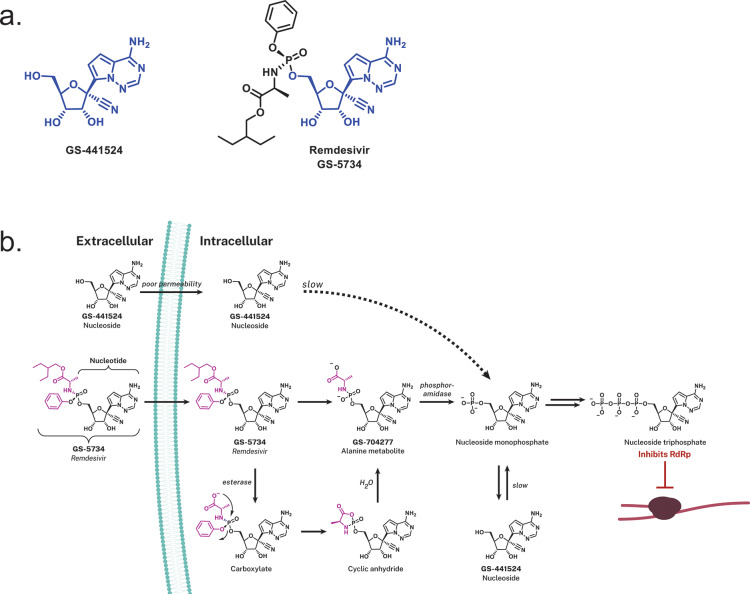
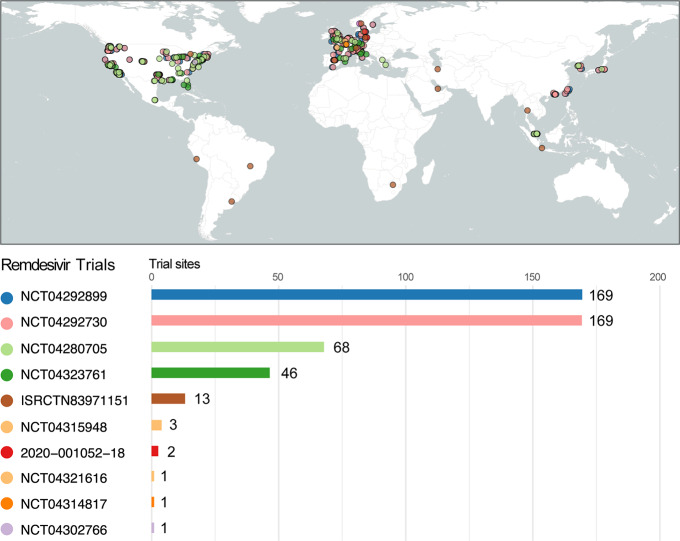
The global pandemic of SARS-CoV-2, the causative viral pathogen of COVID-19, has driven the biomedical community to action-to uncover and develop antiviral interventions. One potential therapeutic approach currently being evaluated in numerous clinical trials is the agent remdesivir, which has endured a long and winding developmental path. Remdesivir is a nucleotide analogue prodrug that perturbs viral replication, originally evaluated in clinical trials to thwart the Ebola outbreak in 2014. Subsequent evaluation by numerous virology laboratories demonstrated the ability of remdesivir to inhibit coronavirus replication, including SARS-CoV-2. Here, we provide an overview of remdesivir's discovery, mechanism of action, and the current studies exploring its clinical effectiveness.


Conflict of interest statement

The authors declare no competing financial interest.

Figures

Figure 1
Figure 1
Life cycle of SARS-CoV-2 in host cells. SARS-CoV-2 primarily infects the respiratory tract (nasal epithelial cells, pneumocytes, and alveolar macrophages) and the gastrointestinal tract (enterocytes). The virus enters though direct interaction between the viral S protein and the cellular receptor angiotensin-converting enzyme 2 (ACE2). Following entry, the viral genome is released and translated into the viral replicase polyproteins PP1a and PP1ab, which are cleaved into functional proteins by viral proteases. Viral genome replication is mediated by the viral replication complex, including the RNA-dependent RNA polymerase (RdRp). Viral nucleocapsids are assembled from the packaged viral genomes and translated viral structural proteins and released through exocytosis. Potential targets and postulated mechanism of action for antiviral interventions are shown: blocking virus/host cell interaction through the use of antibodies/nanobodies (and convalescent plasma therapy) or recombinant ACE2 protein; use of hydroxychloroquine (based on in vitro data) to inhibit endosome maturation; use of protease inhibitors to inhibit viral/endosome membrane fusion or viral polypeptide maturation; nucleoside/nucleotide analogues to inhibit viral genome replication.
Figure 2
Figure 2
SARS-CoV-2 genome and RNA-dependent RNA polymerase structure. (a) Representation of the SARS-CoV-2 RNA genome. As SARS-CoV-2 is a positive-sense RNA virus, the genome serves as a direct template for protein translation. Replication of the viral genome requires a functional viral replication complex, including an RNA-dependent RNA polymerase (RdRp). (b) Domain organization of the SARS-CoV-2 RdRp (encoded by nsp12) domains bound to cofactors nsp7 and dimers of nsp8, that serve as essential cofactors that increase polymerase activity. The rendering was based on the cryo-EM structure at a resolution of 2.9-Å, published by Gao et al, 2020 (PDB: 6M71). The nsp12 RdRp domain is shown in green, nsp7 in purple, nsp8 in cyan, nidovirus RdRp-associated nucleotidyltransferase (NiRAN) domain in yellow, interface in blue, and a newly identified β-hairpin domain is shown in red. Highlighted is RdRp residue S861, which is predicted to sterically interact with the 1′CN substituent of remdesivir inducing delayed chain termination.
Figure 3
Figure 3
Remdesivir and its intracellular conversion. (a) Chemical structures of GS-441524 that compose the nucleoside analogue core (blue) of remdesivir (GS-5734). (b) Intracellular processing of the prodrug remdesivir (GS-5734), the aryloxy phosphoramidate (purple) prodrug of GS-441524 monophosphate. Upon diffusion of remdesivir into the cell, it is metabolized into the nucleoside monophosphate form via a sequence of steps that are presumably initiated by esterase-mediated hydrolysis of the amino acid ester that liberates a carboxylate that cyclizes on to the phosphorus displacing the phenoxide. The unstable cyclic anhydride is hydrolyzed by water to the alanine metabolite GS-704277 whose P–N bond is hydrolyzed by phosphoramidase-type enzymes to liberate the nucleoside monophosphate or nucleotide analog. The artificial nucleoside monophosphate is routed to further phosphorylation events (hijacking the endogenous phosphorylation pathway) yielding the active nucleoside triphosphate analogue form that is utilized by the viral RNA-dependent RNA polymerase (RdRp). Utilization of the GS-441524 nucleoside triphosphate analogue by RdRp inhibits viral replication through inducing delayed chain termination.
Figure 4
Figure 4
Remdesivir global clinical trials. Shown are the locations of the clinical study sites for the ongoing clinical studies of remdesivir for SARS-CoV-2/COVID-19. Number of sites participating for each respective study, if no specific information was given, shown are the countries participating (e.g., ISRCTN83971151). Listed are the number of sites participating for each respective study, if no detailed information was provided; shown are the number of countries participating. NCT04302766 is an expanded access trial with no specific sites listed in the registration. Figure created with R, utilizing the packages rnaturalearth, sf, and ggplot2.

References

    1. Andersen K. G.; Rambaut A.; Lipkin W. I.; Holmes E. C.; Garry R. F. The proximal origin of SARS-CoV-2. Nat. Med. 2020, 26, 450–452. 10.1038/s41591-020-0820-9. - DOI - PMC - PubMed
    1. Fields B. N.; Knipe D. M.; Howley P. M.. Coronavirus. In Fields Virology, 6th ed.; Wolters Kluwer Health/Lippincott Williams and Wilkins, 2013; pp 825–858.
    1. Geller C.; Varbanov M.; Duval R. E. Human coronaviruses: insights into environmental resistance and its influence on the development of new antiseptic strategies. Viruses 2012, 4, 3044–3068. 10.3390/v4113044. - DOI - PMC - PubMed
    1. Song Z. From SARS to MERS, Thrusting Coronaviruses into the Spotlight. Viruses 2019, 11 (1), 59. - PMC - PubMed
    1. Menachery V. D.; Graham R. L.; Baric R. S. Jumping species-a mechanism for coronavirus persistence and survival. Curr. Opin. Virol. 2017, 23, 1–7. 10.1016/j.coviro.2017.01.002. - DOI - PMC - PubMed