. 2020 Oct 15;136(16):1824-1836.
doi: 10.1182/blood.2019004113.

Yap1-Scribble polarization is required for hematopoietic stem cell division and fate

Affiliations

Yap1-Scribble polarization is required for hematopoietic stem cell division and fate

Mark J Althoff et al. Blood. .

Abstract

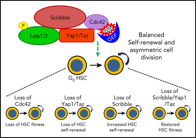
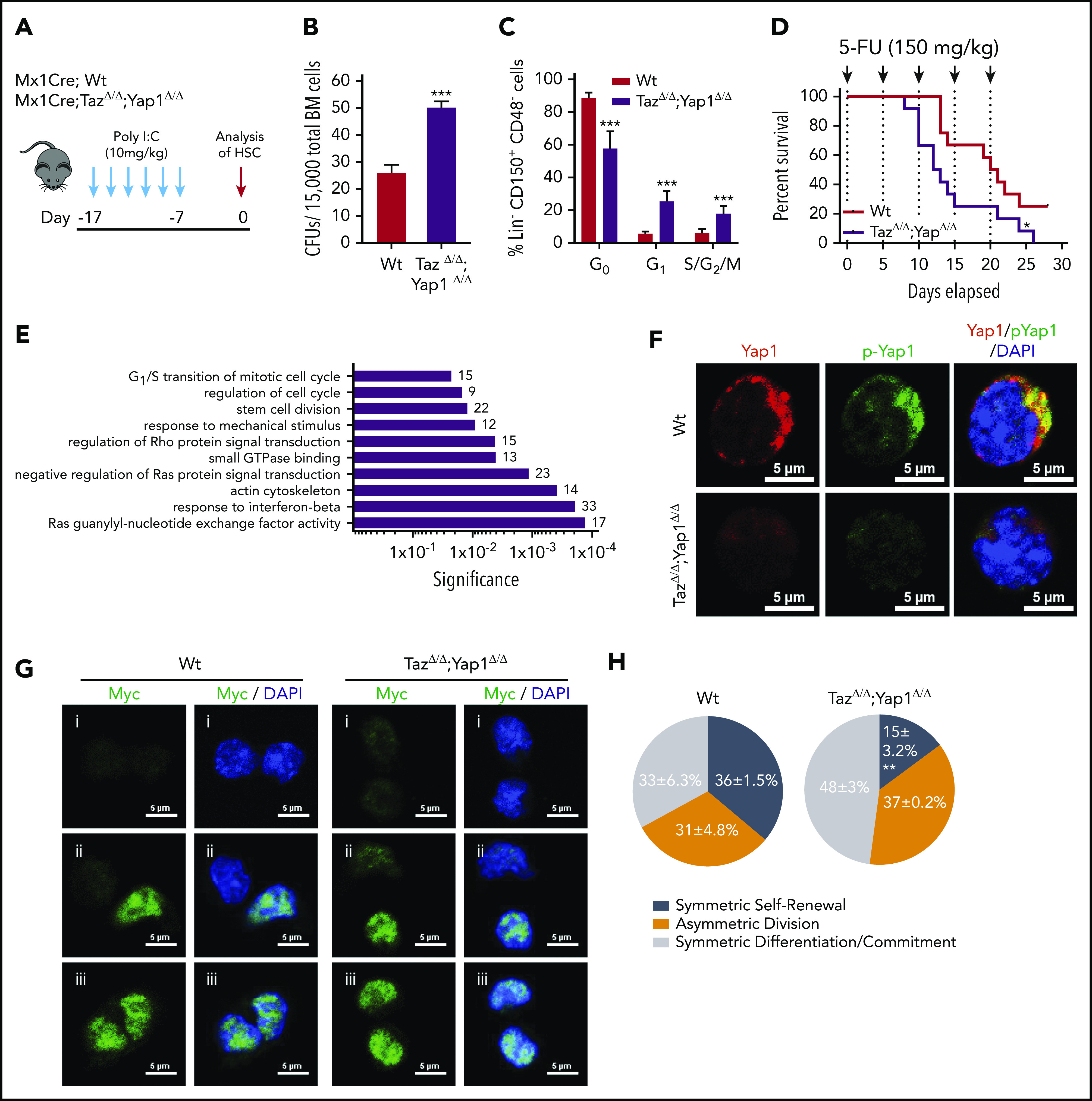
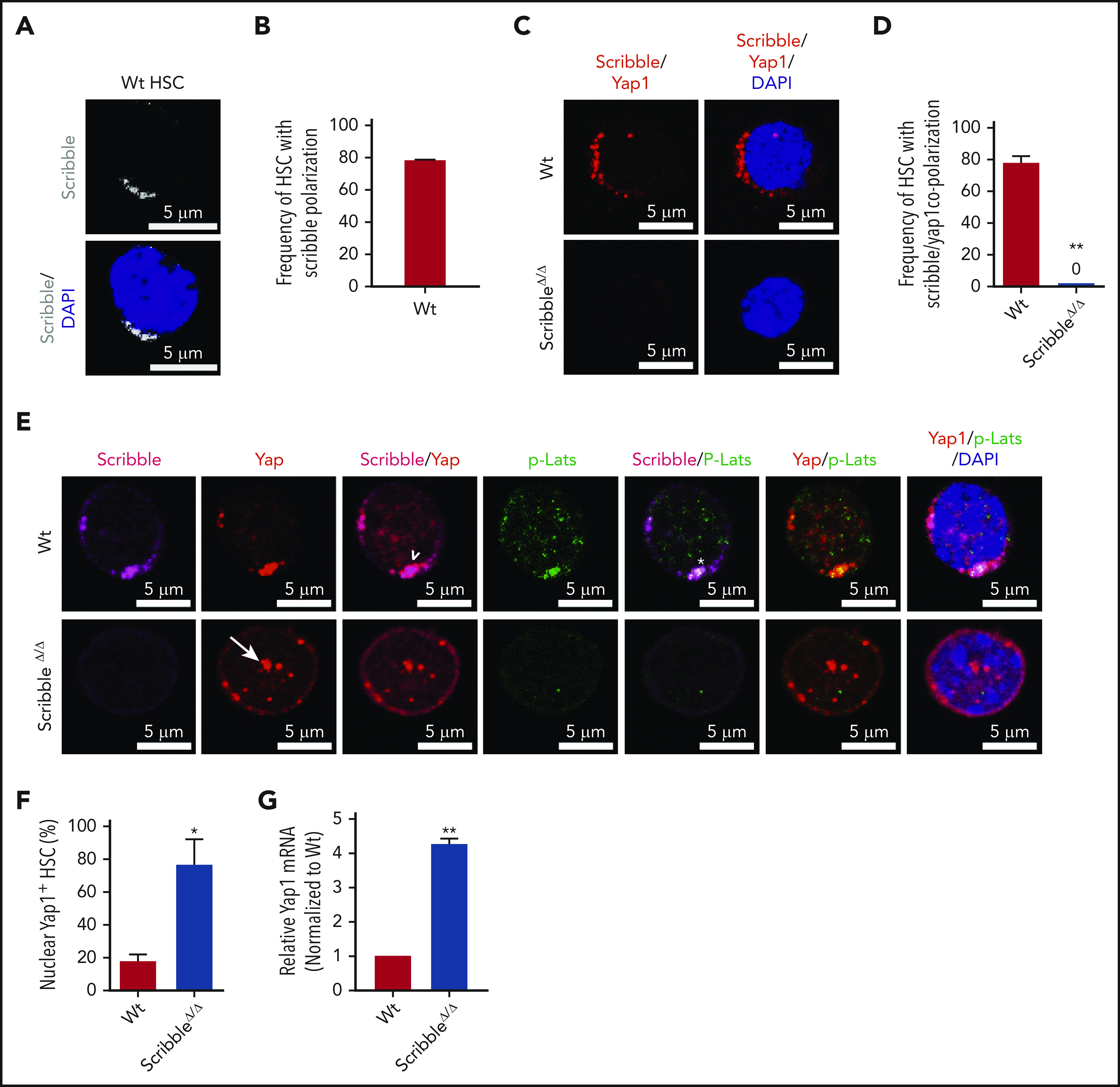
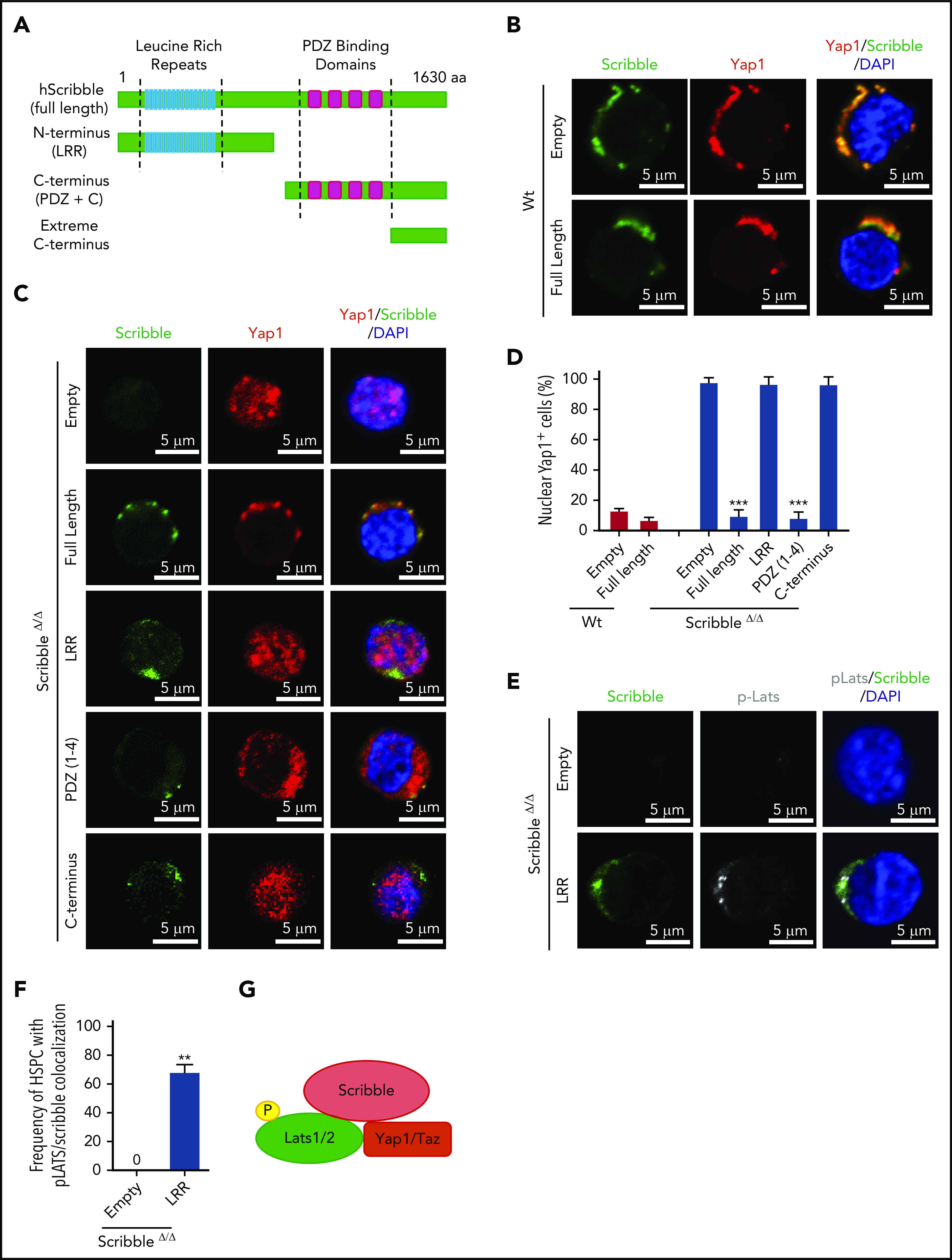
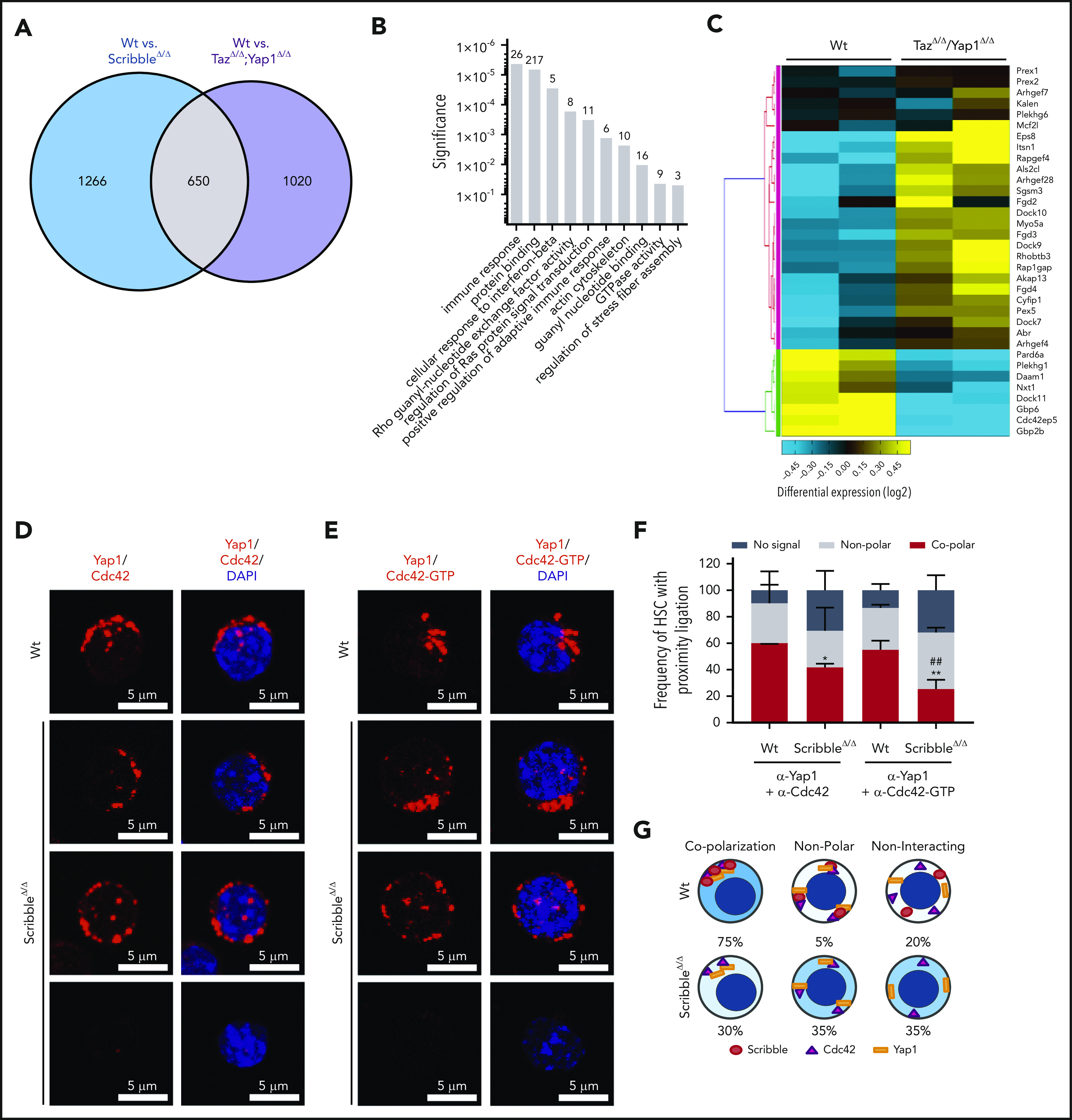
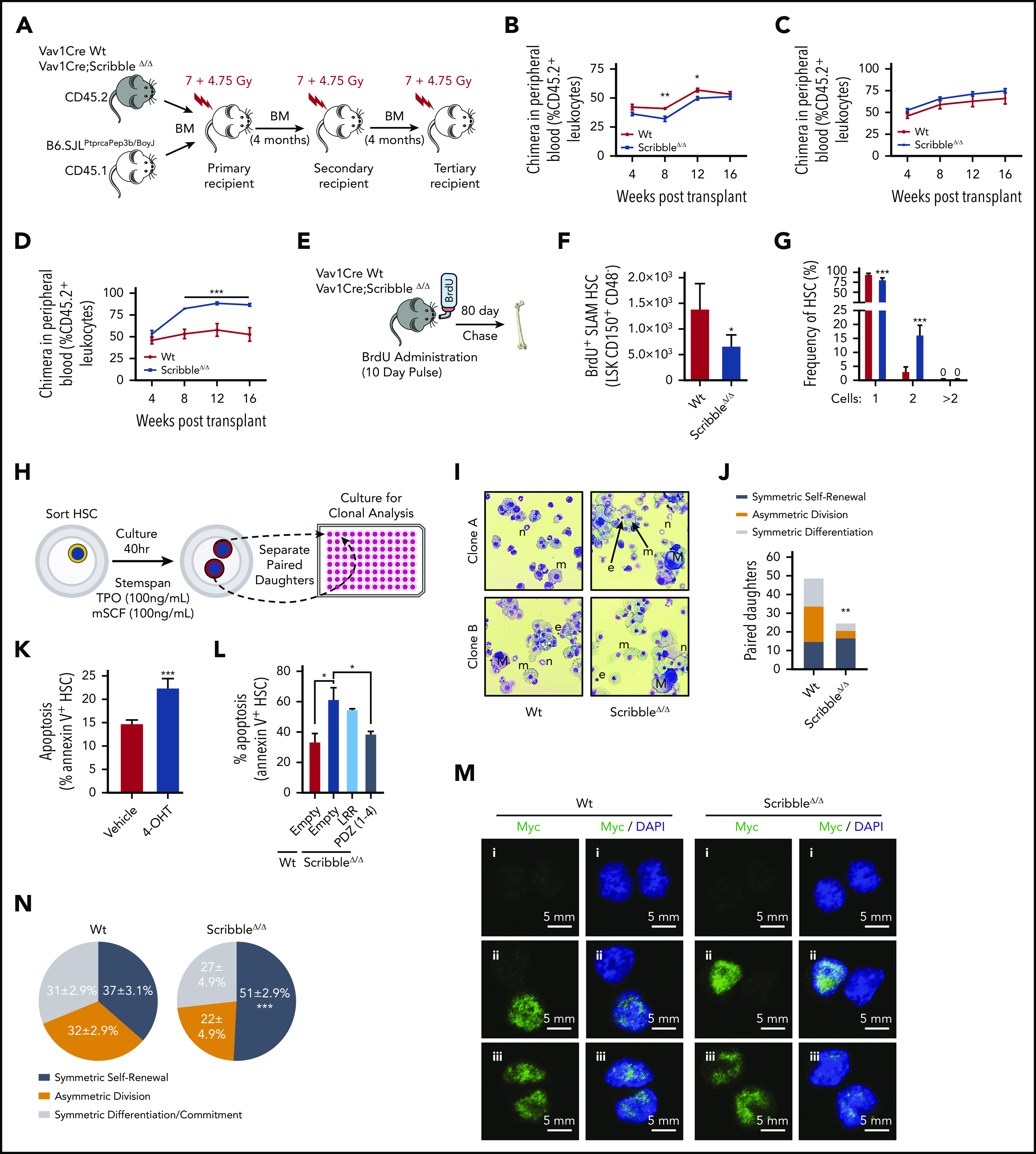
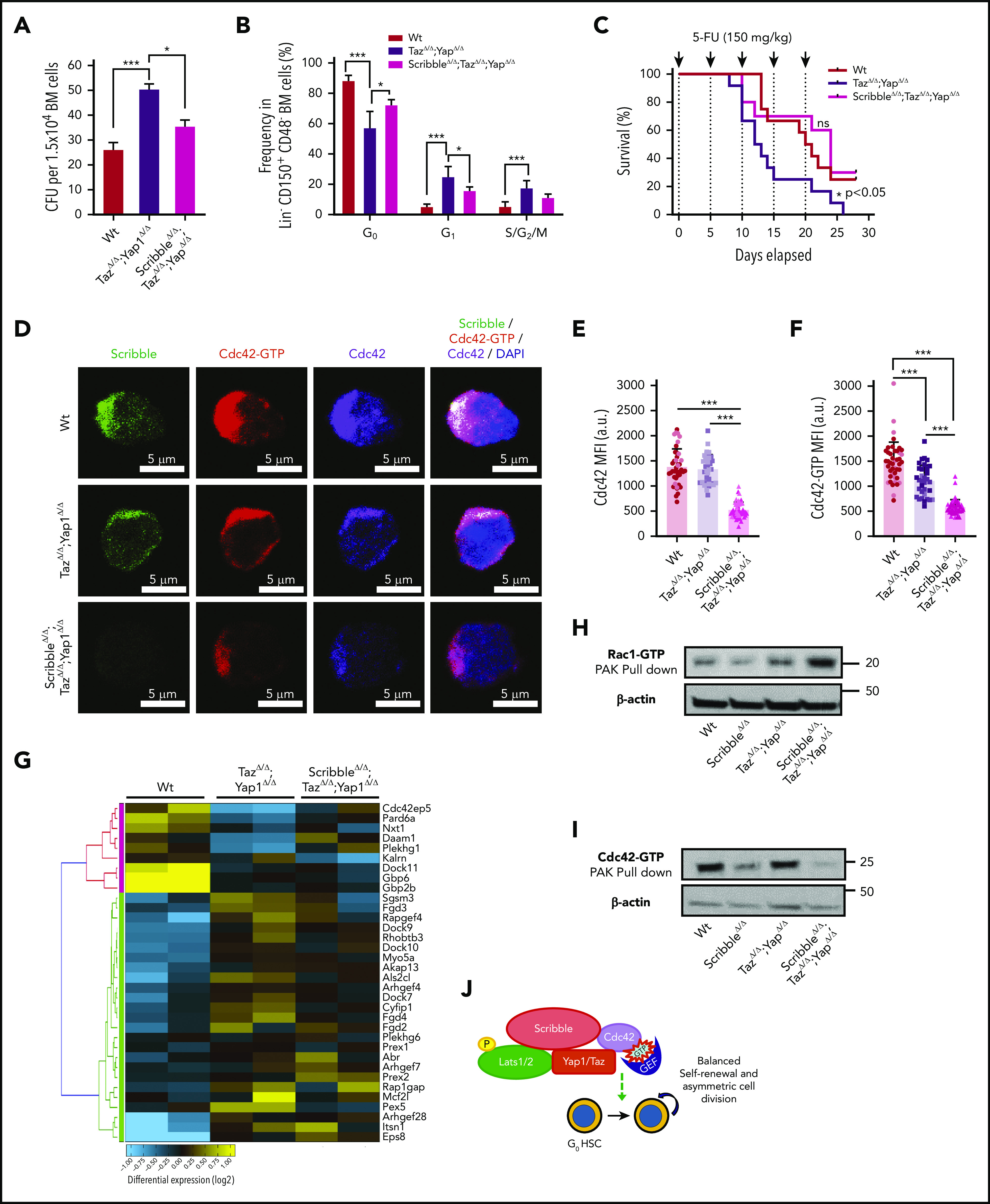
Yap1 and its paralogue Taz largely control epithelial tissue growth. We have identified that hematopoietic stem cell (HSC) fitness response to stress depends on Yap1 and Taz. Deletion of Yap1 and Taz induces a loss of HSC quiescence, symmetric self-renewal ability, and renders HSC more vulnerable to serial myeloablative 5-fluorouracil treatment. This effect depends on the predominant cytosolic polarization of Yap1 through a PDZ domain-mediated interaction with the scaffold Scribble. Scribble and Yap1 coordinate to control cytoplasmic Cdc42 activity and HSC fate determination in vivo. Deletion of Scribble disrupts Yap1 copolarization with Cdc42 and decreases Cdc42 activity, resulting in increased self-renewing HSC with competitive reconstitution advantages. These data suggest that Scribble/Yap1 copolarization is indispensable for Cdc42-dependent activity on HSC asymmetric division and fate. The combined loss of Scribble, Yap1, and Taz results in transcriptional upregulation of Rac-specific guanine nucleotide exchange factors, Rac activation, and HSC fitness restoration. Scribble links Cdc42 and the cytosolic functions of the Hippo signaling cascade in HSC fate determination.


Conflict of interest statement

Conflict-of-interest disclosure: The authors declare no competing financial interests.

Figures

None
Graphical abstract
Figure 1.
Figure 1.
Yap1/Taz are necessary for HSC function. (A) Schematic representing the inducible deletion of Yap1/Taz in the hematopoietic system followed by 1 week of recovery before subjecting mice to experimental testing. (B) Number of colony-forming units (CFUs) from Mx1Cre;Wt and Mx1Cre;TazΔ/Δ;Yap1Δ/Δ total BM cells. (C) Cell-cycle analysis of Lin CD48 CD150+ HSC by fluorescence-activated cell sorting (FACS). (D) Kaplan-Meier survival analysis after serial myeloablation with 5-fluorouracil (150 mg/kg) 5 days apart. (E) Gene ontology (GO) pathway analysis of differentially regulated genes (P < .05) between Mx1Cre;Wt and Mx1Cre;TazΔ/Δ;Yap1Δ/Δ HSC (cutoff, 1.5-fold). Numbers represent the percentage of genes within each GO pathway that are differentially regulated. (F) Immunofluorescence depicting Yap1 protein localization in Wt HSC (immunophenotypically defined as LSK CD150+ CD48) (red pseudo-color) and Yap1 phosphorylation status at Serine 112 (green pseudo-color). Cells are counterstained with 4′,6-diamidino-2-phenylindole (DAPI) and merged images are shown in the right micrographs. Scale bar is 5 µm. (G) Immunofluorescence depicting fate determinant allocation of Myc and the corresponding HSC division mode in Wt and Yap1/TazΔ/Δ paired daughter HSC (nocodazole, 10 nM for 24 hours). Low Myc expression in paired daughter cells represents (i) symmetric self-renewal, (ii) high and low Myc expression between the 2 daughters represents an asymmetric division, whereas (iii) high Myc expression in both cells is indicative of symmetric commitment. (H) Quantification of fate determinant allocation and division mode among Wt and Yap1/TazΔ/Δ paired daughter HSC. *P < .05; **P < .01; ***P < .001.
Figure 2.
Figure 2.
Scribble scaffolds components of the Hippo pathway in HSC and controls Yap1 cytoplasmic localization. (A) Immunofluorescence depicting Scribble protein localization in Wt HSC (immunophenotypically defined as LSK CD150+ CD48) (white areas). Cells are counterstained with DAPI and merged images are shown in the bottom micrographs. Scale bar is 5 µm. (B) Quantification for the frequency of HSC with Scribble polarization. (C) Immunofluorescence depicting a proximity ligation assay (PLA) on Wt and ScribbleΔ/Δ HSC using anti-Scribble and anti-Yap1 primary antibodies subsequently targeted with corresponding probes for oligomerization. The detected dimers are pseudo-colored in red. Nuclei are counterstained with DAPI and merged images are shown in the right micrographs. Scale bar is 5 µm. (D) Quantification of PLA signal. (E) Immunofluorescence showing Scribble polarization and Yap1 colocalization (white arrowheads) in HSC isolated from Wt mice. White asterisks indicate areas of colocalization between Scribble and the activated upstream inhibitory kinase of Yap1, phosphorylated Lats1/2. White arrows denote Yap1 nuclear translocation in ScribbleΔ/Δ HSC. Nuclei are counterstained with DAPI; merged images are shown in the right micrographs. Scale bar is 5 µm. (F) Quantification for the frequency of HSC with Yap1 nuclear foci. (G) Quantitative reverse transcriptase polymerase chain reaction of Yap1 messenger RNA expression from HSC cultured for 40 hours; *P < .05; **P < .01.
Figure 3.
Figure 3.
Cytoplasmic polarization of Yap1 is restored in ScribbleΔ/Δ HSC/P with expression of full-length Scribble or PDZ-containing mutants. (A) Graphical representation of the functional domains in human full-length scribble protein and the truncation mutations incorporated into an Ef1α-IRES-RFP lentivirus. (B) Immunofluorescence showing Scribble polarization and Yap1 colocalization in Lin Sca-1+ c-kit+ (LSK) BM cells isolated from Wt mice and transduced with Ef1α-IRES-RFP (EMPTY) lentivirus or human full-length Scribble as indicated. Nuclei are counterstained with DAPI and merged images are shown in the right micrographs. Scale bar is 5 µm. (C) Immunofluorescence showing Scribble expression and Yap1 localization in LSK BM cells isolated from ScribbleΔ/Δ mice and transduced with EMPTY lentivirus, human full-length Scribble, or structure-function mutants as indicated. Nuclei are counterstained with DAPI and merged images are shown in the right micrographs. Scale bar is 5 µm. (D) Quantifications for the frequency of transduced LSK cells with nuclear Yap1 accumulation. (E) Immunofluorescence showing Scribble and pLats1 expression in LSK BM cells isolated from ScribbleΔ/Δ mice and transduced with EMPTY lentivirus or the LRR mutant. Nuclei are counterstained with DAPI and merged images are shown in the right micrographs. Scale bar is 5 µm. (F) Quantification of pLats1 colocalization with Scribble when the N-terminal portion of Scribble (LRR) is reintroduced into Scribble null LSK cells. (G) Cartoon depicting the ternary complex between Scribble, activated Last1/2, and Yap1 in the cytosol of Wt HSC. **P < .01; ***P < .001.
Figure 4.
Figure 4.
Scribble scaffolds Yap1 and Cdc42 in the cytoplasm of HSC. (A) Venn diagram highlighting the number of differentially regulated genes in common when comparing Mx1Cre;Wt and Mx1Cre;ScribbleΔ/Δ and comparing Mx1Cre;Wt and Mx1Cre;TazΔ/Δ;Yap1Δ/Δ HSC. (B) Gene ontology (GO) pathway analysis of the common differentially regulated genes between 2 independent analysis of Mx1Cre;Wt to Mx1Cre;ScribbleΔ/Δ HSC and of Mx1Cre;Wt to Mx1Cre;TazΔ/Δ;Yap1Δ/Δ HSC. Numbers represent the number of genes within each GO pathway that are differentially regulated. (C) Heat map depicting the differential regulation of common genes between Mx1Cre; ScribbleΔ/Δ and Mx1Cre;TazΔ/Δ;Yap1Δ/Δ HSC that pertain to the Rho guanyl nucleotide exchange factor activity and small GTPase activity gene ontology pathways. Proximity ligation assay (PLA) detection of endogenous (D) Yap1/Cdc42 and (E) Yap1/Cdc42-GTP interactions in HSC. The detected dimers are pseudo-colored in red. Nuclei are counterstained with DAPI and merged images are shown in the right micrographs. Scale bar is 5 µm. (F) Frequency of HSC in which PLA signal depicted in panels D and E was found in relation or not with polarization. *P < .05 and **P < .01 between Vav1Cre;Wt and Vav1Cre;ScribbleΔ/Δ; ##P < .01 between Cdc42 and Cdc42-GTP frequencies. (G) Cartoon representing the overall polarization status between Scribble, Yap1, and Cdc42 in Wt cells emphasizing the loss of copolarization and asymmetry in ScribbleΔ/Δ HSC.
Figure 5.
Figure 5.
ScribbleΔ/Δ hematopoietic reconstitution develops a competitive advantage when serially transplanted by maintaining self-renewal divisions. (A) Schematic representing a serial competitive repopulation assay (CRA). Equal amounts of BM from CD45.2+ Vav1Cre Wt and Scribble null mice mixed with congenic CD45.1+ B6.SJLPtprcaPep3b/BoyJ competitor cells were transplanted at a 1:1 ratio into lethally irradiated B6.SJLPtprcaPep3b/BoyJ recipients. (B-D) PB chimera (CD45.2+ leukocytes) of (B) primary, (C) secondary, and (D) tertiary recipients. (E) Schematic representing a long-term BrdU incorporation assay in which animals freely imbibed water containing BrdU (1 mg/mL) for 10 days. Animals were euthanized after 80 days post-BrdU administration to quantify quiescence within hematopoietic stem and progenitor (HSC/P) populations determined by BrdU retention (BD Pharmingen intracellular staining kit: anti-BrdU, Alexa 488). (F) Absolute BrdU retaining (BrdU+) HSC assessed by FACS analysis of BM from mice as described in panel E. (G) Division of sorted and individually deposited HSC depicting the averages and standard deviations of the relative number (%) of wells containing the indicated number of cells after 20 hours in culture. Four independent experiments, n > 450 HSC. (H) Schematic of an in vitro paired daughter cell assay to assess fate decisions among individually sorted HSC. (I) Representative cytospin images of paired daughters. m, macrophage; n, neutrophil; e, erythrocyte; and M, megakaryocyte. (J) Absolute number of paired daughter cells analyzed for division modality, assessed as the presence or absence of full multilineage differentiation potential among individual paired daughter clones. n of paired daughter separations >200. Chi-squared analysis. (K) Cell death analysis using Annexin V and 7-AAD staining on Rosa26Cre;Scribblefl/fl HSC after 40 hours of culture with 4-OH tamoxifen. (L) Cell death analysis using Annexin V staining in Lin Sca-1+ c-kit+ BM cells isolated from Vav1Cre;Wt or Vav1Cre;ScribbleΔ/Δ mice transduced with EMPTY (Ef1α-IRES-RFP) lentivirus or Scribble structure-function mutants as indicated. (M) Immunofluorescence depicting fate determinant allocation of Myc and the corresponding HSC division mode in Wt and ScribbleΔ/Δ paired daughter HSC (nocodazole, 10 nM for 24 hours). Low Myc expression in (i) paired daughter cells represents symmetric self-renewal, (ii) high and low Myc expression between the 2 daughters represents an asymmetric division, whereas (iii) high Myc expression in both cells is indicative of symmetric commitment. (N) Quantification of fate determinant allocation and division mode among Wt and ScribbleΔ/Δ paired daughter HSC. *P < .05, **P < .01, and ***P < .001.
Figure 6.
Figure 6.
Deficiency of Scribble restores fitness of Yap/TazΔ/Δ HSC and associates with Rac activation. (A) Number of colony-forming units (CFU) from BM cells. (B) Cell-cycle analysis of Lin CD48 CD150+ HSC harvested from the BM of Mx1Cre Wt, TazΔ/Δ;Yap1Δ/Δ, and ScribbleΔ/Δ;TazΔ/Δ;Yap1Δ/Δ mice 1 week after poly I:C. Stages of cell cycle were assessed by FACS analysis with incorporation of DNA binding Hoechst 33342 (2 mg/mL) and nucleotide binding PyroninY (0.25 mg/mL). (C) Kaplan-Meier survival analysis after serial myeloablation with 5-fluorouracil (150 mg/kg) 5 days apart. (D) Immunofluorescence of HSC showing Cdc42 expression and localization along with active Cdc42-GTP expression and localization. Nuclei are counterstained with DAPI and merged images are shown in the right micrographs. Scale bar 5 µm. (E) Quantification of Cdc42 expression measured by MFI. (F) Quantification of Cdc42-GTP expression measured by MFI. (G) Heat map depicting genes within the Rho guanyl nucleotide exchange factor activity and small GTPase activity gene ontology pathways showing differential regulation between Mx1Cre Wt, TazΔ/Δ;Yap1Δ/Δ, and ScribbleΔ/Δ;TazΔ/Δ;Yap1Δ/Δ HSC. (H) Rac1/Cdc42 activation PAK pulldown on lineage-depleted (Lin) BM cells. (I) Cdc42 effector PAK pull-down from on Lin BM cells. (J) Cartoon depicting a complete picture of the Scribble polarity complex and its components in relation with HSC self-renewal and cell-cycle entry. *P < .05 and ***P < .001.

References

    1. Florian MC, Dörr K, Niebel A, et al. . Cdc42 activity regulates hematopoietic stem cell aging and rejuvenation. Cell Stem Cell. 2012;10(5):520-530. - PMC - PubMed
    1. Florian MC, Nattamai KJ, Dörr K, et al. . A canonical to non-canonical Wnt signalling switch in haematopoietic stem-cell ageing. Nature. 2013;503(7476):392-396. - PMC - PubMed
    1. Wang L, Yang L, Debidda M, Witte D, Zheng Y. Cdc42 GTPase-activating protein deficiency promotes genomic instability and premature aging-like phenotypes. Proc Natl Acad Sci USA. 2007;104(4):1248-1253. - PMC - PubMed
    1. Yang L, Wang L, Geiger H, Cancelas JA, Mo J, Zheng Y. Rho GTPase Cdc42 coordinates hematopoietic stem cell quiescence and niche interaction in the bone marrow. Proc Natl Acad Sci USA. 2007;104(12):5091-5096. - PMC - PubMed
    1. Wu M, Kwon HY, Rattis F, et al. . Imaging hematopoietic precursor division in real time. Cell Stem Cell. 2007;1(5):541-554. - PMC - PubMed

Publication types

MeSH terms