Case Reports
. 2020 Nov;297(2):E274-E275.
doi: 10.1148/radiol.2020202197. Epub 2020 Jun 2.

Focal Cerebral Arteriopathy in a Pediatric Patient with COVID-19

Affiliations
Case Reports

Focal Cerebral Arteriopathy in a Pediatric Patient with COVID-19

Seyed Mohammad Mousavi Mirzaee et al. Radiology. 2020 Nov.

Abstract

We present a case of focal cerebral arteriopathy and ischemic stroke in a pediatric patient with coronavirus disease 2019 who presented with seizure, right hemiparesis, and dysarthria with positive findings for severe acute respiratory syndrome coronavirus 2 from nasopharyngeal swab and cerebral spinal fluid.


Figures
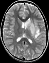
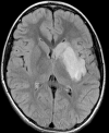
(a–c) Axial T2-weighted (a) and fluid-attenuated inversion recovery (b) MRI scans show diffuse hyperintense signal and edema of caudate nucleus head, putamen, anterior limb of internal capsule, and parts of external capsule and insula on left side, with corresponding low values on axial apparent diffusion coefficient map (c), in keeping with an acute infarction. (d) Maximal intensity projection reformatted image from time-of-flight MR angiography shows focal irregular narrowing and banding of proximal left M1 segment of middle cerebral artery with slightly reduced distal flow in middle cerebral artery (arrows).

References

    1. Li Z, Liu T, Yang N, et al. Neurological manifestations of patients with COVID-19: potential routes of SARS-CoV-2 neuroinvasion from the periphery to the brain. Front Med 2020. 10.1007/s11684-020-0786-5. Published online May 4, 2020. - PMC - PubMed
    1. Wintermark M, Hills NK, DeVeber GA, et al. Clinical and Imaging Characteristics of Arteriopathy Subtypes in Children with Arterial Ischemic Stroke: Results of the VIPS Study. AJNR Am J Neuroradiol 2017;38(11):2172–2179. - PMC - PubMed
    1. Mahase E. Covid-19: concerns grow over inflammatory syndrome emerging in children. BMJ 2020;369:m1710. - PubMed
    1. Castelnovo L, Capelli F, Tamburello A, Faggioli PM, Mazzone A. Symmetric cutaneous vasculitis in COVID-19 pneumonia. J Eur Acad Dermatol Venereol 2020. 10.1111/jdv.16589. Published online May 7, 2020. - PMC - PubMed
    1. Viner RM, Whittaker E. Kawasaki-like disease: emerging complication during the COVID-19 pandemic. Lancet 2020;395(10239):1741–1743. - PMC - PubMed

Publication types