. 2020 Jul 9;48(12):6715-6725.
doi: 10.1093/nar/gkaa473.

Nick-seq for single-nucleotide resolution genomic maps of DNA modifications and damage

Affiliations

Nick-seq for single-nucleotide resolution genomic maps of DNA modifications and damage

Bo Cao et al. Nucleic Acids Res. .

Abstract

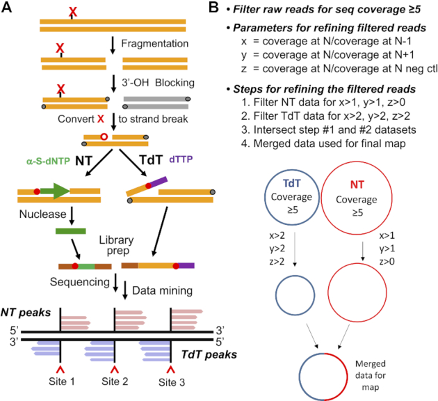
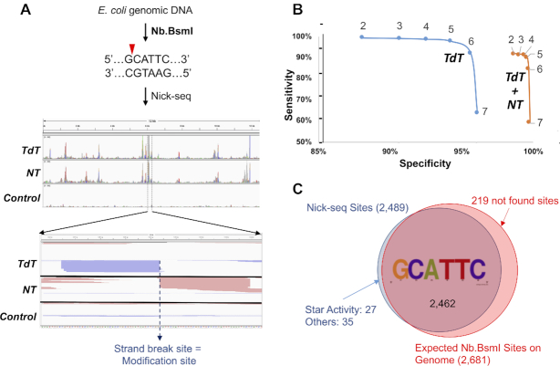
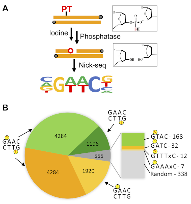
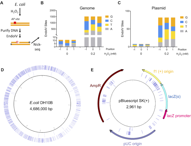
DNA damage and epigenetic marks are well established to have profound influences on genome stability and cell phenotype, yet there are few technologies to obtain high-resolution genomic maps of the many types of chemical modifications of DNA. Here we present Nick-seq for quantitative, sensitive, and accurate mapping of DNA modifications at single-nucleotide resolution across genomes. Pre-existing breaks are first blocked and DNA modifications are then converted enzymatically or chemically to strand-breaks for both 3'-extension by nick-translation to produce nuclease-resistant oligonucleotides and 3'-terminal transferase tailing. Following library preparation and next generation sequencing, the complementary datasets are mined with a custom workflow to increase sensitivity, specificity and accuracy of the map. The utility of Nick-seq is demonstrated with genomic maps of site-specific endonuclease strand-breaks in purified DNA from Eschericia coli, phosphorothioate epigenetics in Salmonella enterica Cerro 87, and oxidation-induced abasic sites in DNA from E. coli treated with a sublethal dose of hydrogen peroxide. Nick-seq applicability is demonstrated with strategies for >25 types of DNA modification and damage.


Figures

Figure 1.
Figure 1.
Overview of Nick-seq and data analysis workflow. (A) Nick-seq library preparation. Briefly, genomic DNA is first subjected to sequencing-compatible fragmentation; the resulting 3′-OH ends are blocked with dideoxyNTPs; the DNA modification is converted to a strand-break by enzymatic or chemical treatment; capture of the 3′- and 5′-ends of resulting strand-breaks using two complementary strategies: one portion of DNA is subjected to nick translation (NT) with α-thio-dNTPs to generate phosphorothioate (PT)-containing oligonucleotides that are resistant to subsequent hydrolysis of the bulk of the genomic DNA by exonuclease III and RecJf. The purified PT-protected fragments are used to generate an NGS library with the modification of interest positioned at the 5′-end of the PT-labeled fragment. A second portion of the same DNA sample is used for terminal transferase (TdT)-dependent poly(dT) tailing of the 3′-end of the strand-break, with the tail used to create a sequencing library by reverse transcriptase template switching (9). Subsequent NGS positions the modification of interest 5′-end of the poly(dT) tail. (B) Processing of the Nick-seq data includes: raw NGS reads are aligned to the reference genome for read coverage calculation; the genome sites with reads coverage ≥5 are then filtered for nick site calling with three parameters: x = the read coverage at position N/coverage at N – 1; y = coverage at position N/coverage at N + 1; z = coverage at position N/coverage at N of negative control sample. The site N is defined as a nick site if its x > 1, y > 1, z > 1 for NT reads and x > 2, y > 2, z > 2 for TdT reads.
Figure 2.
Figure 2.
Nick-seq validation. (A) Mapping single-strand breaks produced by Nb.BsmI in E. coli genomic DNA. Middle panel: Representative view of sequencing reads distributed in one genomic region. Red and green peaks mark reads mapped to forward and reverse strands of the genome, respectively. Lower panel: Amplification of the genomic region surrounding one peak, with read pile ups for TdT and NT sequencing converging on the site of the strand-break. (B) Nb.BsmI mapping data were used to define data processing parameters for accuracy and sensitivity of Nick-seq. Coverage ratios (the ratio of the peaks relative to corresponding sites in an untreated DNA control) were calculated for sequencing data performed with TdT alone (blue line) or the combination of TdT and NT (orange line). The sensitivity and specificity for detection of site-specific strand-breaks was then plotted for ratios ranging from 2 to 7. In general, higher coverage ratios yield greater accuracy but lower sensitivity, and the combination of TdT and NT provided significantly greater specificity. (C) With a coverage ratio of 2, Nick-seq identified 2462 (97.5%) of the 2681 predicted Nb.BsmI sites. Among the 62 (2.5%) ‘false-positive’ sites, 27 (1%) of them occurred in sequences differing from the consensus by one nucleotide. These sites showed lower average sequencing coverage (75 versus 1318) and likely represent Nb.BsmI ‘star’ activity.
Figure 3.
Figure 3.
Mapping PTs across the S. enterica genome by Nick-seq. (A) Schematic showing the iodine cleavage method for converting PTs to strand-breaks. (B) the number of PT modification sites identified by Nick-seq in different sequence motifs. A total of 11 684 breaks were detected at the expected GAAC/GTTC motif, with single- and double-strand PTs denoted by a red ‘S’. The blow up shows minor modification motifs.
Figure 4.
Figure 4.
Application of Nick-seq to quantify abasic sites. (A) generated by H2O2 exposure in E. coli. Cells were treated with a non-lethal dose of H2O2 (0.2 mM) and AP sites in isolated DNA were converted to strand-breaks with EndoIV, followed by Nick-seq mapping. (B) Detection of H2O2-induced EndoIV- sensitive DNA damage sites in E. coli. genomic DNA by Nick-seq. Data for AP sites in the E. coli genome are detailed in Supplementary Table S5. (C) Detection of H2O2-induced EndoIV- sensitive DNA damage sites in a plasmid maintained in this strain of E. coli. Data for AP sites in the E. coli genome are detailed in Supplementary Table S6. (D) The circos plot shows the locations of EndoIV-sensitive sites in E.coli genomic DNA. Outward from the center, circles represent: 0 and 0.2 mM H2O2 induced EndoIV-specific DNA damage sites. (E) The distribution of EndoIV-sensitive sites in the plasmid. Outward from the center, circles represent: 0 and 0.2 mM H2O2 induced EndoIV-specific DNA damage sites.

References

    1. Roos W.P., Thomas A.D., Kaina B.. DNA damage and the balance between survival and death in cancer biology. Nat. Rev. Cancer. 2016; 16:20–33. - PubMed
    1. Chen Y., Hong T., Wang S., Mo J., Tian T., Zhou X.. Epigenetic modification of nucleic acids: from basic studies to medical applications. Chem. Soc. Rev. 2017; 46:2844–2872. - PubMed
    1. Li Q., Hermanson P.J., Springer N.M.. Detection of DNA methylation by Whole-Genome bisulfite sequencing. Methods Mol. Biol. 2018; 1676:185–196. - PubMed
    1. Clark T.A., Spittle K.E., Turner S.W., Korlach J.. Direct detection and sequencing of damaged DNA bases. Genome Integr. 2011; 2:10. - PMC - PubMed
    1. Schibel A.E., An N., Jin Q., Fleming A.M., Burrows C.J., White H.S.. Nanopore detection of 8-oxo-7,8-dihydro-2′-deoxyguanosine in immobilized single-stranded DNA via adduct formation to the DNA damage site. J. Am. Chem. Soc. 2010; 132:17992–17995. - PMC - PubMed

Publication types

MeSH terms