. 2020 Jul 9;5(13):e139388.
doi: 10.1172/jci.insight.139388.

Neutrophil extracellular traps mediate articular cartilage damage and enhance cartilage component immunogenicity in rheumatoid arthritis

Affiliations

Neutrophil extracellular traps mediate articular cartilage damage and enhance cartilage component immunogenicity in rheumatoid arthritis

Carmelo Carmona-Rivera et al. JCI Insight. .

Abstract

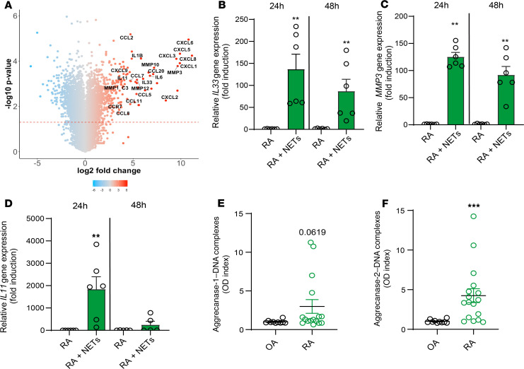
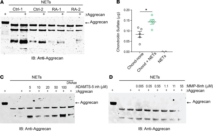
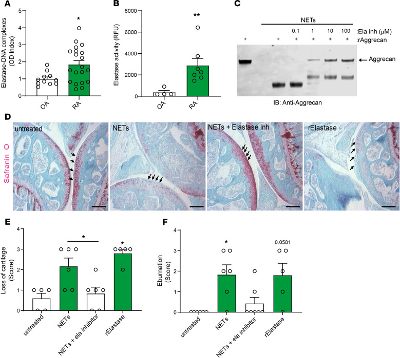
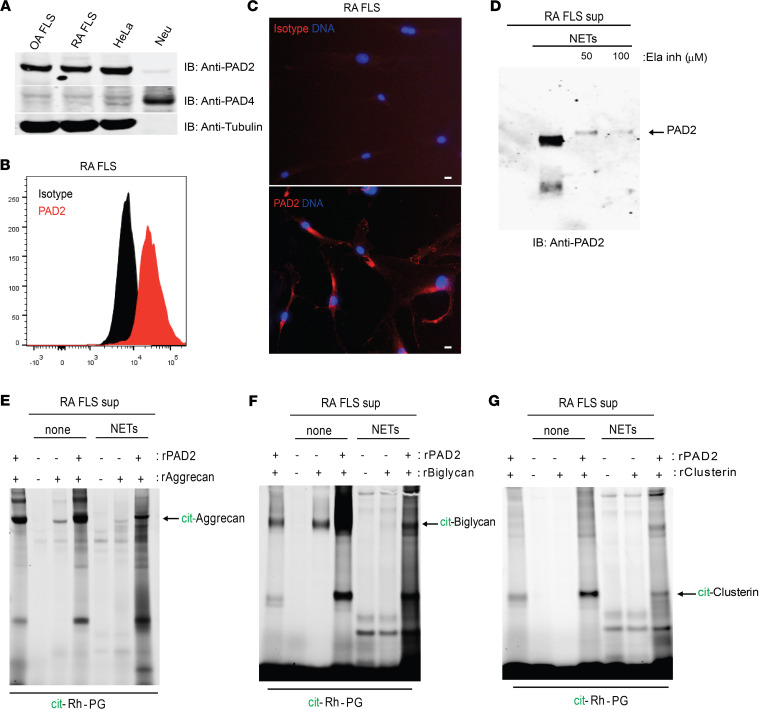
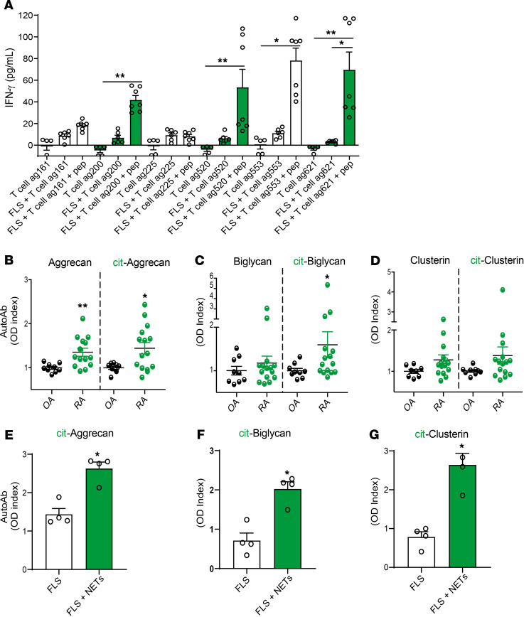
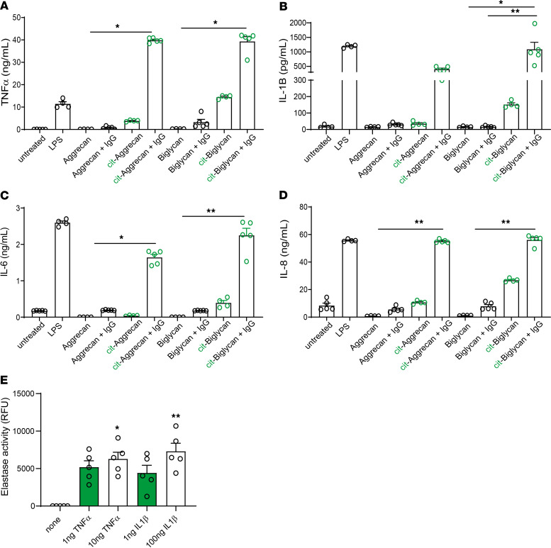
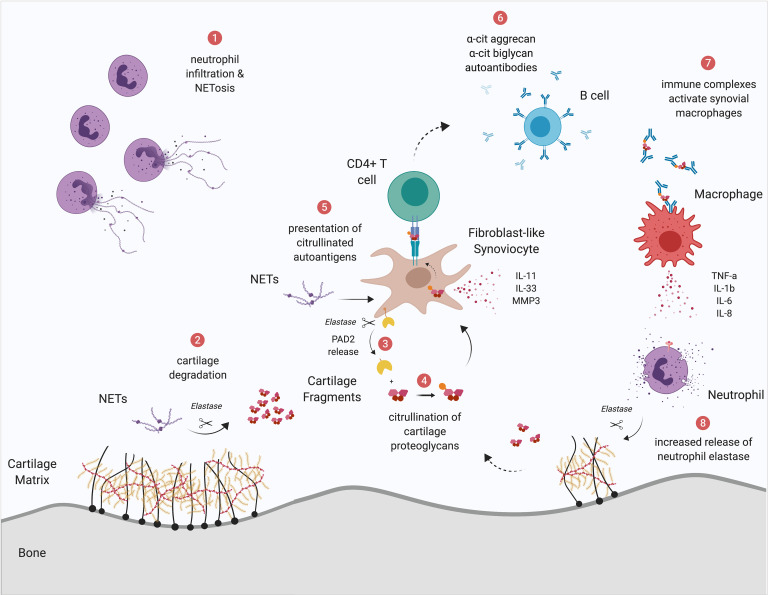
Rheumatoid arthritis (RA) is characterized by synovial joint inflammation, cartilage damage, and dysregulation of the adaptive immune system. While neutrophil extracellular traps (NETs) have been proposed to play a role in the generation of modified autoantigens and in the activation of synovial fibroblasts, it remains unknown whether NETs are directly involved in cartilage damage. Here, we report a new mechanism by which NET-derived elastase disrupts cartilage matrix and induces release of membrane-bound peptidylarginine deiminase-2 by fibroblast-like synoviocytes (FLSs). Cartilage fragments are subsequently citrullinated, internalized by FLSs, and then presented to antigen-specific CD4+ T cells. Furthermore, immune complexes containing citrullinated cartilage components can activate macrophages to release proinflammatory cytokines. HLA-DRB1*04:01 transgenic mice immunized with NETs develop autoantibodies against citrullinated cartilage proteins and display enhanced cartilage damage. Inhibition of NET-derived elastase rescues NET-mediated cartilage damage. These results show that NETs and neutrophil elastase externalized in these structures play fundamental pathogenic roles in promoting cartilage damage and synovial inflammation. Strategies targeting neutrophil elastase and NETs could have a therapeutic role in RA and in other inflammatory diseases associated with inflammatory joint damage.

Keywords: Autoimmunity; Cartilage; Innate immunity; Neutrophils.


Conflict of interest statement

Conflict of interest: The authors have declared that no conflict of interest exists.

Figures

Figure 1
Figure 1. NETs promote a proinflammatory gene signature in FLSs.
(A) Volcano plot of differential gene expression comparing NET-treated and untreated RA FLSs. Genes colored in red are upregulated in NET-treated FLSs, while genes colored in blue are downregulated. Genes involved in inflammation and extracellular remodeling are annotated. Quantitative PCR was performed to support differential gene expression of (B) IL33, (C) MMP3, and (D) IL11 and in RA FLSs (n = 6) in the presence or absence of added NETs for 24 hours and 48 hours. Results are the mean ± SEM. Mann-Whitney U test was used. **P < 0.01. (E) Aggrecanase-1– and (F) aggrecanase-2–dsDNA complexes were measured in synovial fluids (SFs) from osteoarthritis (OA) (n= 10) and RA (n = 17) patients. Results are the mean ± SEM. Mann-Whitney U test was used. ***P < 0.001.
Figure 2
Figure 2. NETs degrade aggrecan.
(A) Western blot analysis to assess NET-mediated degradation of recombinant aggrecan. (B) Quantification of supernatant release of chondroitin sulfate by chondrocytes incubated in the presence or absence of NETs for 24 hours. Results are the mean ± SEM of 5 independent experiments. Mann-Whitney U test was used. *P < 0.05. (C) Western blot analysis to quantify NET-mediated degradation of recombinant aggrecan in the presence of increasing concentrations of ADAMTS-5 inhibitor or DNase I. (D) Western blot analysis to quantify NET-mediated degradation of recombinant aggrecan in the presence of graded concentrations of MMP8 inhibitor.
Figure 3
Figure 3. NET-bound neutrophil elastase mediates cartilage degradation.
(A) Neutrophil elastase–dsDNA complexes and (B) neutrophil elastase activity were measured in OA (n = 10) and RA (n = 17) SF. Results are the mean ± SEM. Mann-Whitney U test was used. (C) Western blot analysis quantifies NET-mediated degradation of recombinant aggrecan in the presence or absence of graded concentrations of neutrophil elastase inhibitor. Results are representative of 3 independent experiments. (D) Safranin-O staining and (E) scores of cartilage loss and (F) eburnation in tibiofemoral mouse explants incubated in the presence or absence of NETs, NETs with neutrophil elastase inhibitor, or recombinant neutrophil elastase. Arrows show cartilage areas. Results are the mean ± SEM of n= 5 per group. Kruskal-Wallis with post hoc Dunn’s test was used. *P < 0.05, and **P < 0.01. Scale bar: 100 μm.
Figure 4
Figure 4. NET-bound neutrophil elastase promotes the release of PAD2 by FLSs and citrullination of extracellular matrix proteins.
(A) Western blot analysis quantifying PAD2 and PAD4 expression in OA and RA FLSs. HeLa cells and neutrophil extracts were used as positive controls for PAD2 and PAD4, respectively. Tubulin was used as a loading control. (B) PAD2 expression in RA FLSs by flow cytometry analysis. (C) Representative immunofluorescence of PAD2 expression in the membrane of RA FLSs. (D) Detection of PAD2 in the supernatants of RA FLSs incubated with NETs in the presence or absence of neutrophil elastase inhibitor. PAD2 activity was measured by detecting citrullinated (E) aggrecan, (F) biglycan, and (G) clusterin using Rh-PG citrulline probe in RA supernatants in the presence or absence of NETs. Recombinant PAD2 was used as a control. Results are representative of 3 independent experiments. Scale bar: 10 μm.
Figure 5
Figure 5. FLSs exposed to NETs present citrullinated cartilage proteins to CD4+ T cells and promote in vitro and in vivo adaptive immune responses to citrullinated cartilage peptides in humans and mice.
(A) IFN-γ was measured in supernatants of RA FLSs cocultured for 5 days with cit-aggrecan–specific CD4+ T cells in the presence or absence of cit-aggrecan peptides. Results are the mean ± SEM of 5 independent experiments. Kruskal-Wallis with post hoc Dunn’s test was used. Autoantibodies against native and citrullinated forms of (B) aggrecan, (C) biglycan, and (D) clusterin were measured in OA (n = 9) and RA (n = 15) SF. Results are the mean ± SEM. Mann-Whitney U test was used. Sera from HLA-DRB1*04:01 transgenic mice immunized with FLSs or FLSs in the presence of NETs were analyzed for the presence of autoantibodies against citrullinated versions of (E) aggrecan, (F) biglycan, and (G) clusterin. Results are the mean ± SEM of n = 5 per group. Mann-Whitney U test was used. *P < 0.05, and **P < 0.01.
Figure 6
Figure 6. Citrullinated cartilage protein–IgG ICs activate macrophages to release proinflammatory cytokines.
M1 macrophages were incubated with native or citrullinated cartilage proteins and ICs for 24–72 hours. Supernatants were analyzed for (A) TNF-α, (B) IL-1β, (C) IL-6, and (D) IL-8. Results are the mean ± SEM of 4–5 independent experiments. Kruskal-Wallis with post hoc Dunn’s test was used. *P < 0.05, and **P < 0.01. (E) Supernatants of control neutrophils were analyzed for elastase activity after incubation with TNF-α or IL-1β. Results are the mean ± SEM of 5 independent experiments. Kruskal-Wallis with post hoc Dunn’s test was used. *P < 0.05, and **P < 0.01.
Figure 7
Figure 7. Schematic representation of the role of neutrophil elastase in synovial cartilage integrity in RA.
(Number 1) Synovial neutrophils display enhanced NET formation. (Number 2) NETs containing elastase cleave cartilage, generating fragments. (Number 3) Also, NET-derived elastase cleaves FLS-PAD2 off the plasma membrane. (Number 4) Released PAD2 citrullinates cleaved cartilage fragments that are taken up by FLSs. (Number 5) Citrullinated cartilage fragments are presented to specific CD4+ T cells and (Number 6) elicit antibody production against citrullinated aggrecan and biglycan by B cells. (Number 7) These autoantibodies form ICs that activate macrophages to release proinflammatory cytokines, such as TNF-α, IL-1β, IL-6, and IL-8. (Number 8) Proinflammatory cytokines activate neutrophils to release more elastase, creating a vicious cycle that is detrimental to the cartilage.

Comment in

References

    1. Holers VM. Autoimmunity to citrullinated proteins and the initiation of rheumatoid arthritis. Curr Opin Immunol. 2013;25(6):728–735. doi: 10.1016/j.coi.2013.09.018. - DOI - PMC - PubMed
    1. Aggarwal A, Srivastava R, Agrawal S. T cell responses to citrullinated self-peptides in patients with rheumatoid arthritis. Rheumatol Int. 2013;33(9):2359–2363. doi: 10.1007/s00296-013-2731-2. - DOI - PubMed
    1. Glant TT, et al. Characterization and localization of citrullinated proteoglycan aggrecan in human articular cartilage. PLoS One. 2016;11(3):e0150784. doi: 10.1371/journal.pone.0150784. - DOI - PMC - PubMed
    1. Law SC, et al. T-cell autoreactivity to citrullinated autoantigenic peptides in rheumatoid arthritis patients carrying HLA-DRB1 shared epitope alleles. Arthritis Res Ther. 2012;14(3):R118. doi: 10.1186/ar3848. - DOI - PMC - PubMed
    1. Markovics A, Ocskó T, Katz RS, Buzás EI, Glant TT, Mikecz K. Immune recognition of citrullinated proteoglycan aggrecan epitopes in mice with proteoglycan-induced arthritis and in patients with rheumatoid arthritis. PLoS One. 2016;11(7):e0160284. doi: 10.1371/journal.pone.0160284. - DOI - PMC - PubMed

Publication types