. 2020 Sep 1;130(9):4587-4600.
doi: 10.1172/JCI137786.

Salt generates antiinflammatory Th17 cells but amplifies pathogenicity in proinflammatory cytokine microenvironments

Affiliations

Salt generates antiinflammatory Th17 cells but amplifies pathogenicity in proinflammatory cytokine microenvironments

Julia Matthias et al. J Clin Invest. .

Abstract

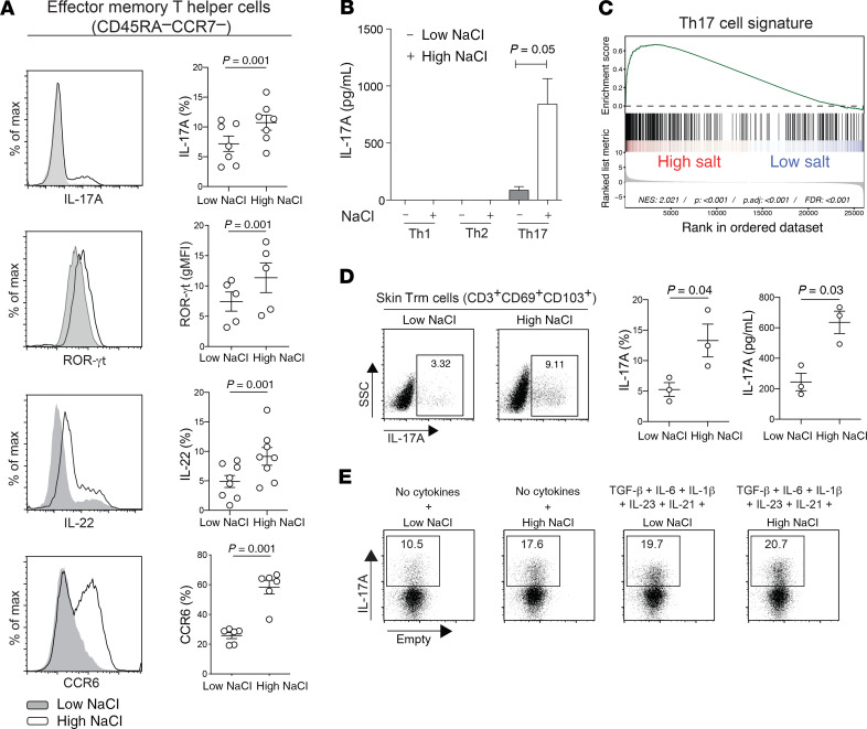
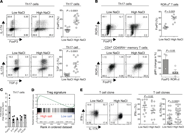
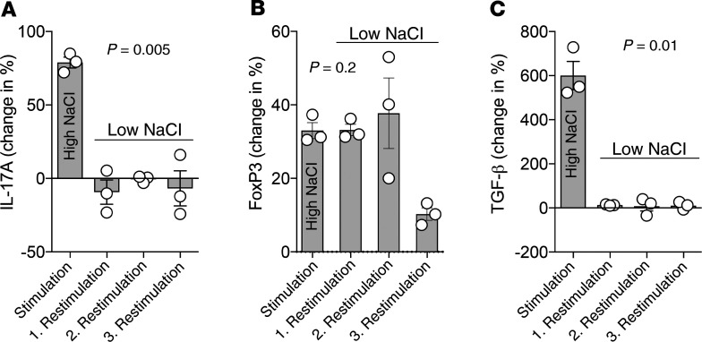
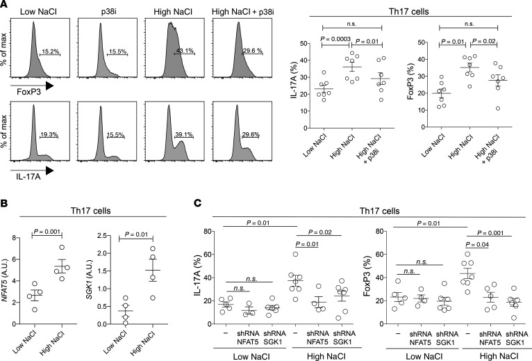
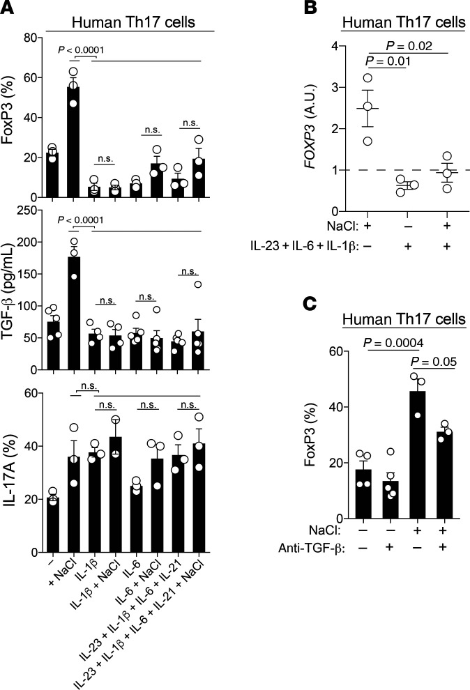
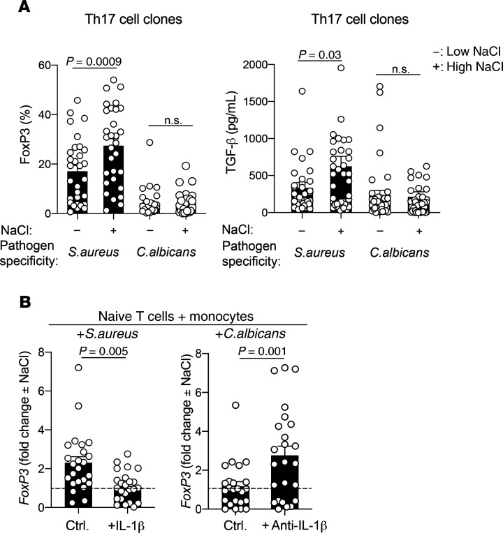
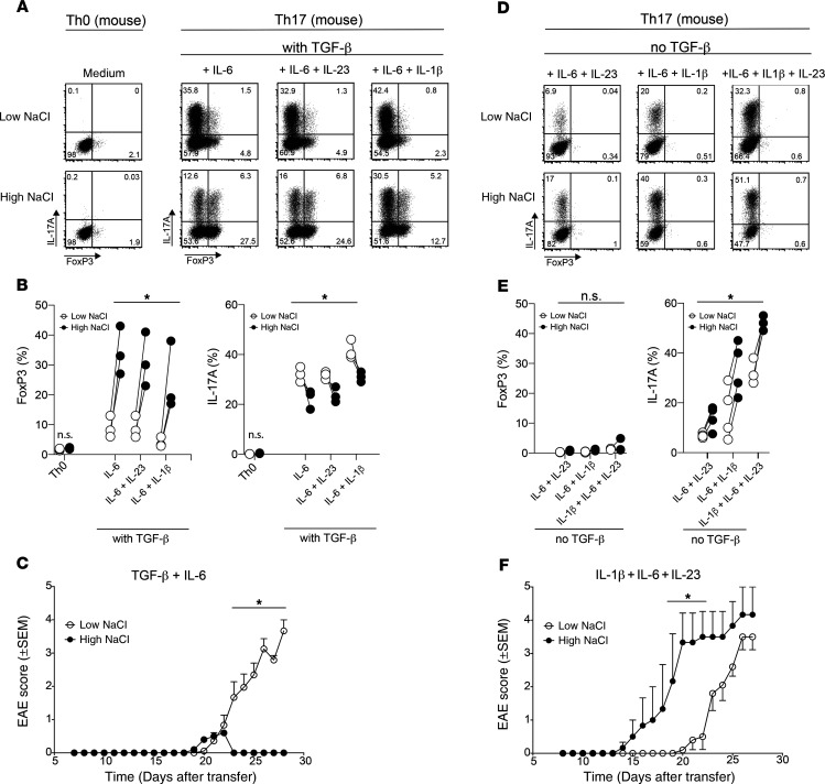
Th cells integrate signals from their microenvironment to acquire distinct specialization programs for efficient clearance of diverse pathogens or for immunotolerance. Ionic signals have recently been demonstrated to affect T cell polarization and function. Sodium chloride (NaCl) was proposed to accumulate in peripheral tissues upon dietary intake and to promote autoimmunity via the Th17 cell axis. Here, we demonstrate that high-NaCl conditions induced a stable, pathogen-specific, antiinflammatory Th17 cell fate in human T cells in vitro. The p38/MAPK pathway, involving NFAT5 and SGK1, regulated FoxP3 and IL-17A expression in high-NaCl conditions. The NaCl-induced acquisition of an antiinflammatory Th17 cell fate was confirmed in vivo in an experimental autoimmune encephalomyelitis (EAE) mouse model, which demonstrated strongly reduced disease symptoms upon transfer of T cells polarized in high-NaCl conditions. However, NaCl was coopted to promote murine and human Th17 cell pathogenicity, if T cell stimulation occurred in a proinflammatory and TGF-β-low cytokine microenvironment. Taken together, our findings reveal a context-dependent, dichotomous role for NaCl in shaping Th17 cell pathogenicity. NaCl might therefore prove beneficial for the treatment of chronic inflammatory diseases in combination with cytokine-blocking drugs.

Keywords: Adaptive immunity; Immunology; Inflammation; T cells.


Conflict of interest statement

Conflict of interest: The authors have declared that no conflict of interest.

Figures

Figure 1
Figure 1. NaCl promotes the Th17 cell program in human effector memory Th cells independently of polarizing cytokines.
(AC) Human effector memory Th cells were FACS sorted from fresh human PBMCs as CD4+CD14CD45RACCR7 T cells and stimulated for a total culture period of 5 days in low- or high-NaCl conditions with CD3 and CD28 mAbs (48 hours plate-bound). (A) Intracellular staining and FACS on day 5 after PMA and ionomycin restimulation for 5 hours. FACS staining of an individual experiment (left) and cumulative data are shown. Each circle indicates an individual donor. gMFI, geometric MFI; max, maximum. (B) ELISA analysis of cell culture supernatants analyzed on day 5 after stimulation with PdBU and CD3 mAb for 8 hours (n = 3). (C) Transcriptome analysis and GSEA (GSE52260) of genes related to the Th17 signature in Th17 cells stimulated as in A (67). (D) Skin CD3+ T cells were isolated from human abdominal skin by overnight collagenase digestion followed by FACS sorting. The cells were stimulated for 48 hours with CD3 and CD28 mAbs in low- or high-NaCl conditions followed by intracellular cytokine staining after PMA and ionomycin restimulation. A representative experiment (left) and cumulative data are shown (middle). ELISA analysis (right) of cell culture supernatants from skin CD3+ T cells restimulated with PdBU and CD3 mAbs for 8 hours after 48 hours of CD3 and CD28 mAb stimulation in low- and high-NaCl conditions. Data were normalized to 20,000 T cells. Each circle indicates an individual donor. (E) FACS analysis performed as in A in the absence or presence of Th17-polarizing cytokines. The data are representative of 3 donors. (A, B, and D) A 2-tailed, paired Student’s t test was performed for comparisons between 2 groups.
Figure 2
Figure 2. Human Th17 cells acquire antiinflammatory functions upon restimulation in high-NaCl conditions.
(A) CD4+CD14CD45RACCR6+CCR4+CXCR3 Th17 cells were sorted by flow cytometry from fresh human PBMCs as T cells and stimulated for a total culture period of 5 days in high- or low-NaCl conditions with CD3 and CD28 mAbs (48 hours plate-bound). Results for intracellular staining and FACS analysis on day 5 following PMA and ionomycin restimulation are shown. A representative FACS plot and cumulative data with each circle representing an individual donor are shown. (B) Intracellular staining for transcription factors followed by FACS analysis of T cells isolated and stimulated for 5 days with CD3 and CD28 mAbs (48 hours plate-bound). Top panel: Circles indicate individual blood donors. Bottom panel: Change in transcription factor expression in T cells stimulated in high- versus low-NaCl conditions (n = 3–4). (C) qRT-PCR analysis of Th17 cells stimulated as in A (n = 3–6). A 2-tailed Student’s t test was performed for comparisons of high- versus low-NaCl conditions; P < 0.05 for each of the transcripts shown. (D) GSEA for Treg-associated genes (GEO GSE18893) in Th17 cells cultured in low- or high-NaCl conditions for 5 days (48 hours CD3/CD28 mAbs). (E) T cell clones were generated from CD4+CD14CD45RACCR6+CCR4+CXCR3 T cells, which were enriched for Th17 cells, and then restimulated in low or high NaCl concentrations, as in A. Intracellular staining and FACS analysis are shown. A representative FACS plot and cumulative data are shown. Each circle represents a separate T cell clone. A 2-tailed Student’s t test was used for comparisons between 2 groups (A–C, and E).
Figure 3
Figure 3. NaCl-induced FoxP3 upregulation in Th17 cells is stably maintained after entering low-NaCl conditions, whereas IL-17A upregulation is transient.
(AC) Human Th17 cells (CCR6+CCR4+CXCR3) were restimulated with CD3 and CD28 mAbs (48 hours plate-bound) in low or high NaCl concentrations for a total culture period of 5 days. Intracellular staining and FACS analysis after PMA and ionomycin restimulation are shown. The Th17 cells were restimulated again with CD3 and CD28 mAbs up to 3 times for 5 days, with 2 additional resting days between the restimulations (n = 3). The change of the geometric mean of FoxP3 expression (B) compared with control Th17 cells that were restimulated once in low-NaCl conditions (C) are shown. One-way ANOVA was used for comparisons among multiple groups.
Figure 4
Figure 4. NFAT5 and SGK1 are engaged in the regulation of FoxP3 and IL-17A expression in high- but not low-NaCl conditions.
(A) Human Th17 cells (CCR6+CCR4+CXCR3) were restimulated in high or low NaCl concentrations in the presence or absence of a pharmacological inhibitor of p38 (p38i, SB202190) for a total culture period of 5 days with CD3 and CD28 mAbs (48 hours plate-bound). Results for intracellular staining and FACS analysis on day 5 after PMA and ionomycin restimulation are shown. Representative FACS data (left) and cumulative data (right) are shown. Gates were set according to the unstained controls. A 2-tailed Student’s t test was used for comparisons between 2 groups. (B) qRT-PCR analysis of Th17 cells on day 5 isolated and stimulated as in A. (C) Intracellular staining and FACS analysis after restimulation in low- or high-NaCl conditions for 5 days in the presence or absence (scrambled shRNA) of shRNA-mediated silencing of NFAT5 and SGK1. Each circle represents an individual blood donor. One-way ANOVA was used for comparisons between multiple groups (FoxP3, P < 0.0004; IL-17A, P < 0.001).
Figure 5
Figure 5. Proinflammatory cytokines abrogate the antiinflammatory switch in the Th17 cell phenotype that is induced in high-NaCl conditions.
(A) FACS analysis (FoxP3, IL-17A) and ELISA (TGF-β) of human Th17 cells stimulated with CD3 and CD28 mAbs for 48 hours in high- or low-NaCl conditions for a total culture period of 5 days. P < 0.0001, by 1-way ANOVA (FoxP3 and TGF-β); P = 0.03, by 1-way ANOVA (IL-17A). (B) qRT-PCR analysis of human Th17 cells on day 5 after stimulation with CD3 and CD28 mAbs for 48 hours in high- or low-NaCl conditions and the presence or absence of Th17-polarizing cytokines. P = 0.01 and P = 0.02, by 1-way ANOVA. (C) FACS analysis performed with cells treated as in B. P = 0.0004 and P = 0.05, by 1-way ANOVA. ±, in the presence or absence of; ± NaCl, in high- or low-NaCl conditions.
Figure 6
Figure 6. The microbial antigen specificity of human Th17 cells determines whether pro- or antiinflammatory effects are promoted by NaCl.
(A) Human Th17 cells were sorted by flow cytometry, labeled with CFSE, and restimulated with autologous monocytes (2:1 ratio) that had been pulsed for 3 hours with S. aureus (DSM799) or C. albicans (SC5314) lysates before glutardehyde fixation as described previously (2). CFSElo Th17 cells were FACS sorted on day 4 and then cloned. The T cell clones were restimulated on days 12 to 14 with CD3 and CD28 for 48 hours and analyzed after a 5-day culture period in low- or high-NaCl conditions before intracellular staining and FACS analysis or ELISA of the culture supernatants. (B) Naive T cells were sorted as CD4+CD45RA+ T cells and stimulated with autologous monocytes that were pulsed with microbial lysates in the presence or absence of endogenously produced or recombinant IL-1β. CFSElo T cells were FACS sorted and cloned on day 9. The T cell clones were restimulated in low- or high-NaCl conditions as in A. The fold change of FoxP3 expression in high- versus low-NaCl conditions is shown. ctrl., control. An unpaired 2-tailed Student’s t test was used for comparisons between 2 groups. Circles indicate individual T cell clones.
Figure 7
Figure 7. TGF-β governs the reciprocal enhancement of pro- versus antiinflammatory Th17 cell functions by NaCl in vitro and in vivo in an EAE mouse model.
(A) Naive murine CD4+ T cells from 2D2 mice were cultured in vitro in neutral (Th0) or various Th17-polarizing conditions (Th17), including TGF-β in low- or high-NaCl conditions, and analyzed by FACS on day 3.5 for IL-17A and FoxP3 expression. Representative dot plots show intracellular IL-17A and FoxP3 expression after restimulation of the cells with PMA and ionomycin. (B) Intracellular IL-17A and FoxP3 expression was quantified as in A. (C) Clinical EAE scores (0–5) were determined after adoptive transfer of T cells (1.5 × 10⁶) that were polarized in vitro for 3.5 days with TGF-β and IL-6 (both 25 ng/mL) in high- or low-NaCl conditions. (D and E) Naive murine CD4+ T cells were cultured as in A in the absence of exogenous TGF-β. Representative dot plots and summary plots are shown. (F) Clinical EAE scores after adoptive transfer of T cells (2 × 10⁶) that were polarized in vitro for 3.5 days with IL-1β, IL-6, and IL-23 in high- versus low-NaCl conditions. *P < 0.05, A 2-tailed, paired Student’s t test was performed for pairwise comparisons of low- and high-NaCl conditions (n = 3–4 mice; data are representative of at least 3 independent experiments) (B and E) and 2-way ANOVA with Bonferroni’s multiple comparisons test (C and F).

References

    1. Zielinski CE, Corti D, Mele F, Pinto D, Lanzavecchia A, Sallusto F. Dissecting the human immunologic memory for pathogens. Immunol Rev. 2011;240(1):40–51. doi: 10.1111/j.1600-065X.2010.01000.x. - DOI - PubMed
    1. Zielinski CE, et al. Pathogen-induced human TH17 cells produce IFN-γ or IL-10 and are regulated by IL-1β. Nature. 2012;484(7395):514–518. doi: 10.1038/nature10957. - DOI - PubMed
    1. Patel DD, Kuchroo VK. Th17 cell pathway in human immunity: lessons from genetics and therapeutic interventions. Immunity. 2015;43(6):1040–1051. doi: 10.1016/j.immuni.2015.12.003. - DOI - PubMed
    1. Sallusto F, Zielinski CE, Lanzavecchia A. Human Th17 subsets. Eur J Immunol. 2012;42(9):2215–2220. doi: 10.1002/eji.201242741. - DOI - PubMed
    1. Stockinger B, Omenetti S. The dichotomous nature of T helper 17 cells. Nat Rev Immunol. 2017;17(9):535–544. doi: 10.1038/nri.2017.50. - DOI - PubMed

Publication types