

Oesophageal foreign bodies in cats: Clinical and anatomic findings
- PMID: 32484841
- PMCID: PMC7266337
- DOI: 10.1371/journal.pone.0233983
Oesophageal foreign bodies in cats: Clinical and anatomic findings
Abstract
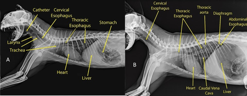
Background: Anatomical feline models can aid in understanding the relationships between clinical findings and anatomical features and the course of foreign bodies passing through the oesophagus. This study has two goals 1) to assess feline oesophageal foreign bodies in feline patients using physical, radiologic and endoscopic examination and, how their location influences treatment plans and complications. 2) How the anatomical sharp angle of the oesophagus contribute to foreign body lodgement. Thirty-five cats were enrolled in this study; 30 of them were clinically ill, and five cats were used for anatomical study.
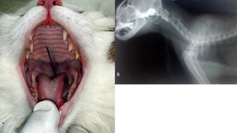
Results: Cats with clinical signs underwent complete clinical and radiologic examination. Endoscopy was performed in only five cases. The site with the highest occurrence of foreign body lodgement was the oesophageal entrance, caudal to the pharynx (63.3%), followed by the thoracic inlet (26.7%) and the mid-cervical region of the oesophagus (10%). Two types of foreign bodies were identified: sewing needles (25/30) and bone (5/30). Radiography was able to identify the location and nature of the foreign body in all 30 affected cats. Therapeutic regimens were applied according to the nature and location of the foreign body and any associated complications. Removal of the foreign body was achieved using Rochester pean artery forceps in 17/30 cases, using full surgical intervention in 8/30 cases, and during endoscopy in 5/30 cases.
Conclusion: The results suggest that the location of the foreign body is strongly related to combination of consumed foreign body type and anatomic features of the cat oesophagus. The feline oesophagus has a variety of sharp angles that facilitate the entrapment of rigid linear and angular foreign bodies. Radiographic imaging remains the most frequently used diagnostic modality for determining the lodgement site and nature of radiopaque foreign bodies. Over all complication rate was low (6/30).
Conflict of interest statement
The authors have declared that no competing interests exist.
Figures


References
-
- Dyce KM, Sack WO, WensingC JG. Textbook of veterinary anatomy 4th Ed Elsevier Health Sciences; 2010
-
- König HE, Liebich HG. Veterinary anatomy of domestic mammals: textbook and color atlas. 6th edTaylor & Francis; 2014
-
- Tams TR. Handbook of small animal gastroenterology. Saunders, Elsevier Science: USA; 2003
-
- Webb BJ. Gastrointestinal and esophageal foreign bodies in the dog and cat. RVT Journal.2014; 38(1):6–10.
-
- Zoran DL. Esophageal foreign bodies and strictures. Standard of care: Emergency and critical care medicine.2005. Available from http://vetnetinfo.com/tudasbazis/files/2016/02/Esophageal-Foreign-Bodies...
MeSH terms
LinkOut - more resources
Full Text Sources
Medical
Miscellaneous
