. 2020 Jul;34(7):9694-9712.
doi: 10.1096/fj.202000667R. Epub 2020 Jun 2.

Adenovirus targets transcriptional and posttranslational mechanisms to limit gap junction function

Affiliations

Adenovirus targets transcriptional and posttranslational mechanisms to limit gap junction function

Patrick J Calhoun et al. FASEB J. 2020 Jul.

Abstract

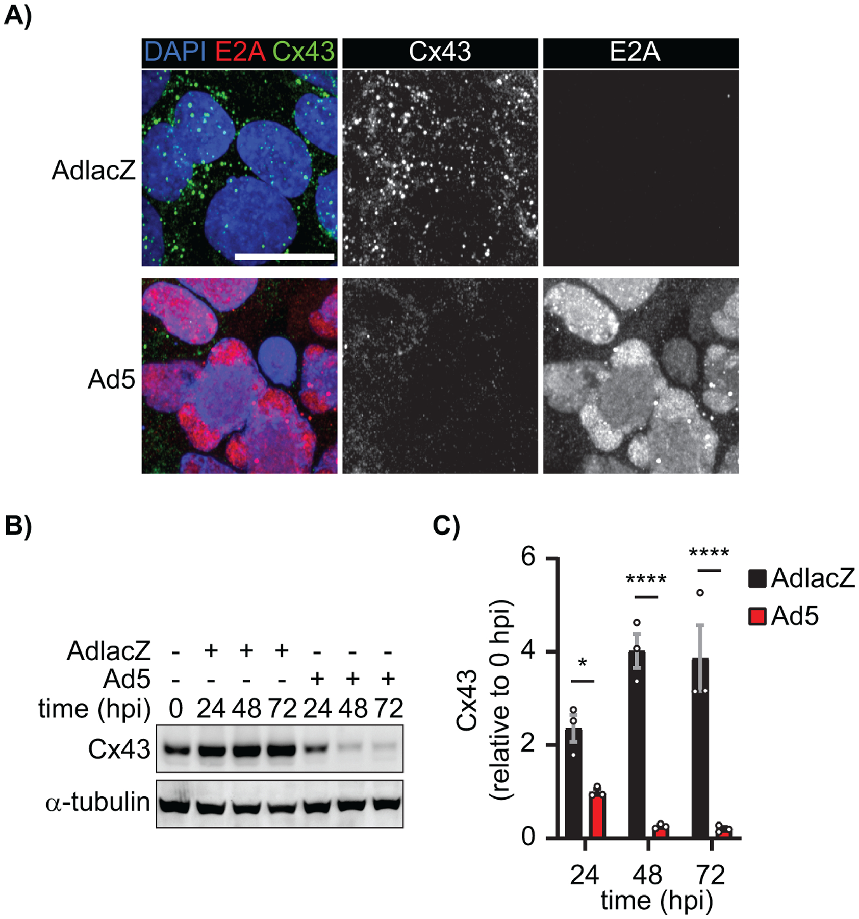
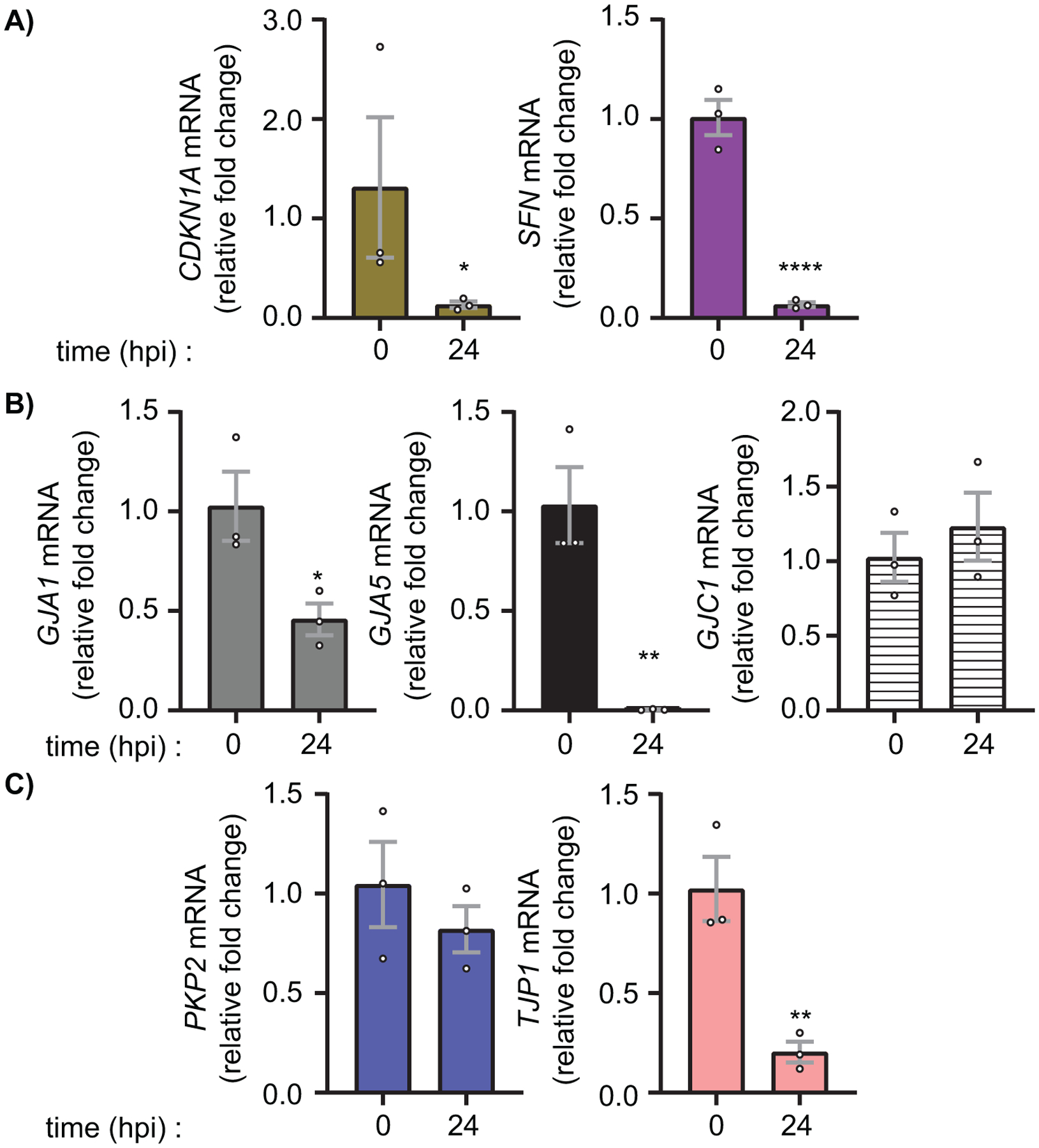
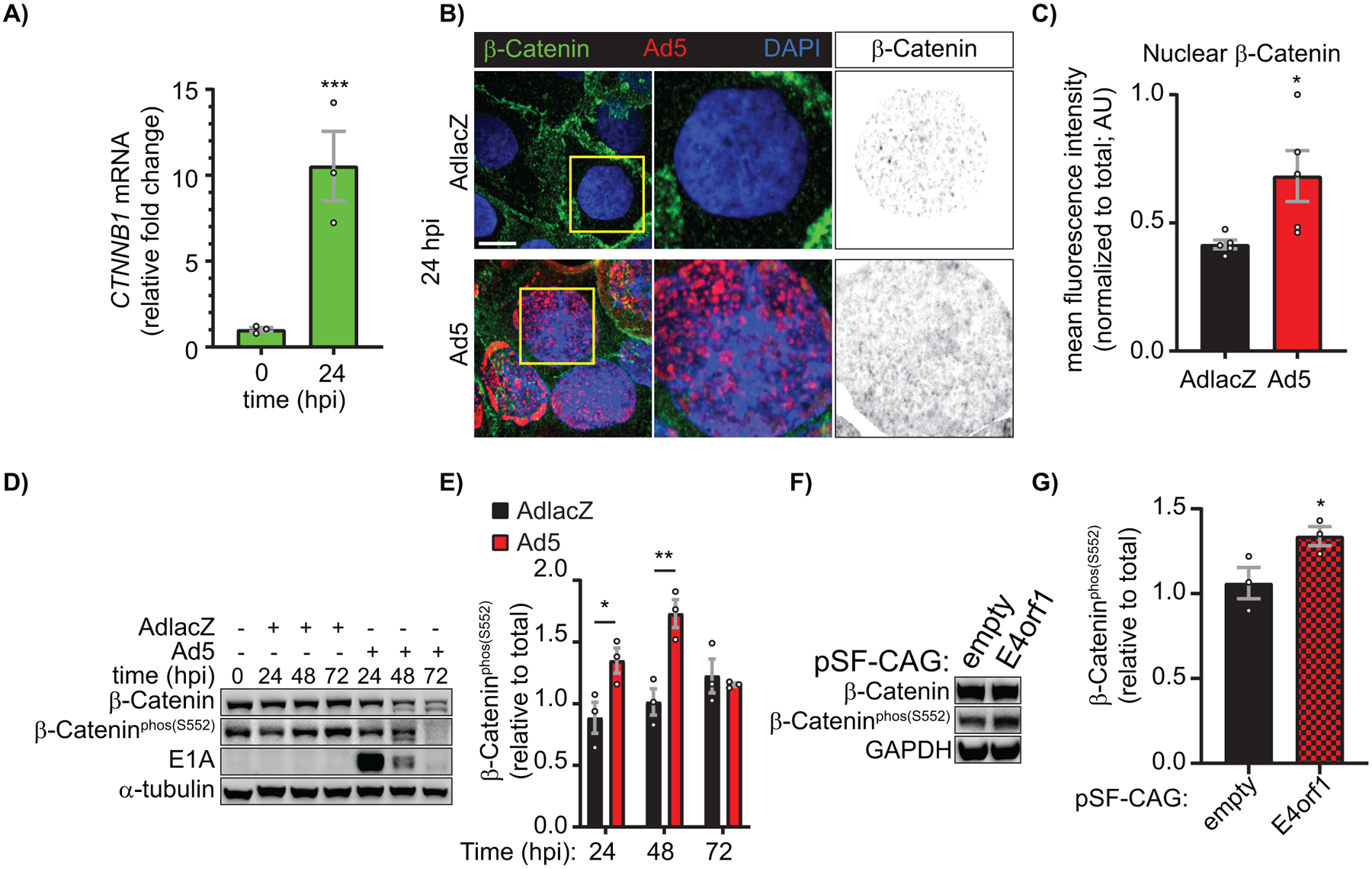
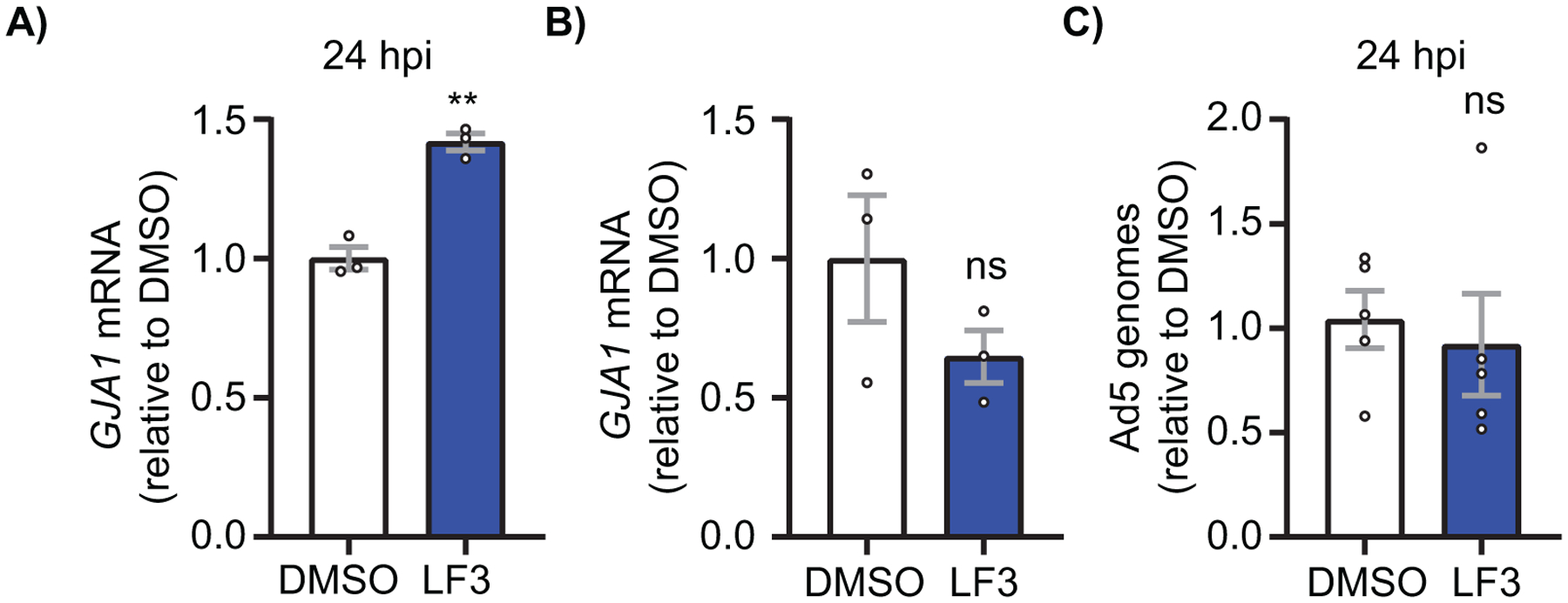
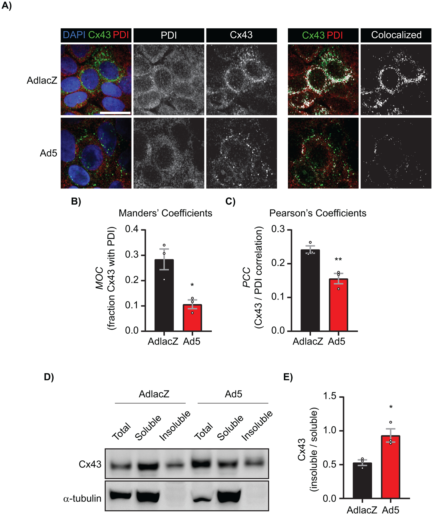
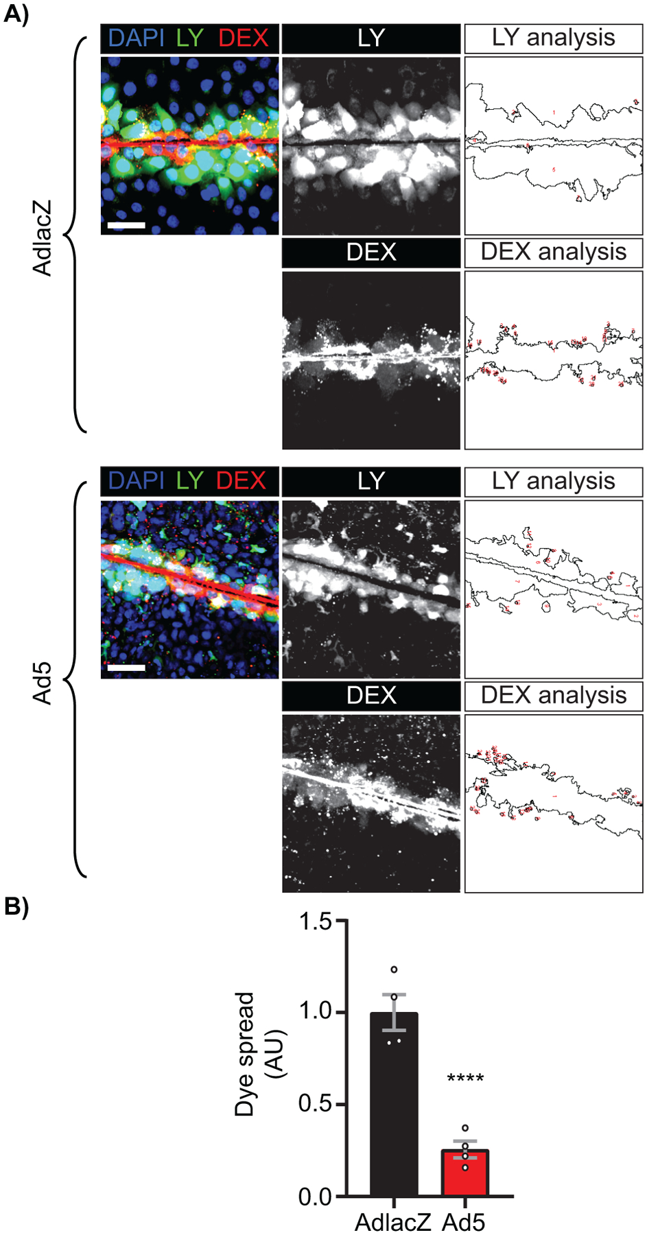
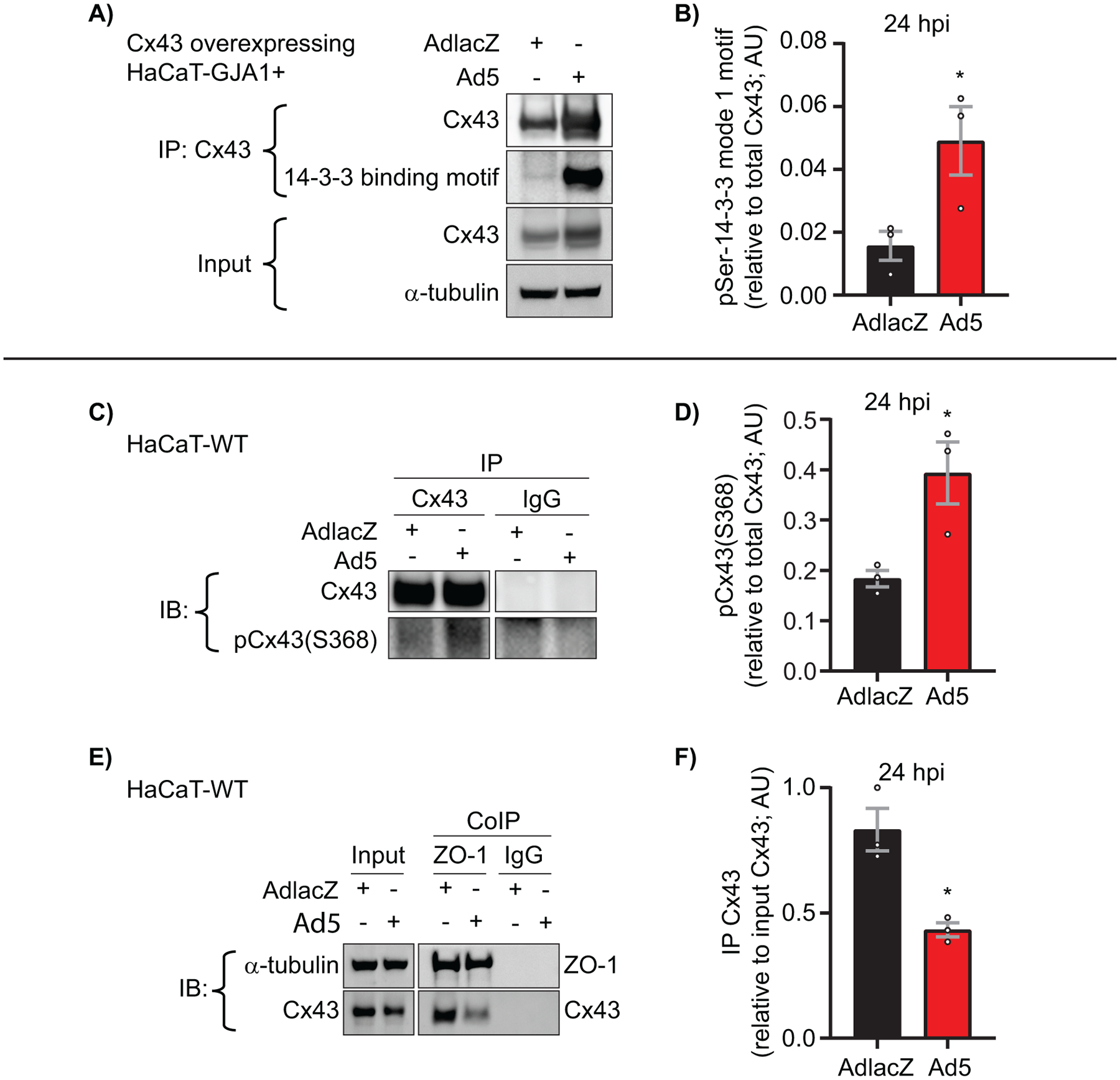
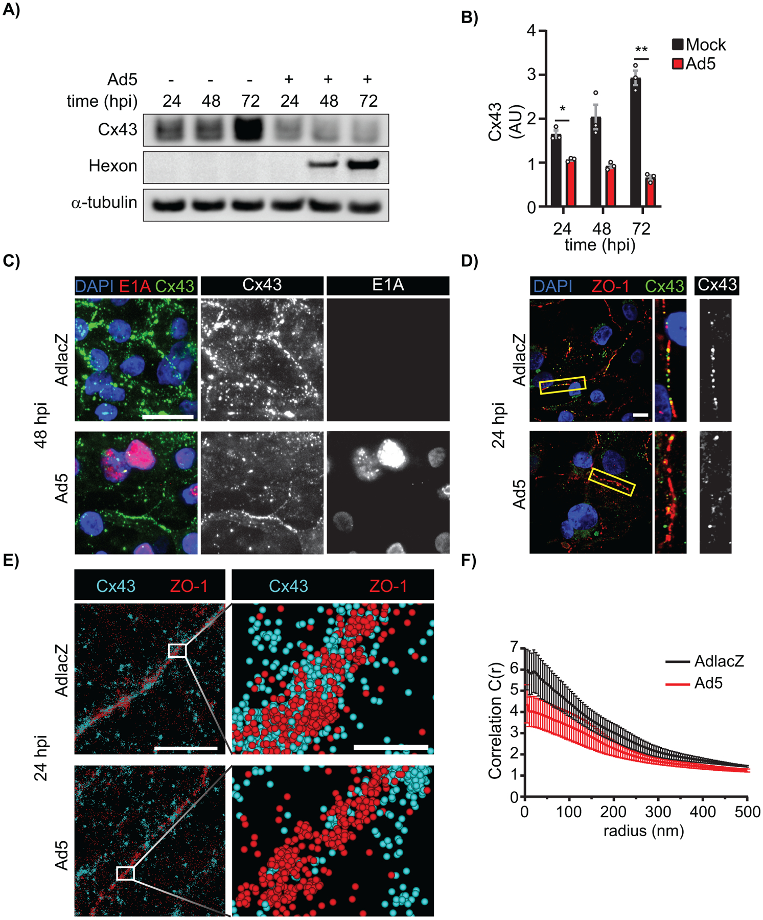
Adenoviruses are responsible for a spectrum of pathogenesis including viral myocarditis. The gap junction protein connexin43 (Cx43, gene name GJA1) facilitates rapid propagation of action potentials necessary for each heartbeat. Gap junctions also propagate innate and adaptive antiviral immune responses, but how viruses may target these structures is not understood. Given this immunological role of Cx43, we hypothesized that gap junctions would be targeted during adenovirus type 5 (Ad5) infection. We find reduced Cx43 protein levels due to decreased GJA1 mRNA transcripts dependent upon β-catenin transcriptional activity during Ad5 infection, with early viral protein E4orf1 sufficient to induce β-catenin phosphorylation. Loss of gap junction function occurs prior to reduced Cx43 protein levels with Ad5 infection rapidly inducing Cx43 phosphorylation events consistent with altered gap junction conductance. Direct Cx43 interaction with ZO-1 plays a critical role in gap junction regulation. We find loss of Cx43/ZO-1 complexing during Ad5 infection by co-immunoprecipitation and complementary studies in human induced pluripotent stem cell derived-cardiomyocytes reveal Cx43 gap junction remodeling by reduced ZO-1 complexing. These findings reveal specific targeting of gap junction function by Ad5 leading to loss of intercellular communication which would contribute to dangerous pathological states including arrhythmias in infected hearts.

Keywords: adenovirus; connexin; gap junction; myocarditis; β-catenin.


Conflict of interest statement

DECLARATION OF INTERESTS

The authors declare no competing interests.

Figures

Figure 1)
Figure 1). Connexin43 protein levels are reduced during adenovirus type 5 infection.
HaCaT cells were infected with Ad5 or replication-incompetent AdlacZ at a multiplicity of infection (MOI) of 10 iu/cell and fixed at 48 hpi or protein harvested every 24 hpi for 72 h. A) Immunofluorescence confocal microscopy of HaCaT cells at 48 hpi probed for Cx43 (green) and adenovirus E2A (red) with nuclei identified using DAPI (blue). Original magnification: X100. Scale bar: 20μm. B) Western blot probed for Cx43 expression in HaCaT cells. Detection of α-tubulin serves as loading control. C) Densitometry analysis of B. Statistical analysis was performed with two-way analysis of variance (ANOVA) with Sidak’s multiple comparisons test. (n=3). *p≤0.05, **p≤0.01, ***p≤0.001, ****p<0.0001. Data are represented as mean ±SEM. See also Figure S1.
Figure 2)
Figure 2). Specific targeting of gap junction gene transcription during adenovirus type 5 infection.
HaCaT cells were infected with Ad5 at a MOI of 10 iu/cell and RNA was harvested at 24 hpi. A) RT-qPCR analysis of CDKN1A (p21) and SFN (14-3-3σ) to confirm active viral infection and known altered gene transcription. B) RT-qPCR analysis of gap junction family genes GJA1 (Cx43), GJA5 (Cx40), and GJC1 (Cx45). C) RT-qPCR analysis of junctional protein genes PKP2 (Plakophilin-2) and TJP1 (ZO-1). Statistical analysis performed by one-way ANOVA with Dunnett’s multiple comparisons test. (n=3). *p≤0.05 **p≤0.01 ***p≤0.001 ****p<0.0001. Data are represented as mean ±SEM. See also Figure S1.
Figure 3)
Figure 3). Early adenoviral factors induce β-catenin transcriptional activity through growth factor signaling.
HaCaT cells were infected with Ad5 or replication-incompetent AdlacZ at a MOI of 10 iu/cell and RNA and protein were harvested or cells were fixed for immunofluorescence over a 72 h time course. A) RT-qPCR analysis of CTNNB1 (β-catenin) relative fold change from 0 hpi. (n=3). B) Immunofluorescence confocal microscopy for β-catenin (green) and Ad5 (red) with nuclei identified with DAPI (blue). Greyscale images in right panels identify nuclear β-catenin levels through DAPI binary mask multiplication from a single Z-slice. Original magnification: X100. Scale bar: 10μm. C) Quantification of nuclear β-catenin represented in B. (n=5). D) Western blot of total β-catenin and β-catenin phospho-Ser552 (transcriptionally active) also probed for Ad5-E1A to confirm infection and α-tubulin for loading control. E) Quantification of D by densitometry. (n=3). HaCaT cells were transfected with pSF-CAG-empty vector or -E4orf1 and protein harvested 24 hours post transfection. F) Western blot of total β-catenin and β-catenin phospho-Ser552 (transcriptionally active) also probed for GAPDH for loading control. G) Quantification of F by densitometry. (n=3). Statistical analyses performed using the unpaired Student’s t-test (A, C, G) or two-way ANOVA with Sidak’s multiple comparisons test (E). *p≤0.05 **p≤0.01 ***p≤0.001 ****p<0.0001. Data are represented as mean ±SEM. See also Figure S2, S3.
Figure 4)
Figure 4). β-catenin transcriptional activity is necessary to reduce GJA1 mRNA during adenoviral infection.
HaCaT cells were treated with the β-catenin transcriptional inhibitor LF3 1 hpi or LF3 alone and harvested for RNA and DNA at indicated time points. A) RT-qPCR analysis of GJA1 mRNA at 24 hpi in cells treated with LF3 or vehicle and infected at a MOI of 10 iu/cell. (n=3). B) RT-qPCR analysis of uninfected HaCaT cells treated with vehicle or LF3 for 24 h. (n=3). C) qPCR analysis of viral genomes in HaCaT cells infected with Ad5 and treated with LF3 or vehicle and harvested 24 hpi. (n=5). ns: not significant. Statistical analyses performed using the unpaired Student’s t test. p≤0.05 is considered statistically significant. **p≤0.01. Data are represented as mean ±SEM.
Figure 5)
Figure 5). Cx43 protein occurs in reduced levels at the endoplasmic reticulum and is primarily junctional during adenovirus infection.
HaCaT cells were infected with AdlacZ or Ad5 at a MOI of 10 iu/cell prior to fixation or protein harvesting at 24 hpi. A) AdlacZ- or Ad5-infected HaCaT cells were immunolabeled against PDI (red) to localize ER and against Cx43 (green). Cells were stained using DAPI to identify nuclei (blue). Colocalized Cx43 / PDI signal was determined with ImageJ (white). Original magnification: X100. Scale bar: 20 μm. B) Manders’ Coefficients calculations determined using fraction Cx43 with PDI. Data points represent averages of 8 images from 3 separate experiments. C) Pearson’s Coefficients calculations determined for Cx43 and PDI correlation. Data points represent averages of 8 images from 3 separate experiments. D) AdlacZ- or Ad5-infected HaCaT cells were lysed in 1% Triton X-100 solubility assay buffer prior to fractionation and western blotting. Membrane probed against Cx43 (top panel) and α-tubulin (bottom panel) for loading control. E) Quantification of D. Statistical analysis was performed with Student’s t-test. (n=3). *p≤0.05, **p≤0.01. Data are represented as mean ±SEM.
Figure 6)
Figure 6). Adenovirus inhibits gap junction intercellular communication prior to reduction of Cx43 total protein levels during infection.
HaCaT cells were plated to confluence on glass bottomed dishes and infected with Ad5 or replication-incompetent AdlacZ at a MOI of 10 iu/cell. The Lucifer yellow (LY) scrape-load dye-transfer assay was performed at 12 hpi with 10,000 MW Dextran-AlexaFluor647 (Dex-647) as a gap junction non-permeable control. A) Representative confocal microscopy images of LY (green) and Dex-647 (red) transfer with nuclei identified with DAPI (blue). Middle greyscale panels show LY and Dex-647 alone and right panels illustrate quantification of each dye spread. Original magnification: X20. Scale bar: 50μm. B) Quantification of dye spread in A. Statistical analysis was performed with the unpaired Student’s t-test. (n=4). ****p<0.0001. Data are represented as mean ±SEM. See also Figure S4.
Figure 7)
Figure 7). Direct targeting of Cx43 gap junctions through phosphorylation during adenovirus type 5 infection.
HaCaT-GJA1+ cells or wild-type HaCaT cells were infected with Ad5 or replication-incompetent AdlacZ at a MOI of 10 iu/cell and protein was harvested at 24 hpi. A) Immunoprecipitation (IP) of Cx43 followed by western blot probed for phosphoSerine-14-3-3 mode-1 binding motif to detect phosphorylation of Cx43-Ser373 at 24 hpi. α-tubulin serves as loading control. B) Densitometry analysis of A. C) IP of Cx43 followed by western blot for Cx43 phosphorylation at Ser368. Cx43 IP and IgG IP are from same membrane. D) Densitometry analysis of C. E) HaCaT cells were infected with Ad5 or AdlacZ at a MOI of 10 iu/cell and protein harvested 24 hpi. Co-immunoprecipitation (CoIP) was performed for ZO-1 and Cx43 followed by western blotting. α-tubulin serves as loading control. F) Densitometry analysis of E. Statistical analysis performed with the unpaired Student’s t-test. (n=3). *p≤0.05 **p≤0.01 ***p≤0.001 ****p<0.0001. Data are represented as mean ±SEM. See also Figure S5.
Figure 8)
Figure 8). Ad5 targets Cx43 in cardiomyocytes and induces Cx43-gap junction remodeling.
HiPSC-CMs were infected with Ad5 or replication-incompetent AdlacZ at a MOI of 10 iu/cell and protein harvested or fixed for immunolabeling over a 72 h time course. A) Western blot probed for Cx43 expression in HiPSC-CMs. Detection of Ad5-Hexon protein expression serves to confirm infection and α-tubulin serves as loading control. B) Densitometry analysis of A. C) Immunofluorescence confocal microscopy of HiPSC-CMs at 48 hpi probed for Cx43 (green) and adenovirus E1A (red) with nuclei identified using DAPI (blue). Original magnification: X100. Scale bar: 20μm. D) Immunofluorescence confocal microscopy of HiPSC-CMs 24 hpi probed for Cx43 (green) and ZO-1 (red) with nuclei identified using DAPI (blue). Original magnification: X100. Scale bar: 10μm. Images representative of 3 separate experiments. E) Super resolution stochastic optical reconstruction microscopy (STORM) derived point-cloud localizations of Cx43 (cyan) and ZO-1 (red) in Ad5- and AdlacZ-infected HiPSC-CMs 24 hpi. Zoomed out panels (left) scale bar: 2μm. Zoomed in panels (right) scale bar: 200nm. Sphere size: 30nm. F) Cross-Pair correlation functions for Cx43/ZO-1 complexing in E. (n=10). Statistical analysis was performed with two-way analysis of variance (ANOVA) with Sidak’s multiple comparisons test (B) (n=3). *p≤0.05, **p≤0.01, ***p≤0.001, ****p<0.0001. Data are represented as mean ±SEM.

References

    1. Unwin PN, and Zampighi G (1980) Structure of the junction between communicating cells. Nature 283, 545–549 - PubMed
    1. Vozzi C, Dupont E, Coppen SR, Yeh HI, and Severs NJ (1999) Chamber-related differences in connexin expression in the human heart. J Mol Cell Cardiol 31, 991–1003 - PubMed
    1. Gutstein DE, Morley GE, Tamaddon H, Vaidya D, Schneider MD, Chen J, Chien KR, Stuhlmann H, and Fishman GI (2001) Conduction slowing and sudden arrhythmic death in mice with cardiac-restricted inactivation of connexin43. Circulation research 88, 333–339 - PMC - PubMed
    1. Kanagaratnam P, Rothery S, Patel P, Severs NJ, and Peters NS (2002) Relative expression of immunolocalized connexins 40 and 43 correlates with human atrial conduction properties. Journal of the American College of Cardiology 39, 116–123 - PubMed
    1. Poelzing S, and Rosenbaum DS (2004) Altered connexin43 expression produces arrhythmia substrate in heart failure. Am J Physiol Heart Circ Physiol 287, H1762–1770 - PubMed

Publication types

LinkOut - more resources