. 2020 Jun 18;78(6):1178-1191.e6.
doi: 10.1016/j.molcel.2020.05.001. Epub 2020 Jun 1.

ERK2 Phosphorylates PFAS to Mediate Posttranslational Control of De Novo Purine Synthesis

Affiliations

ERK2 Phosphorylates PFAS to Mediate Posttranslational Control of De Novo Purine Synthesis

Eunus S Ali et al. Mol Cell. .

Abstract

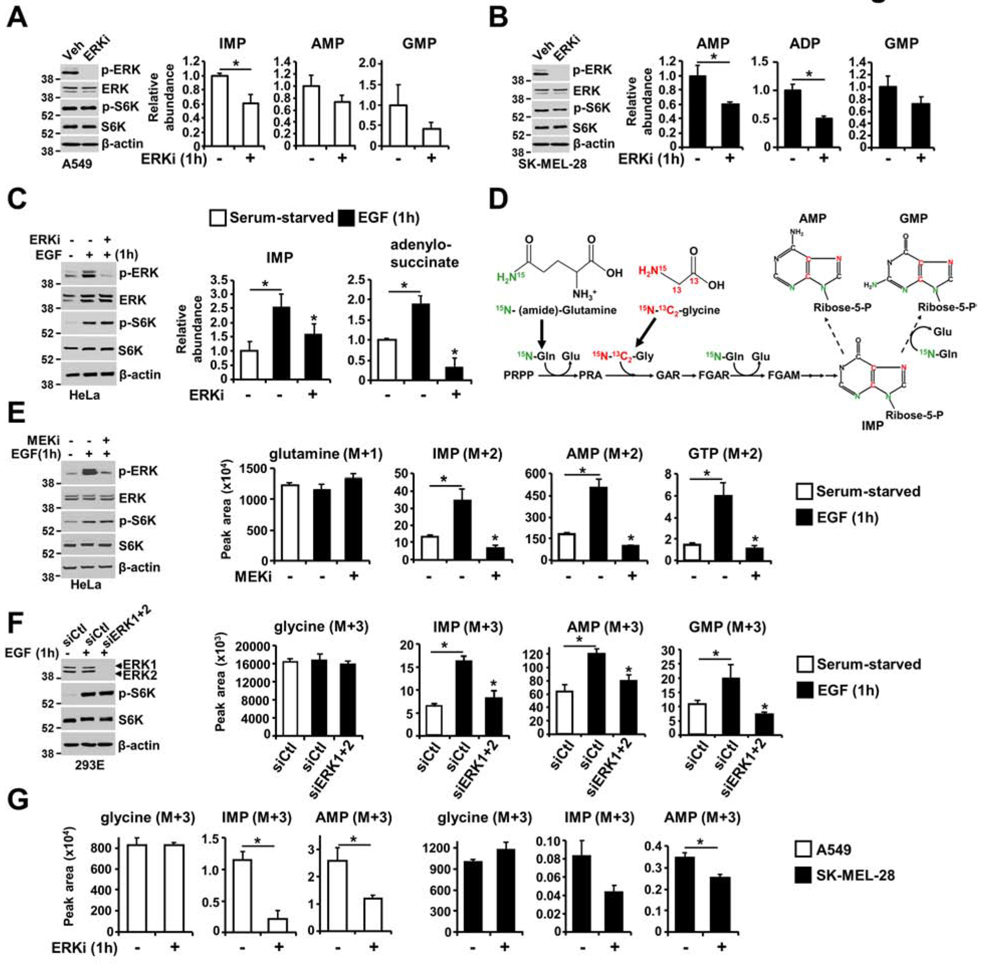
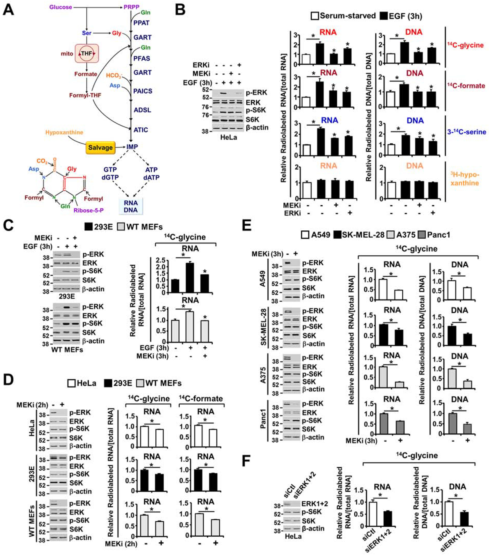
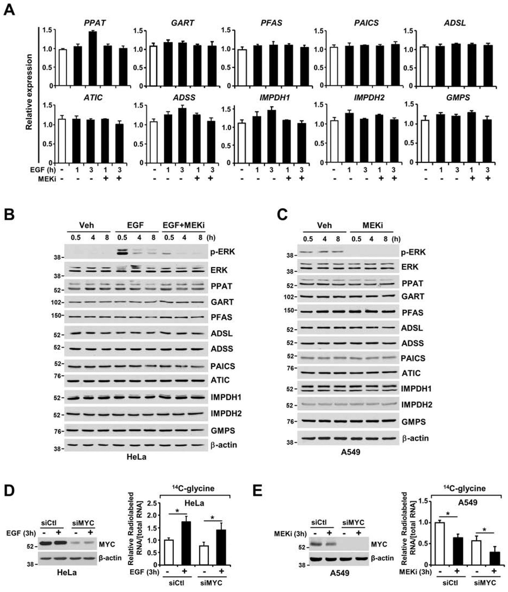
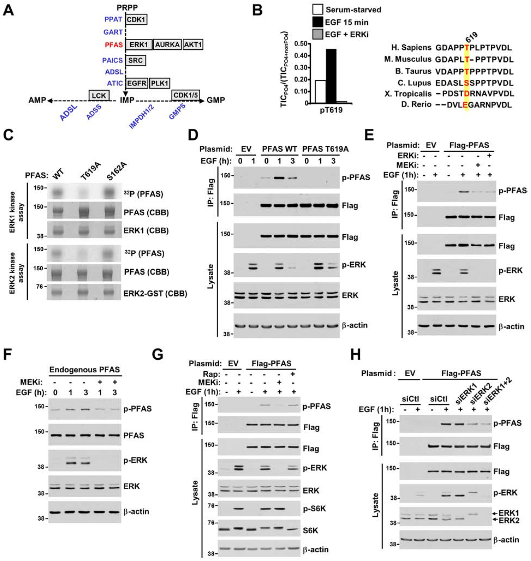
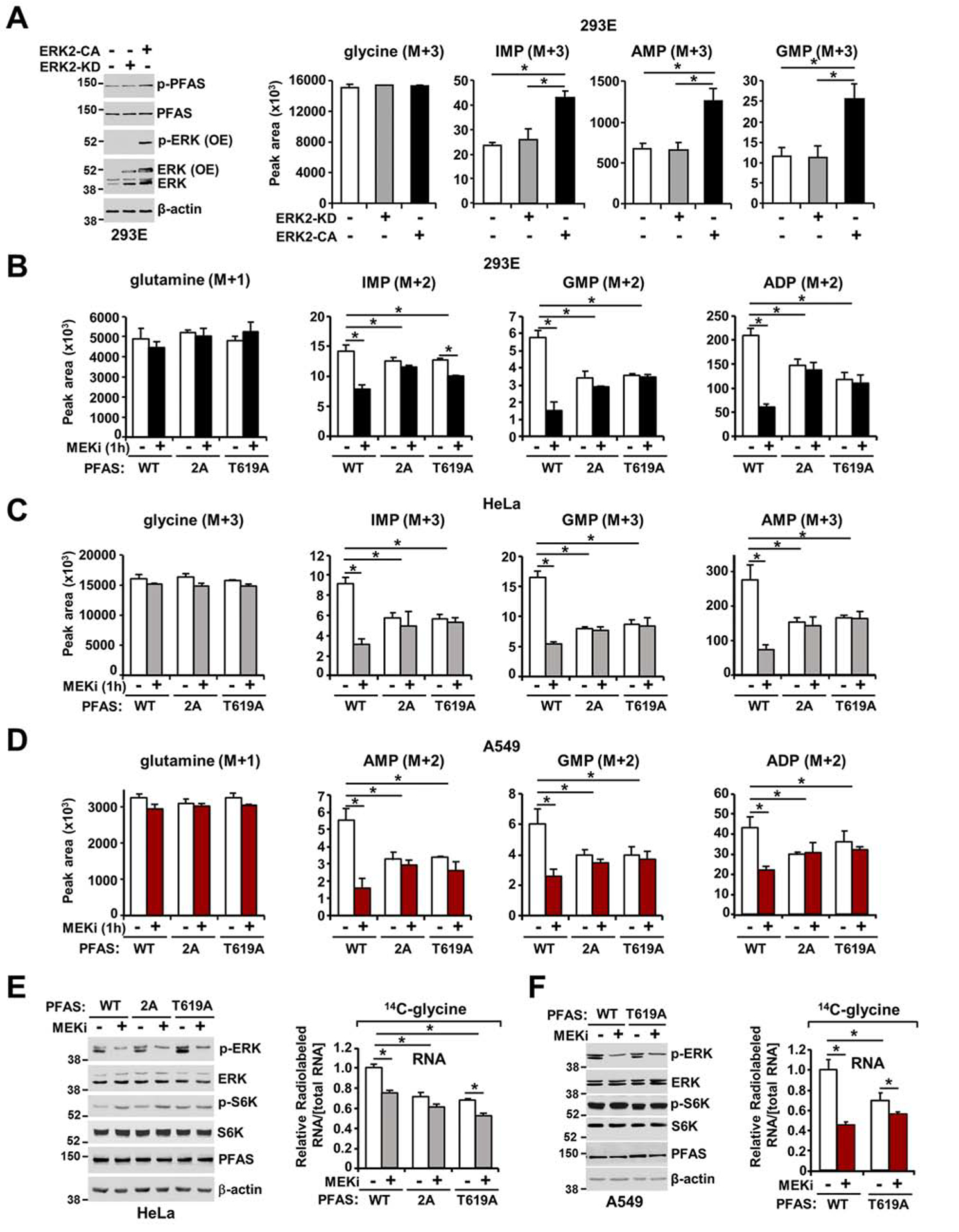
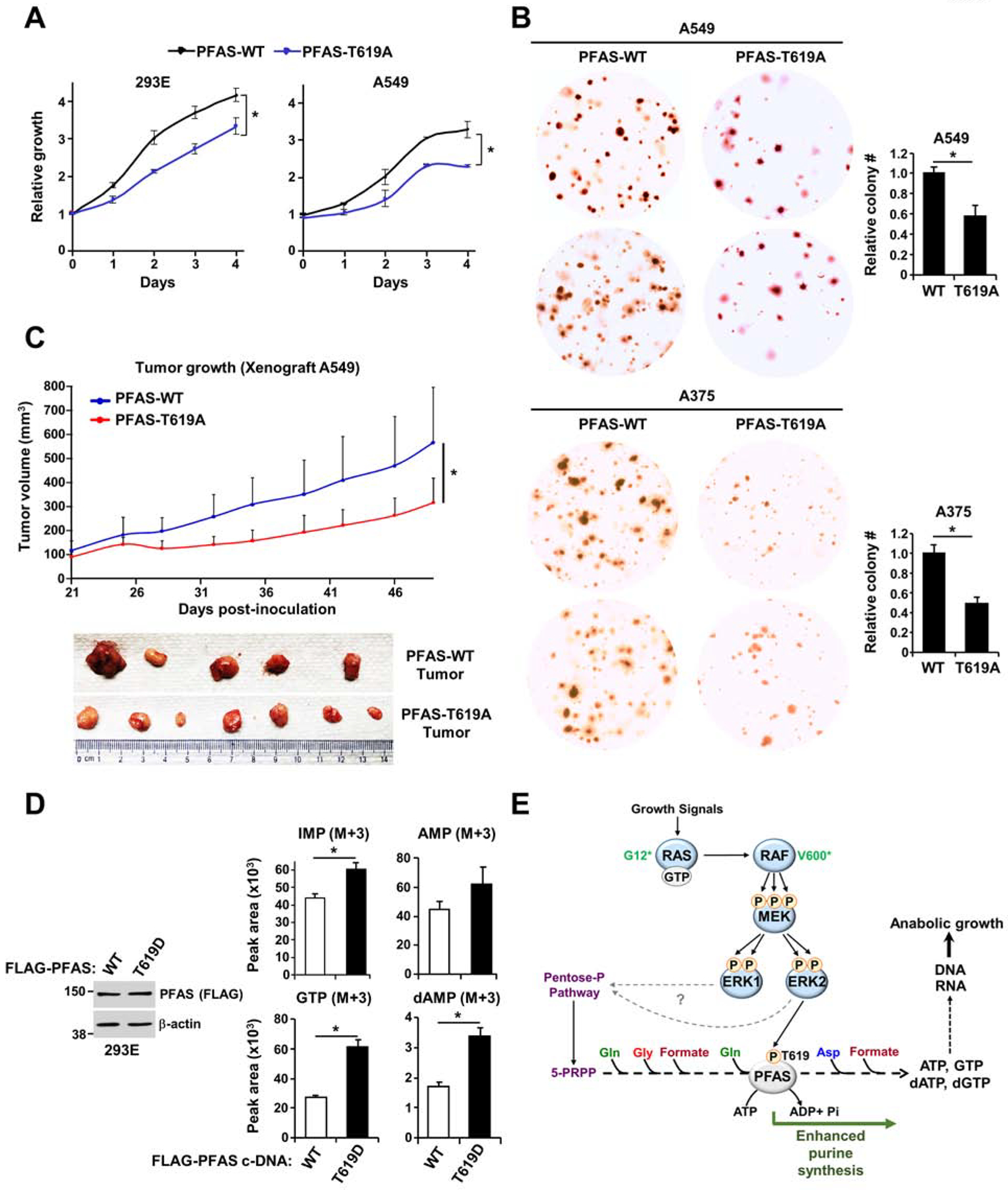
The RAS-ERK/MAPK (RAS-extracellular signal-regulated kinase/mitogen-activated protein kinase) pathway integrates growth-promoting signals to stimulate cell growth and proliferation, at least in part, through alterations in metabolic gene expression. However, examples of direct and rapid regulation of the metabolic pathways by the RAS-ERK pathway remain elusive. We find that physiological and oncogenic ERK signaling activation leads to acute metabolic flux stimulation through the de novo purine synthesis pathway, thereby increasing building block availability for RNA and DNA synthesis, which is required for cell growth and proliferation. We demonstrate that ERK2, but not ERK1, phosphorylates the purine synthesis enzyme PFAS (phosphoribosylformylglycinamidine synthase) at T619 in cells to stimulate de novo purine synthesis. The expression of nonphosphorylatable PFAS (T619A) decreases purine synthesis, RAS-dependent cancer cell-colony formation, and tumor growth. Thus, ERK2-mediated PFAS phosphorylation facilitates the increase in nucleic acid synthesis required for anabolic cell growth and proliferation.

Keywords: ERK; FGAM; MAPK; PFAS; RAS; cancer; nucleotide synthesis; posttranslational modification; purine metabolism; tumor growth.


Conflict of interest statement

Declaration of Interests The authors declare no competing interests.

Figures

Figure 1.
Figure 1.. Immediate stimulation of de novo purine synthesis by ERK signaling.
(A, B) The effects of acute ERK inhibition on the steady-state levels of purine intermediates, as measured via LC-MS/MS, in A549 (A) and SK-MEL-28 (B) cells following 15 hours of serum starvation and 1 hour of treatment with SCH772984 (ERKi, 1 μM) or DMSO. (C) Purine intermediates were measured as in (A, B) in HeLa cells serum starved for 15 hours and treated with vehicle or SCH772984 (ERKi, 1 μM) for 30 min prior to 1 hour of stimulation with EGF (50 ng/ml). (D) Schematic of the incorporation of nitrogen and carbon from glutamine and glycine into the purine ring. (E) Normalized peak areas of 15N-labeled purine intermediates, as measured by targeted LC-MS/MS, in HeLa cells serum starved for 15 hours and pretreated with vehicle or U0126 (MEKi, 10 μM) before stimulation with EGF (50 ng/ml) and labeling with 15N-(amide)-glutamine for 1 hour (see also Figure S1B). (F) Normalized peak areas of labeled glycine and purine intermediates HeLa cells transfected with either nontargeting control siRNA (siCtl), or siRNA against ERK1 and ERK2 (siERK1+2) for 48 hours and were treated as in (E) and labeled with 15N-13C2-glycine for the last hour prior to metabolite extraction (see also Figure S1C). (G) A549 and SK-MEL-28 cells serum-starved for 15 hours and treated with vehicle or SCH772984 (ERKi, 1 μM) before labeling with 15N-13C2-glycine for 1 hour (see also Figure S1D). The data are presented as the means ± SDs of biological triplicates and are representative of two independent experiments (A-F). * P<0.05 by two-tailed Student’s t test for pairwise comparisons (A, B, G) and one-way ANOVA with Tukey’s post hoc test for multiple pairwise comparisons (C, E, F).
Figure 2.
Figure 2.. ERK signaling promotes the integration of newly synthesized purines into nucleic acids.
(A) Schematic of the purine synthesis pathways, including the pentose phosphate pathway, serine biosynthesis pathway and the mitochondrial THF cycles providing carbon and nitrogen for de novo purine nucleotide synthesis. Premade nucleobases, such as hypoxanthine, can sustain nucleotide synthesis via the purine salvage pathway. (B) Immunoblots assessing ERK and mTORC1 signaling; the relative incorporation of 14C from glycine, formate, and serine; and the relative incorporation of 3H from hypoxanthine into RNA and DNA. Labeling was performed for 3 hours under serum-free or EGF (50 ng/ml, 3 hours) stimulation conditions in the presence or absence of U0126 (MEKi, 10 μM) or SCH772984 (ERKi, 1 μM), reflecting de novo purine synthesis (14C-glycine, 14C-formate), one-carbon metabolism into purine nucleotides (3-14C-serine) and purine salvage pathway activity (3H-hypoxanthine) (see also Figure S2B and S2C). (C) HEK293E cells and MEFs were treated as in (B) but labeled with only 14C-glycine for 3 hours. The relative levels of incorporation of 14C from glycine into RNA are shown. Immunoblots performed in parallel to the radio-tracing experiments are shown. (D) As in (B, C), but the cells were cultured in 10% dialyzed serum and treated with vehicle or U0126 (MEKi, 10 μM, 2 hours) and labeled with 14C-glycine for 2 hours. (E) Relative incorporation of 14C-glycine into RNA, with labeling for 3 hours under serum-free conditions in the given cancer cell lines with high levels of ERK signaling (A549, SK-MEL-28, A375, and Panc1), treated with vehicle or U0126 (MEKi, 10 μM) for 3 hours (see also Figure S2D and S2E). (F) As in (D), HeLa cells were transfected with ERK1 and ERK2 siRNAs or nontargeting controls (siCtl) for 48 hours. Cells were cultured in 10% dialyzed serum for 15 hours and were then treated with vehicle or U0126 (MEKi, 10 μM, 2 hours) and concurrently labeled with 14C-glycine for 2 hours. The data are graphed as the means ± SDs of biological triplicates and are representative of at least two independent experiments (B-F). * P<0.05 by one-way ANOVA with Tukey’s post hoc test for multiple pairwise comparisons (B, C) and two-tailed Student’s t test for pairwise comparisons (D, E, F).
Figure 3.
Figure 3.. ERK signaling regulation does not alter the transcript and protein levels of purine enzymes.
(A) Purine enzyme mRNA levels, as measured by qRT-PCR, in serum-starved HeLa cells (15 hours) and stimulated or not with EGF (50 ng/ml) in the presence or absence of U0126 (MEKi, 10 μM) for 1 or 3 hours. (B) Immunoblot showing all purine enzymes measured in (A). HeLa cells were serum starved for 15 hours and stimulated or not with EGF (50 ng/ml) over a time course (0.5, 4 or 8 hours) in the presence or absence of U0126 (MEKi, 10 μM). (C) Immunoblot showing all the purine enzymes measured in (A) and (B). A549 cells were serum starved for 15 hours and treated with vehicle or U0126 (MEKi, 10 μM) over a time course (0.5, 4 or 8 hours). (D, E) c-MYC knockdown did not abolish EGF- and MEK-dependent regulation of de novo purine synthesis in HeLa (D) and A549 (E) cells. Relative incorporation of radiolabels from 14C-glycine (3 hours of labeling) into RNA from HeLa and A549 cells 48 hours after transfection with c-MYC siRNAs or nontargeting controls (siCtl). Cells were serum starved (15 hours) and stimulated or not with EGF (50 ng/ml, 3 hours) (D) or were serum starved and treated with vehicle or MEKi (U0126, 10 μM) for 3 hours (E). The data are presented as the means±SEMs relative to unstimulated serum-starved HeLa cells (A). The data are plotted as the means ± SDs of biological triplicates (D, E). * P<0.05 by two-tailed Student’s t test for pairwise comparisons. The data are representative of at least three independent experiments (A-E).
Figure 4.
Figure 4.. PFAS is a direct substrate of ERK2.
(A) Diagram depicting the purine enzymes predicted to be phosphorylated by canonical kinases based on a computational analysis with Scansite 4.0 software (see also Figure S3A). (B) Effects of EGF and SCH772984 on PFAS phosphorylation. FLAG-PFAS was immunopurified from serum-starved (15 hours) HEK293E cells treated for 30 min with DMSO or SCH772984 (ERKi, 1 μM) prior to stimulation with EGF (15 min, 50 ng/ml). The ratios of phosphorylated T619 peptides on PFAS to the total peptide levels, as measured by the total ion current (TIC) with LC-MS/MS, are plotted. Alignment showing the sequence conservation of T619 among PFAS orthologs (see also Figures S3B, S3C, and S3D). (C) In vitro kinase assays with active ERK1, ERK2 and PFAS variants (wild-type and mutant (T619A, S162A)) were performed with a 10 min reaction time and analyzed by autoradiography (see also Figures S3E, S3F, and S3G). (D) HeLa cells expressing empty vector (EV) or wild-type (WT) or T619A versions of FLAG-PFAS were serum-starved (15 hours) and stimulated with EGF (1 hour, 3 hours, 50 ng/ml). FLAG-immunoprecipitates were immunoblotted with a phospho-PFAS-T619 antibody (see also Figure S4A and S4B). (E) Cells were treated as in (D) and pretreated for 30 min with U0126 (MEKi, 10 μM) or SCH772984 (ERKi, 1 μM) prior to EGF stimulation (1 hour, 50 ng/ml). (F) HeLa cells were serum-starved (15 hours) and pretreated for 30 min with U0126 (MEKi, 10 μM), prior to 1-hour or 3-hour stimulation with EGF (50 ng/mL) (see also Figure S4C). (G) Cells were treated as in (D) and pretreated for 30 min with U0126 (MEKi, 10 μM) or rapamycin (Rap, 20 nM) prior to EGF stimulation (1 hour, 50 ng/ml) (see also Figures S4D and S4E). (H) Cells were treated as in (D), but were transfected with siRNAs targeting ERK1, ERK2, or both, or nontargeting controls (siCtl) (see also Figure S4F).
Figure 5.
Figure 5.. The T619 site in PFAS is required for ERK-dependent stimulation of the de novo purine synthesis pathway.
(A) Immunoblots and normalized peak areas of 15N-13C-labeled purine intermediates measured in HEK293E cells transfected with either empty vector, constitutive active ERK2 (ERK2-CA), or catalytically inactive ERK2 (ERK2-KD) (see also Figure S5A). (B) Normalized peak areas of 15N-labeled metabolites measured in HEK293E ΔPFAS cells stably reconstituted with PFAS-WT, PFAS-S215A/T619A (2A) or PFAS-T619A cultured in dialyzed serum for 15 hours, isotopically labeled with 15N-(amide)-glutamine for 1 hour, and treated with vehicle (DMSO) or U0126 (MEKi, 10 μM). (C) Normalized peak areas of 15N-13C-labeled metabolites measured in HeLa ΔPFAS cells stably reconstituted with PFAS-WT, PFAS-2A or PFAS-T619A cultured in dialyzed serum for 15 hours, isotopically labeled with 15N-13C2-glycine for 1 hour and treated as in (B) (see also Figures S5E and S5F). (D) A549 ΔPFAS cells stably reconstituted with PFAS-WT, PFAS-S215A/T619A (2A) or PFAS-T619A cultured without serum for 15 hours, isotopically labeled with 15N-(amide)-glutamine for 1 hour, and treated as in (B). (E) HeLa ΔPFAS cells stably reconstituted with PFAS-WT, PFAS-S215A/T619A (2A) or PFAS-T619A were cultured in dialyzed serum, treated with vehicle or U0126 (MEKi, 10 μM) and concurrently labeled with 14C-glycine for 2 hours. Incorporation of the specific radiolabel into RNA was measured and normalized to the total concentration of RNA (see also Figure S5G). (F) A549 ΔPFAS cells stably reconstituted with PFAS-WT or PFAS-T619A were serum starved for 15 hours and treated as in (E) (see also Figure S5H). (B-D) CRISPR-mediated PFAS knockout validated in Figures S5B, S5C and S5D. The data are plotted as the means ± SDs of biological triplicates. * P<0.05 by one-way ANOVA with Tukey’s post hoc test for multiple pairwise comparisons (A-F). (A-F) The data are representative of at least two independent experiments.
Figure 6.
Figure 6.. ERK2-mediated PFAS phosphorylation stimulates RAS-dependent tumor growth.
(A) Cell proliferation was measured in HEK293E and A549 ΔPFAS cells stably reconstituted with PFAS-WT or PFAS-T619A (see also Figures S6A and S6B). (B) Soft agar colony formation assay with A549 and A375 ΔPFAS cells stably reconstituted with PFAS-WT or PFAS-T619A. Cell images are at 3x magnification. The relative colony counts are presented as the means ± SDs of biological triplicates. (C) A549 ΔPFAS cells (5 × 10⁶) stably-reconstituted with either WT or T619A-PFAS were injected subcutaneously into athymic nude mice (n = 5 per group). After tumor onset (100 mm3), tumor growth was monitored over time. (D) Normalized peak areas of 15N-13C-labeled purine intermediates measured in HEK293E ΔPFAS cells reconstituted with PFAS-WT or PFAS-T619D cultured in the absence of serum for 15 hours and isotopically labeled with 15N-13C2-glycine for 1 hour (see also Figures S6D, S6E and S6F). (E) Model of purine synthesis stimulation by the RAS-ERK signaling pathway. * P<0.05 by two-tailed Student’s t test for pairwise comparisons (A-D). The data are representative of at least two independent experiments.

Similar articles

Cited by

References

    1. Barbie DA, Tamayo P, Boehm JS, Kim SY, Moody SE, Dunn IF, Schinzel AC, Sandy P, Meylan E, Scholl C, et al. (2009). Systematic RNA interference reveals that oncogenic KRAS-driven cancers require TBK1. Nature 462, 108–112. - PMC - PubMed
    1. Ben-Sahra I, Howell JJ, Asara JM, and Manning BD (2013). Stimulation of de novo pyrimidine synthesis by growth signaling through mTOR and S6K1. Science (New York, N.Y.) 339, 1323–1328. - PMC - PubMed
    1. Ben-Sahra I, Hoxhaj G, Ricoult SJH, Asara JM, and Manning BD (2016). mTORC1 induces purine synthesis through control of the mitochondrial tetrahydrofolate cycle. Science 351, 728–733. - PMC - PubMed
    1. Bodoky G, Timcheva C, Spigel DR, La Stella PJ, Ciuleanu TE, Pover G, and Tebbutt NC (2012). A phase II open-label randomized study to assess the efficacy and safety of selumetinib (AZD6244 [ARRY-142886]) versus capecitabine in patients with advanced or metastatic pancreatic cancer who have failed first-line gemcitabine therapy. Investigational new drugs 30, 1216–1223. - PubMed
    1. Brunet A, Roux D, Lenormand P, Dowd S, Keyse S, and Pouyssegur J (1999). Nuclear translocation of p42/p44 mitogen-activated protein kinase is required for growth factor-induced gene expression and cell cycle entry. The EMBO journal 18, 664–674. - PMC - PubMed

Publication types

MeSH terms

Substances

LinkOut - more resources