. 2020 Jul 8;28(1):104-116.e4.
doi: 10.1016/j.chom.2020.05.002. Epub 2020 Jun 1.

During Aspergillus Infection, Monocyte-Derived DCs, Neutrophils, and Plasmacytoid DCs Enhance Innate Immune Defense through CXCR3-Dependent Crosstalk

Affiliations

During Aspergillus Infection, Monocyte-Derived DCs, Neutrophils, and Plasmacytoid DCs Enhance Innate Immune Defense through CXCR3-Dependent Crosstalk

Yahui Guo et al. Cell Host Microbe. .

Abstract

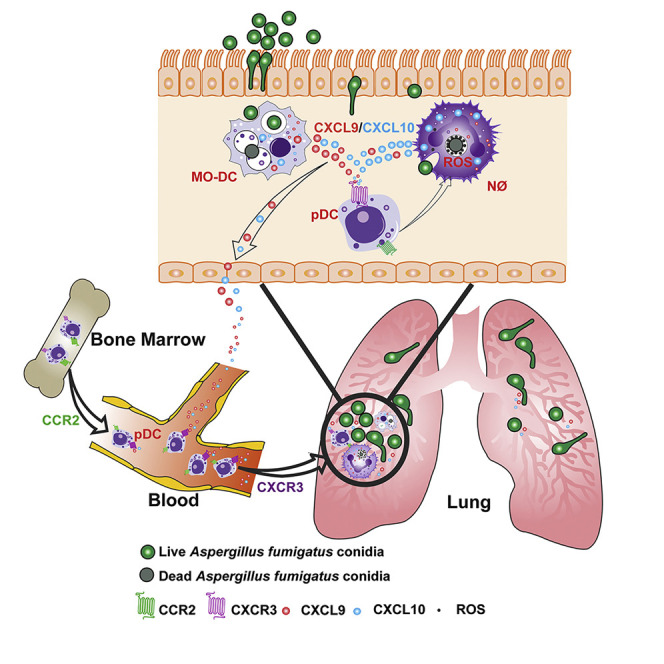
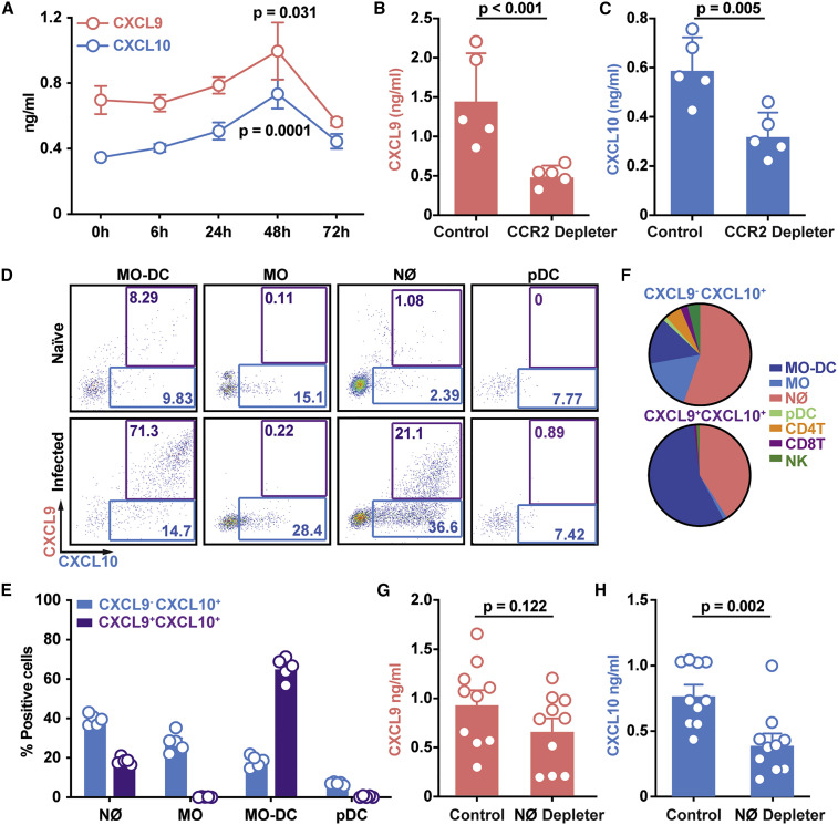
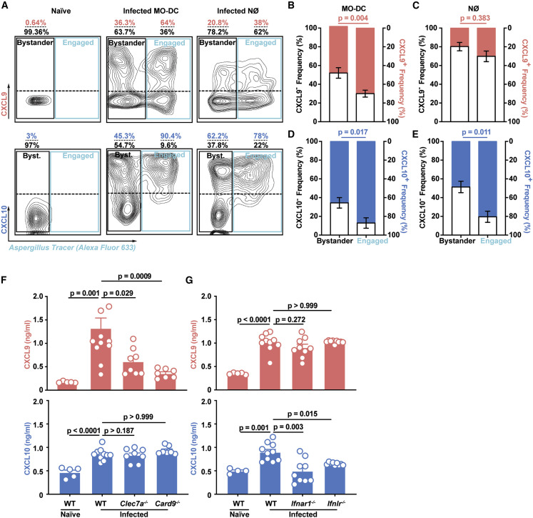
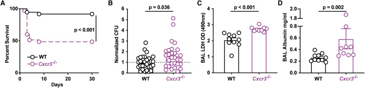
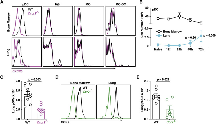
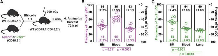
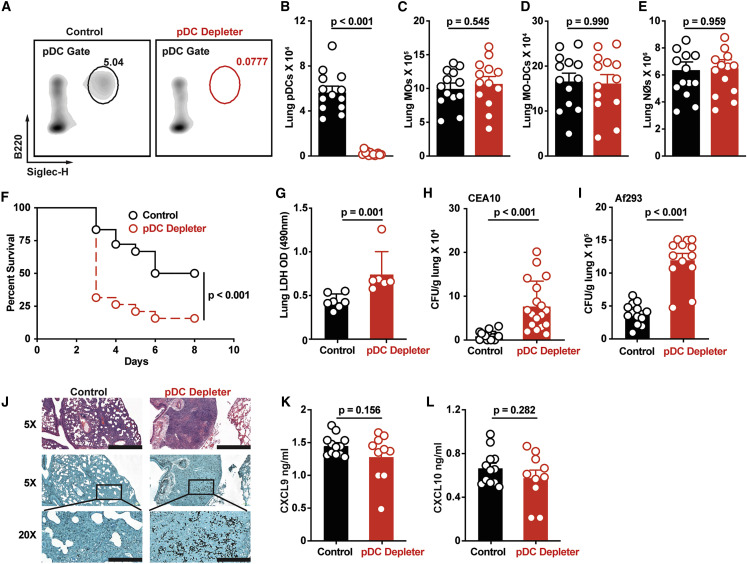
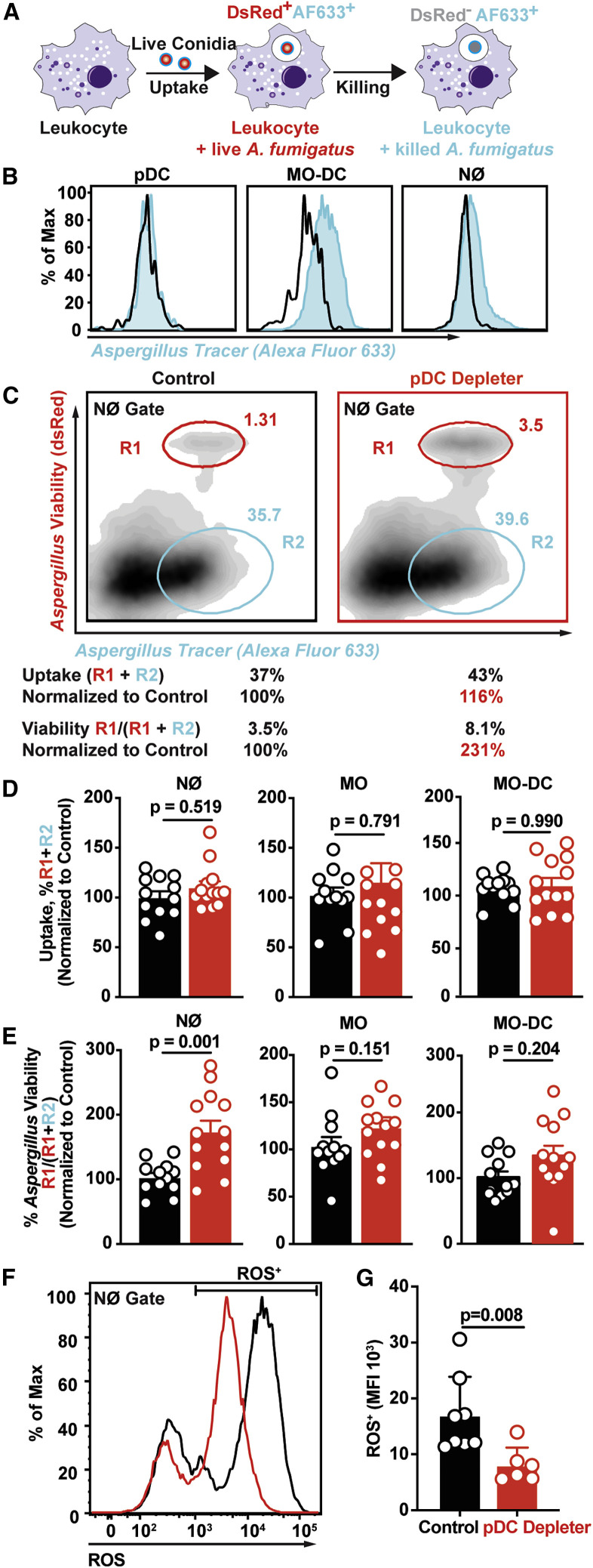
Aspergillus fumigatus, a ubiquitous mold, is a common cause of invasive aspergillosis (IA) in immunocompromised patients. Host defense against IA relies on lung-infiltrating neutrophils and monocyte-derived dendritic cells (Mo-DCs). Here, we demonstrate that plasmacytoid dendritic cells (pDCs), which are prototypically antiviral cells, participate in innate immune crosstalk underlying mucosal antifungal immunity. Aspergillus-infected murine Mo-DCs and neutrophils recruited pDCs to the lung by releasing the CXCR3 ligands, CXCL9 and CXCL10, in a Dectin-1 and Card9- and type I and III interferon signaling-dependent manner, respectively. During aspergillosis, circulating pDCs entered the lung in response to CXCR3-dependent signals. Via targeted pDC ablation, we found that pDCs were essential for host defense in the presence of normal neutrophil and Mo-DC numbers. Although interactions between pDC and fungal cells were not detected, pDCs regulated neutrophil NADPH oxidase activity and conidial killing. Thus, pDCs act as positive feedback amplifiers of neutrophil effector activity against inhaled mold conidia.

Keywords: Aspergillus fumigatus; CXCL10; CXCL9; CXCR3; fungus; innate immunity; lung; monocyte-derived dendritic cell; neutrophil; plasmacytoid DC.


Conflict of interest statement

Declaration of Interests The authors declare no competing interests.

Figures

None
Graphical abstract
Figure 1
Figure 1
CCR2+ Mo-DCs and Neutrophils Produce CXCL9 and CXCL10 during A. fumigatus Challenge (A–C) Lung CXCL9 and CXCL10 levels in (A) C57BL6 mice (n = 5) at 0–72 h pi or (B and C) in DT-treated CCR2-DTR+/− and non-Tg (CCR2-DTR−/−) littermates (n = 5) at 48 h pi with 3 × 107 CEA10 conidia. (D) Representative plots of RFP (CXCL9) and BFP (CXCL10) expression in indicated lung leukocytes isolated from Rex3 Tg → C57BL6.SJL BM chimeric mice at baseline (naive, top row) and 48 h pi with 3 × 107 CEA10 conidia (infected, bottom row). The blue and purple gates indicate the frequency of BFP+ (CXCL9+) and BFP+RFP+ (CXCL9+ CXCL10+) cells, respectively. (E and F) (E) The graphs indicate the frequency of CXCL9 CXCL10+ and CXCL9+ CXCL10+ neutrophils, monocytes, Mo-DCs, and pDCs and (F) the cellular identity of all CXCL9 CXCL10+ (top) and CXCL9+ CXCL10+ lung leukocytes (bottom) at 48 h pi. (G and H) Lung (G) CXCL9 and (H) CXCL10 levels in DT-treated ROSA26-iDTRMrp8-Cre → C57BL6.SJL (neutrophil depleter) or non-Cre ROSA26-iDTR littermates → C57BL 6.SJL (control) BM chimeric mice (n = 10) at 48 h pi with 3–4 × 107 heat-killed swollen CEA10 conidia. Data from two independent experiments were pooled. (A–C, G, and H) Dots represent individual mice and data are presented as mean ± SEM. Statistical analysis: (A) Kruskal-Wallis test, time points compared with t = 0 h, (B, C, G, and H) Mann-Whitney test. See also Figure S1.
Figure 2
Figure 2
Fungal Uptake and Dectin-1 and Card9 and IFN Signaling Regulate Lung CXCL9 and CXCL10 Levels during A. fumigatus Infection (A–E) (A) Representative flow cytometry plots and bar graphs (B–E) that indicate the frequency of RFP+ (CXCL9+; red bar in B and C), RFP (CXCL9; gray bar in B and C), BFP+ (CXCL10+; blue bar, in D and E), and BFP (CXCL10; gray bar in D and E) bystander and fungus-engaged leukocytes isolated from (A, left column) naive or (A, middle and right column, B, and E) infected Rex3 Tg → C57BL6.SJL BM chimeric mice (n = 7) with 3 × 107 AF633-labeled CEA10 conidia. The solid black gates in (A) indicate bystander neutrophils, whereas the solid blue gates indicate fungus-engaged leukocytes. The dashed line in the black and blue gate indicate CXCL9+ and CXCL9 (top quadrants) leukocytes and CXCL10+ and CXCL10 (bottom quadrants) leukocytes. (F) Lung CXCL9 and (G) CXCL10 levels in naive wild-type (WT, n = 5) and in WT (n = 10), Clec7a−/− (n = 8), Card9−/− (n = 7), Ifnar1−/− (n = 9), and Ifnlr−/− (n = 9) mice 48 h pi with 3 × 107 CEA10 conidia. Data were pooled from two independent experiments. (B–G) Dots represent individual mice and data are presented as mean ± SEM. Statistical analysis: Mann-Whitney test. See also Figure S2.
Figure 3
Figure 3
CXCR3 Is Critical for Anti-Aspergillus Defense (A) Kaplan-Meier survival of C57BL6 (n = 37) and Cxcr3−/− (n = 35) mice challenged with 4–8 × 107 CEA10 conidia. Data from three experiments were pooled. (B–D) (B) Normalized lung fungal burden, (C) BALF LDH level, and (D) BALF albumin levels in C57BL6 and Cxcr3−/− mice 48 h pi with 3 × 107 CEA10 conidia. (A–D) Dots represent individual mice and data were pooled from 2–4 experiments and presented as mean ± SEM. Statistical analysis: Mann-Whitney test. See also Figure S3.
Figure 4
Figure 4
CXCR3+ pDCs Traffic to A. fumigatus-Infected Lungs (A) CXCR3 surface expression in BM (top row) and lung (bottom row) leukocytes isolated from WT (black lines) or Cxcr3−/− mice (purple lines). (B) Lung (blue filled dots) and BM (open black dots) pDC numbers at baseline and indicated times pi with 3 × 107 CEA10 conidia (n = 5). (C) Lung pDC numbers in WT (open black dots) and Cxcr3−/− mice (open purple dots) at 72 h pi (n = 10). (D) CCR2 surface expression in BM and lung pDCs in WT (black lines) or Ccr2−/− mice (green lines). (E) Lung pDC numbers in WT (open black dots) and Ccr2−/− mice (open green dots) at 72 h pi (n = 10). (B, C, and E) Data from 2–3 experiments were pooled and expressed as mean ± SEM. Statistical analysis: Mann-Whitney test. See also Figure S4.
Figure 5
Figure 5
Sequential CCR2- and CXCR3-Dependent Signals Mediate Lung pDC Recruitment (A) Experimental scheme to generate mixed BM chimeric mice and compare the trafficking of Cxcr3−/−, Ccr2−/−, and WT pDCs during A. fumigatus infection. (B and C) Relative frequencies of (B) Cxcr3−/− (open purple bars) and Cxcr3+/+ (WT, open black bars) and (C) Ccr2−/− (open green bars) and Ccr2+/+ (WT, open black bars) pDCs in the BM, blood, and lung of mixed BM chimeric mice 72 h pi. Data from 2–3 experiments were pooled and expressed as mean ± SEM. Statistical analysis: Mann-Whitney test. See also Figure S5.
Figure 6
Figure 6
pDCs Are Critical for Anti-Aspergillus Defense (A–E) (A) Representative flow cytometry plots of B220+Siglec-H+ pDCs and (B) lung pDC, (C) lung monocyte, (D) lung Mo-DC, and (E) lung neutrophil numbers in DT-treated pDC depleter mice (BDCA2-DTR; open red symbols) and non-Tg littermate controls (open black symbols) at 72 h pi with 3 × 107 CEA10 conidia. (F) Kaplan-Meier survival of DT-treated pDC depleter (n = 20) and non-Tg littermate control mice (n = 19) infected with 3–6 × 107 CEA10 conidia. (G and H) (G) BALF LDH levels and (H) fungal burden of DT-treated pDC depleter (open red symbols; n = 6–18) or non-Tg littermates (control, open black symbols; n = 6–17) at 72 h pi with 3 × 107 CEA10 conidia. (I) Lung fungal burden at 72 h pi with 3 × 107 Af293 conidia. (J) Lung histopathology (top row, hematoxylin and eosin stain, scale bar: 800 μm; middle row, Gomori methenamine silver stain, scale bar: 800 μm; bottom row, GMS, scale bar: 200 μm) of DT-treated pDC depleter (right column; n = 6) or non-Tg littermates (Control, left column; n = 6) at 72 h pi with 3 × 107 CEA10 conidia. (K and L) (K) Lung CXCL9 and (L) lung CXCL10 levels in DT-treated pDC depleter mice (red symbols) and non-Tg littermate controls (black symbols) at 72 h pi with 3 × 107 CEA10 conidia. (B–L) Data were pooled from 2–3 independent experiments and expressed as mean ± SEM. Statistical analysis: Mann-Whitney test. See also Figure S6.
Figure 7
Figure 7
Lung pDCs Enhance Neutrophil Fungicidal Activity (A) Schematic of FLARE strain and changes in fluorescence emission following fungal uptake and killing by host leukocytes. (B) AF633 fluorescence intensity in indicated lung leukocytes 72 h pi with FLARE (blue line) or AF633-unlabeled conidia. (C) Representative plots that display dsRed and AF633 fluorescence intensity of lung neutrophils in DT-treated pDC depleter (right panel) and non-Tg littermates (left panel) 72 h pi with 3 × 107 Af293 FLARE conidia. R1 denotes neutrophils that contain live conidia, R2 denotes neutrophils that contain killed conidia. (D and E) The plots show neutrophil, monocyte, and Mo-DC (D) conidial uptake (R1 + R2) ± SEM and (E) conidial viability (R1/ (R1 + R2) ± SEM in indicated lung leukocytes isolated from DT-treated pDC depleter (red symbols) and non-Tg littermates (black symbols) 72 h pi with 3 × 107 FLARE conidia. (F and G) (F) Representative plot and (G) mean ± SEM neutrophil ROS production in cells isolated from DT-treated pDC depleter (right panel) and non-Tg littermates (left panel) 72 h pi with 3 × 107 CEA10 conidia. (C–E) Data from 2 experiments were pooled. (F and G) Data are representative of two experiments. (D, E, and G) Dots represent individual mice and data are expressed as mean ± SEM. Statistical analysis: Mann-Whitney test. See also Figure S7.

Comment in

References

    1. Ang D.K.Y., Oates C.V.L., Schuelein R., Kelly M.K., Sansom F.M., Bourges D., Boon L., Hertzog P.J., Hartland E.L., van Driel I.R. Cutting edge: pulmonary Legionella pneumophila is controlled by plasmacytoid dendritic cells but not type I IFN. J. Immunol. 2010;184:5429–5433. - PubMed
    1. Assil S., Coléon S., Dong C., Décembre E., Sherry L., Allatif O., Webster B., Dreux M. Plasmacytoid dendritic cells and infected cells form an interferogenic synapse required for antiviral responses. Cell Host Microbe. 2019;25:730–745.e6. - PubMed
    1. Bianchi M., Hakkim A., Brinkmann V., Siler U., Seger R.A., Zychlinsky A., Reichenbach J. Restoration of NET formation by gene therapy in CGD controls aspergillosis. Blood. 2009;114:2619–2622. - PMC - PubMed
    1. Blasius A.L., Giurisato E., Cella M., Schreiber R.D., Shaw A.S., Colonna M. Bone marrow stromal cell antigen 2 is a specific marker of type I IFN-producing cells in the naive mouse, but a promiscuous cell surface antigen following IFN stimulation. J. Immunol. 2006;177:3260–3265. - PubMed
    1. Bonnett C.R., Cornish E.J., Harmsen A.G., Burritt J.B. Early neutrophil recruitment and aggregation in the murine lung inhibit germination of Aspergillus fumigatus conidia. Infect. Immun. 2006;74:6528–6539. - PMC - PubMed

Publication types

MeSH terms