

Diagnostic impact of bedside chest X-ray features of 2019 novel coronavirus in the routine admission at the emergency department: case series from Lombardy region
- PMID: 32485335
- PMCID: PMC7250080
- DOI: 10.1016/j.ejrad.2020.109092
Diagnostic impact of bedside chest X-ray features of 2019 novel coronavirus in the routine admission at the emergency department: case series from Lombardy region
Abstract
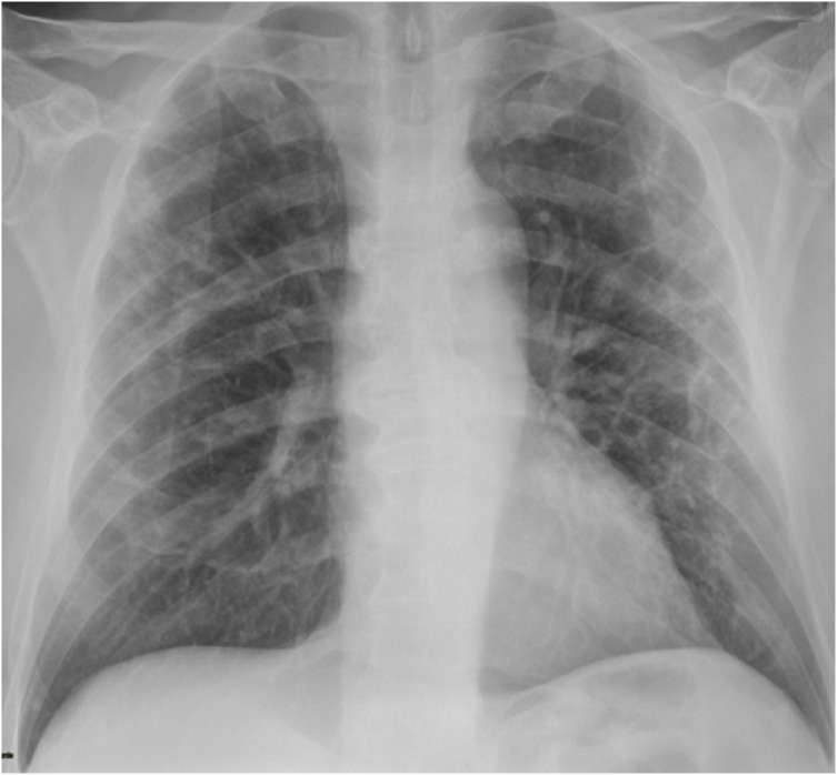
Purpose: To evaluate the diagnostic accuracy and the imaging features of routine admission chest X-ray in patients suspected for novel Coronavirus 2019 (SARS-CoV-2) infection.
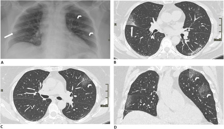
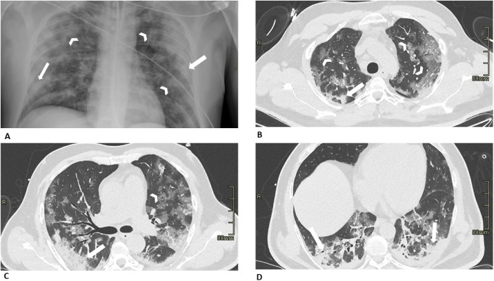
Method: We retrospectively evaluated clinical and X-ray features in all patients referred to the emergency department for suspected SARS-CoV-2 infection between March 1st and March 13th. A single radiologist with more than 15 years of experience in chest-imaging evaluated the presence and extent of alveolar opacities, reticulations, and/or pleural effusion. The percentage of lung involvement (range <25 % to 75-100 %) was also calculated. We stratified patients in groups according to the time interval between symptoms onset and X-ray imaging (≤ 5 and > 5 days) and according to age (≤ 50 and > 50 years old).
Results: A total of 518 patients were enrolled. Overall 314 patients had negative and 204 had positive RT-PCR results. Lung lesions in patients with SARS-Cov2 pneumonia primarily manifested as alveolar and interstitial opacities and were mainly bilateral (60.8 %). Lung abnormalities were more frequent and more severe by symptom duration and by increasing age. The sensitivity and specificity of chest X-ray at admission in the overall cohort were 57 % (95 % CI = 47-67) and 89 % (83-94), respectively. Sensitivity was higher for patients with symptom onset > 5 days compared to ≤ 5 days (76 % [62-87] vs 37 % [24-52]) and in patients > 50 years old compared to ≤ 50 years (59 % [48-69] vs 47 % [23-72]), at the expense of a slightly lower specificity (68 % [45-86] and 82 % [73-89], respectively).
Conclusions: Overall chest X-ray sensitivity for SARS-CoV-2 pneumonia was 57 %. Sensitivity was higher when symptoms had started more than 5 days before, at the expense of lesser specificity, while slightly higher in older patients in comparison to younger ones.
Keywords: Coronavirus; Infections; Radiography; Tomography; X-ray computed.
Copyright © 2020 Elsevier B.V. All rights reserved.
Conflict of interest statement
Declaration of Competing Interest All authors declare no conflicts-of-interest related to this article.
Figures
Similar articles
-
Chest X-ray features of SARS-CoV-2 in the emergency department: a multicenter experience from northern Italian hospitals.Respir Med. 2020 Aug-Sep;170:106036. doi: 10.1016/j.rmed.2020.106036. Epub 2020 May 22. Respir Med. 2020. PMID: 32469732 Free PMC article.
-
Thoracic imaging tests for the diagnosis of COVID-19.Cochrane Database Syst Rev. 2020 Sep 30;9:CD013639. doi: 10.1002/14651858.CD013639.pub2. Cochrane Database Syst Rev. 2020. Update in: Cochrane Database Syst Rev. 2020 Nov 26;11:CD013639. doi: 10.1002/14651858.CD013639.pub3. PMID: 32997361 Updated.
-
Frequency and Distribution of Chest Radiographic Findings in Patients Positive for COVID-19.Radiology. 2020 Aug;296(2):E72-E78. doi: 10.1148/radiol.2020201160. Epub 2020 Mar 27. Radiology. 2020. PMID: 32216717 Free PMC article.
-
Imaging and clinical features of patients with 2019 novel coronavirus SARS-CoV-2.Eur J Nucl Med Mol Imaging. 2020 May;47(5):1275-1280. doi: 10.1007/s00259-020-04735-9. Epub 2020 Feb 28. Eur J Nucl Med Mol Imaging. 2020. PMID: 32107577 Free PMC article.
-
Imaging findings in COVID-19 pneumonia.Clinics (Sao Paulo). 2020 Jun 22;75:e2027. doi: 10.6061/clinics/2020/e2027. eCollection 2020. Clinics (Sao Paulo). 2020. PMID: 32578826 Free PMC article. Review.
Cited by
-
Descriptive analysis of a comparison between lung ultrasound and chest radiography in patients suspected of COVID-19.Ultrasound J. 2021 Feb 26;13(1):11. doi: 10.1186/s13089-021-00215-9. Ultrasound J. 2021. PMID: 33635443 Free PMC article.
-
Sextus chest radiograph severity score correlates to clinical outcomes in patients with COVID-19: A cross-sectional study.Medicine (Baltimore). 2021 Nov 12;100(45):e27663. doi: 10.1097/MD.0000000000027663. Medicine (Baltimore). 2021. PMID: 34766569 Free PMC article.
-
Pulmonary Ultrasound in the Diagnosis and Monitoring of Coronavirus Disease (COVID-19): A Systematic Review.Ultrasound Med Biol. 2021 Aug;47(8):1997-2005. doi: 10.1016/j.ultrasmedbio.2021.04.011. Epub 2021 Apr 20. Ultrasound Med Biol. 2021. PMID: 34024680 Free PMC article.
-
Thoracic imaging tests for the diagnosis of COVID-19.Cochrane Database Syst Rev. 2021 Mar 16;3(3):CD013639. doi: 10.1002/14651858.CD013639.pub4. Cochrane Database Syst Rev. 2021. Update in: Cochrane Database Syst Rev. 2022 May 16;5:CD013639. doi: 10.1002/14651858.CD013639.pub5. PMID: 33724443 Free PMC article. Updated.
-
Thoracic imaging tests for the diagnosis of COVID-19.Cochrane Database Syst Rev. 2022 May 16;5(5):CD013639. doi: 10.1002/14651858.CD013639.pub5. Cochrane Database Syst Rev. 2022. PMID: 35575286 Free PMC article.
References
-
- Wong K.T., Antonio G.E., Hui D.S., Lee N., Yuen E.H., Wu A., Leung C.B., Rainer T.H., Cameron P., Chung S.S., Sung J.J., Ahuja A.T. Severe acute respiratory syndrome: radiographic appearances and pattern of progression in 138 patients. Radiology. 2003;228(August 2):401–406. - PubMed
Publication types
MeSH terms
LinkOut - more resources
Full Text Sources
Medical
Miscellaneous
