Review
. 2020 May 29;12(6):1400.
doi: 10.3390/cancers12061400.

Urinary Biomarkers in Bladder Cancer: Where Do We Stand and Potential Role of Extracellular Vesicles

Affiliations
Review

Urinary Biomarkers in Bladder Cancer: Where Do We Stand and Potential Role of Extracellular Vesicles

Manuel Castanheira de Oliveira et al. Cancers (Basel). .

Abstract

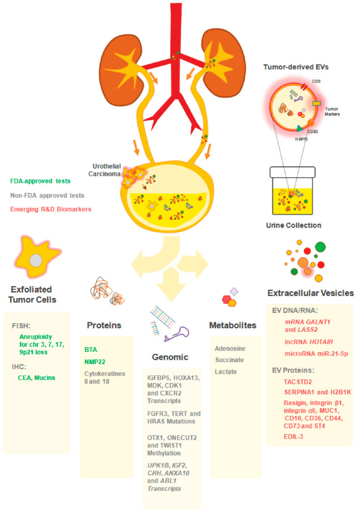
Extracellular vesicles (EVs) are small membrane vesicles released by all cells and involved in intercellular communication. Importantly, EVs cargo includes nucleic acids, lipids, and proteins constantly transferred between different cell types, contributing to autocrine and paracrine signaling. In recent years, they have been shown to play vital roles, not only in normal biological functions, but also in pathological conditions, such as cancer. In the multistep process of cancer progression, EVs act at different levels, from stimulation of neoplastic transformation, proliferation, promotion of angiogenesis, migration, invasion, and formation of metastatic niches in distant organs, to immune escape and therapy resistance. Moreover, as products of their parental cells, reflecting their genetic signatures and phenotypes, EVs hold great promise as diagnostic and prognostic biomarkers. Importantly, their potential to overcome the current limitations or the present diagnostic procedures has created interest in bladder cancer (BCa). Indeed, cystoscopy is an invasive and costly technique, whereas cytology has poor sensitivity for early staged and low-grade disease. Several urine-based biomarkers for BCa were found to overcome these limitations. Here, we review their potential advantages and downfalls. In addition, recent literature on the potential of EVs to improve BCa management was reviewed and discussed.

Keywords: biomarkers; bladder cancer; extracellular vesicles; liquid biopsy; microvesicles; urothelial cancer.


Conflict of interest statement

MHV and HRC are members of the research team of a project financed by Celgene. MHV is member of the team of a grant co-financed by AMGEN. These companies had no role in the decision to publish nor were they involved in the writing of this manuscript.

Figures

Figure 1
Figure 1
Urine biomarkers for bladder cancer (BCa) diagnosis and follow up. Illustration of the distinct available approaches for the detection of urothelial cancer cells in patients’ urine. The close interaction between the bladder tumor and the urine makes this body fluid a reliable source of cancer biomarkers. A plethora of non-invasive assays exploring distinct analytes (exfoliated tumor cells; proteins; genes; metabolites and extracellular vesicles) in patients’ urine allows the longitudinal analysis of tumor progression. Some of the commercially-available tests includes FDA-approved (UroVysion™: aneuploidy of chromosomes 3; 7; 17 and the loss of 9p21 by FISH; ImmunoCyt/uCyt+ test: detection of carcinoembrionary antigen (CEA) and mucins by immunohistochemistry (IHC); bladder tumor antigen (BTA) TRAK/BTA Stat and NMP22 BC test kit); non-FDA approved (CxBladder™: IGFBP5, HOXA13, MDK, CDK1 and CXCR2 by RT-qPCR; Assure MDx™: FGFR3, TERT and HRAS (mutations), OTX1, ONECUT2 and TWIST1 (methylation); XPert Bladder Cancer Monitor™: UPK1B, IGF2, CRH, ANXA10 and ABL1 by RT-qPCR; and UBC™: cytokeratins 8 and 18 by ELISA) and the emerging extracellular vesicles (EV)-based biomarkers (not commercialized yet).
Figure 2
Figure 2
Schematic representation of extracellular vesicles biogenesis. Extracellular and plasma membrane molecules are engulfed by plasma membrane endocytosis, creating the early endosomes. These are converted into late endosomes called multivesicular bodies (MVB) containing intraluminal vesicles (ILV). The MVBs may either fuse with the plasma membrane and empty their ILVs by exocytosis, termed exosomes, or may be converted into lysosomes and degrade their components. The process of microvesicle formation is calcium dependent and comes from direct shedding from outward cellular membrane budding; thereby carrying membrane markers of the parent cell. Apoptotic bodies are produced by secreting cells undergoing programmed cell death. Extracellular vesicle uptake by recipient cells may occur via fusion of the vesicle membrane with the cell membrane or by endocytosis. The vesicle may also transduce an intracellular signal by ligand binding to a receptor on the recipient cell. Abbreviations: MHC—major hystocompatibility complex; ER—endoplasmic reticulum; MVB—multivesicular bodies; ILV—intraluminal vesicles.
Figure 3
Figure 3
Routes of EV delivery to target cells and their potential role in bladder cancer progression. (a) Transfer of EV-enclosed nucleic acids derived urothelial carcinoma cells to nearby naïve fibroblasts can induce their transformation into Cancer-Associated Fibroblasts (CAFs) with altered pro-tumoral secretome. (b) Likewise, transfer of EV-associated lipids and/or oncoproteins to neighbor or distant cells modulate targeted immune cells into an immunosuppressive phenotype and/or facilitate the transformation of healthy epithelial cells. (c) Many of the signaling molecules that integrate the EVs membrane can act directly on the surface receptors of target cells and trigger their own cell signaling pathways without the need of EV internalization. (d) The uptake of EVs derived from drug resistant tumor cells by drug sensitive tumor cells (or even supporting stroma) can mediate the transfer of functional receptors/molecules to recipient cells, thus allowing a similar phenotypic behavior in cells that originally lacked those receptors/molecules (e.g., transfer of functional drug efflux pumps).

References

    1. Babjuk M., Böhle A., Burger M., Capoun O., Cohen D., Compérat E.M., Hernández V., Kaasinen E., Palou J., Rouprêt M., et al. EAU Guidelines on Non-Muscle-Invasive Urothelial Carcinoma of the Bladder. Uptdate 2016. Eur. Urol. 2017;71:447–461. doi: 10.1016/j.eururo.2016.05.041. - DOI - PubMed
    1. Bray F., Ferlay J., Soerjomataram I., Siegelm R.L., Torre L.A., Jemal A. Global cancer statistics 2018: GLOBOCAN estimates of incidence and mortality worldwide for 36 cancers in 185 countries. CA Cancer J. Clin. 2018;68:394–424. doi: 10.3322/caac.21492. - DOI - PubMed
    1. DeGeorge K.C., Holt H.R., Hodges S.C. Bladder Cancer: Diagnosis and Treatment. Am. Fam. Physician. 2017;96:507–514. - PubMed
    1. Bladder cancer: Diagnosis and management of bladder cancer: © NICE (2015) Bladder cancer: Diagnosis and management of bladder cancer. BJU Int. 2017;120:755–765. doi: 10.1111/bju.14045. - DOI - PubMed
    1. Burger M., Catto J.W.F., Dalbagni G., Grossman H.B., Herr H., Karakiewicz P., Kassouf W., Kiemeney L.A., La Vecchia C., Shariat S., et al. Epidemiology and risk factors of urothelial bladder cancer. Eur. Urol. 2013;63:234–241. doi: 10.1016/j.eururo.2012.07.033. - DOI - PubMed

LinkOut - more resources