

Gastric Cancer Staging: Is It Time for Magnetic Resonance Imaging?
- PMID: 32485933
- PMCID: PMC7352169
- DOI: 10.3390/cancers12061402
Gastric Cancer Staging: Is It Time for Magnetic Resonance Imaging?
Abstract
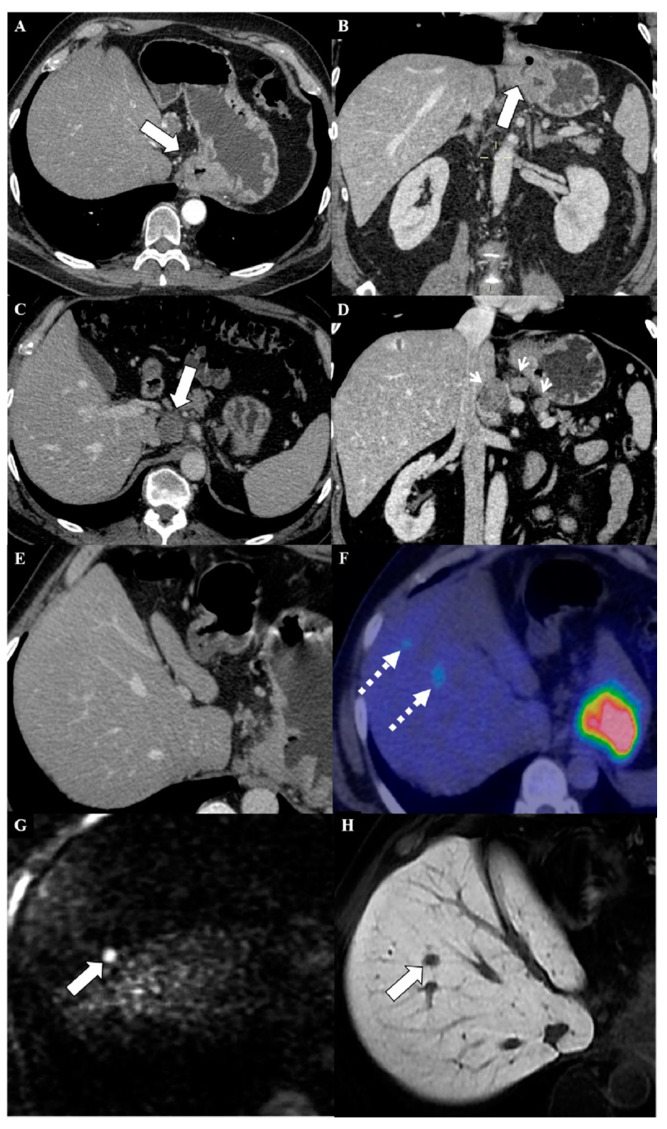
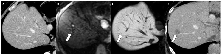
Gastric cancer (GC) is a common cancer worldwide. Its incidence and mortality vary depending on geographic area, with the highest rates in Asian countries, particularly in China, Japan, and South Korea. Accurate imaging staging has become crucial for the application of various treatment strategies, especially for curative treatments in early stages. Unfortunately, most GCs are still diagnosed at an advanced stage, with the peritoneum (61-80%), distant lymph nodes (44-50%), and liver (26-38%) as the most common metastatic locations. Metastatic disease is limited to the peritoneum in 58% of cases; in nonperitoneal distant metastases, the most involved GC metastasization site is the liver (82%). The eighth edition of the tumor-node-metastasis staging system is the most commonly used system for determining GC prognosis. Endoscopic ultrasonography, computed tomography, and 18-fluorideoxyglucose positron emission tomography are historically the most accurate imaging techniques for GC staging. However, studies have recently shown renewed interest in magnetic resonance imaging (MRI) as a useful tool in GC staging, especially for distant metastasis assessment. The technical improvement of diffusion-weighted imaging and the increasing use of hepatobiliary contrast agents have been shown to increase the diagnostic performance of MRI, particularly for detecting peritoneal and liver metastasis. However, no principal oncological guidelines have included the use of MRI as a first-line technique for distant metastasis evaluation during the GC staging process, such as the National Comprehensive Cancer Network Guidelines. This review analyzed the role of the principal imaging techniques in GC diagnosis and staging, focusing on the potential role of MRI, especially for assessing peritoneal and liver metastases.
Keywords: diagnosis; gastric cancer; magnetic resonance imaging; treatment.
Conflict of interest statement
The authors declare no conflicts of interest.
Figures
References
-
- Borggreve A.S., Goense L., Brenkman H.J.F., Mook S., Meijer G.J., Wessels F.J., Verheij M., Jansen E.P.M., van Hillegersberg R., van Rossum P.S.N., et al. Imaging strategies in the management of gastric cancer: Current role and future potential of MRI. Br. J. Radiol. 2019;92:20181044. doi: 10.1259/bjr.20181044. - DOI - PMC - PubMed
Publication types
LinkOut - more resources
Full Text Sources
Miscellaneous
