. 2020 May 29;12(6):358.
doi: 10.3390/toxins12060358.

The Procoagulant Snake Venom Serine Protease Potentially Having a Dual, Blood Coagulation Factor V and X-Activating Activity

Affiliations

The Procoagulant Snake Venom Serine Protease Potentially Having a Dual, Blood Coagulation Factor V and X-Activating Activity

Zorica Latinović et al. Toxins (Basel). .

Abstract

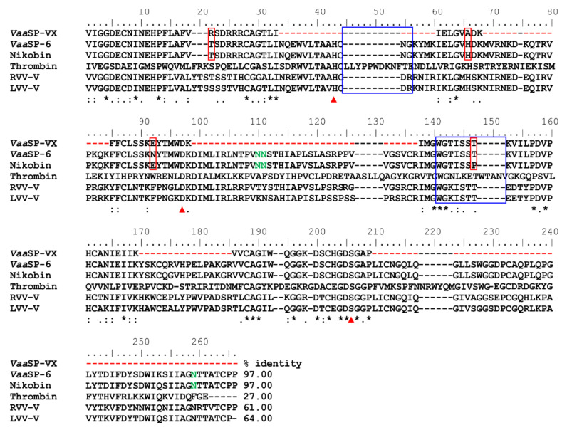
A procoagulant snake venom serine protease was isolated from the venom of the nose-horned viper (Vipera ammodytes ammodytes). This 34 kDa glycoprotein, termed VaaSP-VX, possesses five kDa N-linked carbohydrates. Amino acid sequencing showed VaaSP-VX to be a chymotrypsin-like serine protease. Structurally, it is highly homologous to VaaSP-6 from the same venom and to nikobin from the venom of Vipera nikolskii, neither of which have known functions. VaaSP-VX does not affect platelets. The specific proteolysis of blood coagulation factors X and V by VaaSP-VX suggests that its blood-coagulation-inducing effect is due to its ability to activate these two blood coagulation factors, which following activation, combine to form the prothrombinase complex. VaaSP-VX may thus represent the first example of a serine protease with such a dual activity, which makes it a highly suitable candidate to replace diluted Russell's viper venom in lupus anticoagulant testing, thus achieving greater reliability of the analysis. As a blood-coagulation-promoting substance that is resistant to serpin inhibition, VaaSP-VX is also interesting from the therapeutic point of view for treating patients suffering from hemophilia.

Keywords: FV activator; FX activator; procoagulant; serine protease; snake venom.


Conflict of interest statement

The authors declare no conflict of interest. The funders had no role in the design of the study; in the collection, analyses, or interpretation of data; in the writing of the manuscript, or in the decision to publish the results.

Figures

Figure 1
Figure 1
The purification and basic characterization of VaaSP-VX. (A) Size-exclusion chromatography of the raw Vaa venom was carried out on a Sephacryl S-200 superfine column. The elution of proteins was monitored at 280 nm (A280). (B) Fraction B1 from the size-exclusion chromatography was sub-fractionated on a Mono S 5/50 GL ion-exchange fast protein liquid chromatography (FPLC) column. The retained proteins were eluted using a linear gradient of NaCl (dotted line) in 11 fractions. (C) Fractions were tested for their effect on blood coagulation. Three clinical blood coagulation assays were used: a prothrombin time assay (PT), an activated partial thromboplastin time assay (aPTT), and a thrombin time assay (TT). The table shows the effects of each fraction on aPTT, PT, and TT in terms of the percent change relative to the value obtained in the control experiment performed without the addition of a fraction. Fraction 10, which displayed a pronounced procoagulant effect, contained pure VaaSP-VX (values in bold). (D) Under reducing conditions, SDS-PAGE electrophoresis revealed VaaSP-VX to be a monomeric protein of about 34 kDa. Incubation of VaaSP-VX with peptide N-glycosidase F (PNGF) at 37 °C overnight followed by SDS-PAGE analysis displayed a lower molecular mass of about 29 kDa. VaaSP-VX was thus found to be an N-glycosylated protein.
Figure 2
Figure 2
Effects of VaaSP-VX on platelets and fibrinogen. (A) Platelet-rich plasma was pre-incubated with 1 μM VaaSP-VX at 37 °C. The agonists of platelet aggregation (ADP and collagen) or agglutination (ristocetin) were then added. Platelet aggregation/agglutination was followed by monitoring the optical density of the samples. Control values, obtained in the absence of the venom protein, were regarded as a 100% change of optical density. Evidently, VaaSP-VX did not affect the platelet function. (B) VaaSP-VX, in concentrations from 0.1 to 10 µM, was added to human fibrinogen. Thrombin was used as a positive control. The formation of fibrin clots was tracked using continuous measurements of the absorbance at 405 nm (A405). In the control experiment, thrombin induced a transformation of fibrinogen to fibrin, as indicated by the rise in A405 (red line). For all concentrations tested, VaaSP-VX did not stimulate clotting (black lines). (C) Human fibrinogen, consisting of α, β, and γ chains, was incubated with VaaSP-VX in a mass ratio of 100:1. In the control experiment, no VaaSP-VX was added. Following incubation at 37 °C for 60 min, the samples were analyzed on SDS-PAGE under reducing conditions and stained with PageBlue®. Only a slight degradation of the α-chain was apparent; therefore, the pronounced procoagulant effect of VaaSP-VX could not be due to its action on fibrinogen.
Figure 3
Figure 3
Clotting of fresh or coagulation factor-depleted plasmas in the presence of VaaSP-VX. (A) To fresh human plasma, VaaSP-VX was added at indicated concentrations and incubated at 37 °C for 10 min. The activated partial thromboplastin (aPTT) was then measured as described under Section 5: Materials and Methods. (B) To assess the plasma re-calcification time, the same procedure as in (A) was used, except the addition of the aPTT SA reagent was replaced by the addition of PBS. (C) To human plasma depleted in FVII, VaaSP-VX was added at the indicated concentrations. After 2 min of incubation at 37 °C, 25 mM CaCl2 was added and the formation of fibrin clotting was observed by measuring the absorbance at 405 nm (A405).
Figure 4
Figure 4
Hydrolysis of FV and FX by VaaSP-VX. Bovine FV (A), (or human FX (B)), was incubated with or without VaaSP-VX. An aliquot of the respective reaction mixture (10 µL) was taken immediately (0 h) and then after 1, 3, 6, and 24 h of incubation at 37 °C; it was then analyzed on SDS-PAGE under reducing conditions. The gels were stained with PageBlue® to visualize the protein bands. As products of degradation by VaaSP-VX, the protein bands of FV or FX (arrows) were electroblotted to the PVDF membrane and N-terminally sequenced to define the cleavage positions. VaaSP-VX activated FV into FVa and FX into FXa. Schematic presentations of the domain structures of FV (C) and FX (D), with the indicated sites of activation by the following physiological activators: FXa, thrombin (FIIa), FVIIa, and FIXa, and RVV-V (below), and VaaSP-VX (above). Abbreviations: A1, A2, A3, B, C1, and C2: structural domains of FV; AP: activation peptide; EGF, GLA, and SP: epidermal growth factor-like, γ-carboxyglutamic acid-rich, and serine protease domain, respectively; LC, HC, and HC’: light, heavy, and C-terminally truncated heavy chain, respectively.
Figure 5
Figure 5
Partial amino acid sequence of VaaSP-VX and its comparison with sequences of some relevant serine proteases (SPs). VaaSP-VX was sequenced using a combination of Edman degradation and tandem MS. Based on its established partial structure, VaaSP-VX was recognized as an isoform of VaaSP-6, whose complete cDNA sequence was determined and deposited at the National Center for Biotechnology Information (NCBI, MG958495). Nikobin, an SP isolated from Vipera nikolskii venom, is a protein with the same extent of structural identity to the sequenced parts of VaaSP-VX as VaaSP-6 (97.0%). Amino acid residues, where differences between VaaSP-VX and VaaSP-6 or nikobin occurred, are in red boxes. Red arrows at the bottom of the aligned sequences point to three amino acid residues that constituted the catalytic triad in SPs (His57, Asp102, and Ser195, according to chymotrypsin numbering). The undetermined parts of the VaaSP-VX sequence are denoted by red dashes, while predicted gaps are denoted by black ones. Amino acid residues that may be N-glycosylated in VaaSP-VX (if present) are printed in green. Other SPs that were aligned are human thrombin, which is the main physiological activator of FV, and two snake venom FV activators, namely RVV-V from Daboia siamensis and LVV-V from Macrovipera lebetina. Except for thrombin, the two loops, 44-loop and 148-loop (in blue squares), are absent in all presented FV activators from snake venoms. This was established as the reason why snake venom SPs were resistant to inhibition by antithrombin. Below the sequences, an asterisk (*) designates a position that is strictly conserved in all aligned sequences, a colon (:) denotes a position in which only conservative substitutions were detected, and a dot (.) denotes a position harboring semi-conservative substitutions. On all other sites, the substitutions were non-conservative. The percentages of identity between the sequence of VaaSP-VX and the corresponding sequences in VaaSP-6, nikobin, thrombin, RVV-V, and LVV-V are presented.

Similar articles

Cited by

References

    1. Kang T.S., Georgieva D., Genov N., Murakami M.T., Sinha M., Kumar R.P., Kaur P., Kumar S., Dey S., Sharma S., et al. Enzymatic toxins from snake venom: Structural characterization and mechanism of catalysis. FEBS J. 2011;278:4544–4576. doi: 10.1111/j.1742-4658.2011.08115.x. - DOI - PubMed
    1. Sajevic T., Leonardi A., Križaj I. Haemostatically active proteins in snake venoms. Toxicon. 2011;57:627–645. doi: 10.1016/j.toxicon.2011.01.006. - DOI - PubMed
    1. Alvarez-Flores M.P., Faria F., de Andrade S.A., Chudzinski-Tavassi A.M. Snake venom components affecting the coagulation system. In: Gopalakrishnakone P., Inagaki H., Vogel C.W., Mukherjee A.K., Rahmy T.R., editors. Snake Venoms. Springer; Dordrecht, The Netherlands: 2017. pp. 417–436.
    1. Tans G., Rosing J. Snake venom activators of factor X: An overview. Haemostasis. 2001;31:225–233. doi: 10.1159/000048067. - DOI - PubMed
    1. Rawlings N.D., Barrett A.J., Bateman A. Merops: The database of proteolytic enzymes, their substrates and inhibitors. Nucleic Acids Res. 2012;40:343–350. doi: 10.1093/nar/gkr987. - DOI - PMC - PubMed

Publication types