Review
. 2020 May 31;9(6):1361.
doi: 10.3390/cells9061361.

Current View on EpCAM Structural Biology

Affiliations
Review

Current View on EpCAM Structural Biology

Aljaž Gaber et al. Cells. .

Abstract

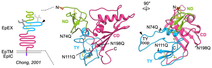
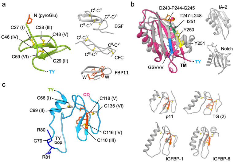
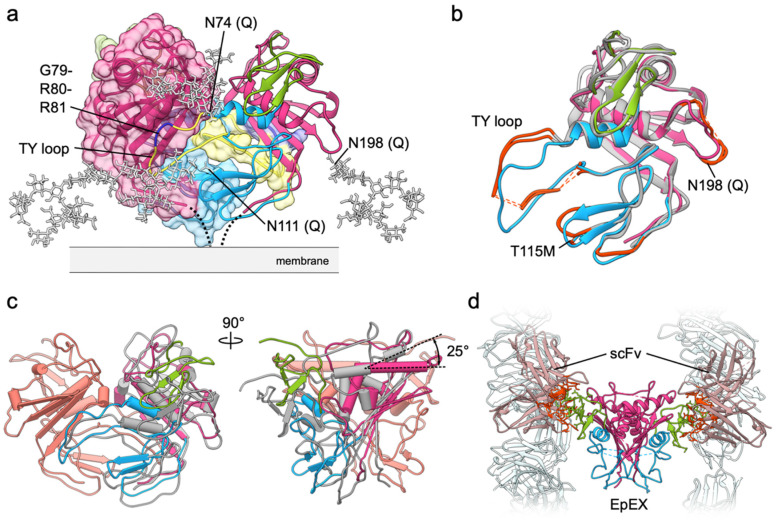
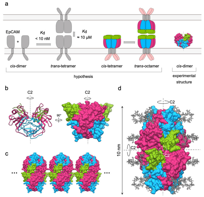
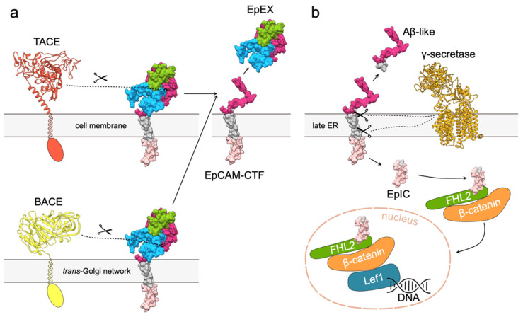
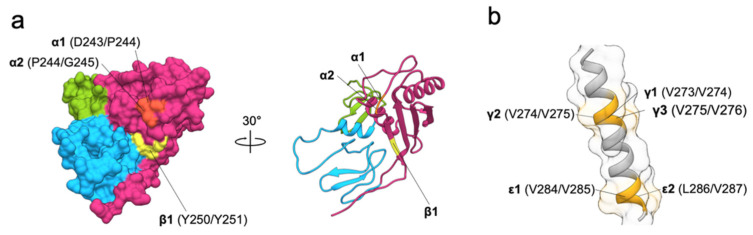
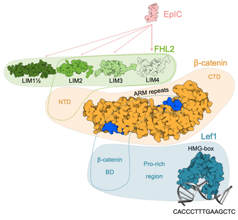
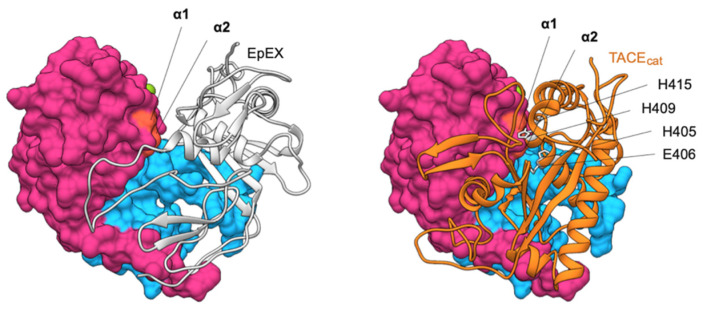
EpCAM, a carcinoma cell-surface marker protein and a therapeutic target, has been primarily addressed as a cell adhesion molecule. With regard to recent discoveries of its role in signaling with implications in cell proliferation and differentiation, and findings contradicting a direct role in mediating adhesion contacts, we provide a comprehensive and updated overview on the available structural data on EpCAM and interpret it in the light of recent reports on its function. First, we describe the structure of extracellular part of EpCAM, both as a subunit and part of a cis-dimer which, according to several experimental observations, represents a biologically relevant oligomeric state. Next, we provide a thorough evaluation of reports on EpCAM as a homophilic cell adhesion molecule with a structure-based explanation why direct EpCAM participation in cell-cell contacts is highly unlikely. Finally, we review the signaling aspect of EpCAM with focus on accessibility of signaling-associated cleavage sites.

Keywords: Keywords: EpCAM; adhesion; dimer; disease; regulated intramembrane proteolysis; signaling; structure; transmembrane protein.


Conflict of interest statement

The authors declare no conflict of interest.

Figures

Figure 1
Figure 1
Structure of EpCAM extracellular part (EpEX). Left, adaptation of EpCAM model as presented by Chong et al. [15]; the N74 and N111 are shown as partially and fully glycosylated (gray and black, respectively), and the main protease-sensitive site marked by an arrowhead. Middle and right, crystal structure of EpEX (PDB 4MZV) in ribbon representation depicts the three domains (ND, TY and CD), disulfide linkages (yellow spheres), N-terminal pyroglutamate residue (orange-red sticks), and three glycosylation sites where asparagine was mutated to glutamine to abolish glycosylation and thereby achieve a homogenous protein sample for structure determination (mutations N74Q, N111Q, N198Q; dark gray sticks). Polypeptide chain is color-coded according to the domains and the same color coding is used throughout the paper. This and all other structural figures were prepared using UCSF Chimera version 1.14 (University of California San Francisco, Resource for Biocomputing, Visualization, and Informatics, San Francisco, USA) [16].
Figure 2
Figure 2
Domains of the EpCAM extracellular part. (a) ND (green ribbon) and its disulfide-packed core (yellow sticks); N-terminal pyroglutamate residue is shown in red sticks. Examples of other small cysteine-rich domains are shown on the right (gray ribbons): 9th EGF-like domain of human EGF (PDB 1JL9), CFC domain of murine Crypto (PDB ID 2J5H), and WW domain of human FBP11 (PDB 1YWJ) with characteristic tryptophan and tyrosine residues (orange-red sticks); disulfide bonds with connectivity order are shown as yellow sticks. (b) CD (deep pink ribbon) of EpCAM superimposed on the structure of the SEA domain of human MUC1 (gray; PDB 2ACM) which has an auto-proteolytic motif GSVVV (blue). Right, the sea urchin sperm (SEA) domains of human receptor-type tyrosine-protein phosphatase IA-2 (PDB 2QT7) and Notch receptor (PDB 3ETO), both without an auto-proteolytic activity, shown in the same orientation. Identified cleavage sites within CD by TACE (D243–P244–G245) and BACE (Y250–Y251) are shown in orange and yellow, respectively, and the AGR2-binding region is shown in dark green (overlapping the BACE cleavage site). The superposition was done using UCSF Chimera [16]. RMSD values range from 1.75 (IA-2 and EpCAM pair) to 3.31 Å (IA-2 and NOTCH1 pair) with an overall RMSD of 2.79 Å. (c) TY domain of EpCAM (left) with indicated disulfide bridges (yellow) and the protease-sensitive site GRR (dark blue). Homologous TY domains from p41 invariant chain (PDB 1ICF), thyroglobulin (TG, 2nd TY type-1 domain; PDB 6SCJ), and IFGBP-1 and -6 (PDB 1ZT3 and 1RMJ) are shown in gray. TY-characteristic CWCV sequence motif is shown in orange-red.
Figure 3
Figure 3
EpEX cis-dimer. (a) The EpEX cis-dimer is mostly stabilized by interactions between TY loop (yellow) and concave β-sheet of the CD. Both subunits are shown as ribbons, with one covered by molecular surface color-coded in the same manner. Modeled high-mannose glycans are shown as gray sticks. The protease-sensitive site within TY loop is shown in dark blue. (b) Superposition of one EpEX subunit from EpEX-only structure (PDB 4MZV; color-coded by domains) and of the two subunits from the EpEX-scFv structure (PDB 6I07; gray). Significant structural differences are marked with orange-red. Missing segments in the EpEX-scFv structure are shown as dotted lines. (c) Superposition of the cis-dimers (calculated over one subunit) from the EpEX-only and scFv-EpEX structures. For the superposed subunits only one is shown (salmon), for the other subunit the color coding is the same as in (b). (d) In EpEX-scFv structure (PDB 6I07) the scFv molecules (light gray-pink) interact via several residues (orange-red) with NDs of the EpEX dimer (color coded by domains, ND in green). Few other symmetry-related scFv molecules in the crystal are shown in light gray.
Figure 4
Figure 4
Models of EpCAM homo-oligomerization and formation of adhesion units. (a) The initial cis-dimer/trans-tetramer and cis-tetramer/trans-octamer hypotheses proposed in 2001 (left and center, respectively) and the experimental structure of EpCAM dimer from 2014 (right). (b) The symmetry of EpCAM cis-dimer in two different orientations, depicted as ribbons and molecular surface. Axis of rotation (C2) is shown as a dashed line. (c) Potential clustering of EpCAM adhesion units due to inherent symmetry implying lateral extension in both directions (dots). (d) Adaptation of the trans-tetramer model presented by Pavšič et al. in 2014 [11] with three C2 axes indicated by black dotted lines or a white dot.
Figure 5
Figure 5
EpCAM signaling via RIP. (a) Cleavage of EpCAM extracellular part by either TACE or BACE results in release of soluble EpEX and membrane bound EpCAM-CTF. Extracellular part of TACE was modeled using Robetta [53,54], and structure extracellular part of BACE was obtained from the Protein Data Bank (PDB 2WJO). Transmembrane and cytosolic regions of both proteases are depicted schematically. EpCAM is presented as molecular surface; transmembrane region and intracellular domain are shown in gray and light pink, respectively. (b) Cleavage of EpCAM-CTF by γ -secretase complex (PDB 5A63) results in release of Aβ-like peptide and EpIC that is recruited in the EpIC–FHL2–β-catenin–Lef1 signaling complex. FHL (green), β-catenin (orange) and Lef1 (blue) are depicted by shapes corresponding to their relative sizes.
Figure 6
Figure 6
Structural details of RIP cleavages. (a) TACE (⍺1, ⍺2, orange) and BACE (β1, yellow) cleavage sites on EpEX shown in two different orientations. (b) γ-secretase (γ1–γ3 and ε1, ε2; light orange) cleavage sites in EpCAM transmembrane region.
Figure 7
Figure 7
Schematic model of EpIC–FHL2–β-catenin–Lef1 signaling complex. EpIC was modeled using MODELLER [64]. Binding of EpIC to FHL2 is indicated by dotted lines (light pink; width is related to importance of interaction). First and a half, second, third and fourth domain of FHL2 are depicted based on corresponding NMR structures (PDB 2MIU, 1X4K, 2D8Z, and 1X4L respectively). Binding of FHL2 to β-catenin N-terminal domain is indicated by a green dotted outline. β-catenin is represented by structure of ARM repeats with bound part of Lef1 β-catenin BD (PDB 3OUW) and relative positions of N- and C-terminal domains (NTD and CTD, respectively), the structures of which are yet unknown. Position of β-catenin BD is indicated by blue dotted outline. Structure of Lef1, except for the C-terminal HMG-BOX bound to its target DNA sequence (PDB 2LEF), is not known. β-catenin BD and Pro-rich region are indicated at their relative position.
Figure 8
Figure 8
Comparison of EpEX cis-dimer structure (left) and model of EpEX–TACEcat complex (right). One subunit of the dimer (gray ribbon) covers the cleavage site within the other subunit (molecular surface, cleavage site in orange). This cleavage site is in EpEX monomer easily accessible as shown by the complex of one subunit with a catalytic domain of TACE (TACEcat; orange ribbon). The model was generated using HADDOCK [67] with α1 and α2 cleavage sites on EpEX (orange surface) or TACE active and zinc binding site (H405, E406, H409, and H415; gray side chains) used as interaction restraints.

References

    1. Herlyn M., Steplewski Z., Herlyn D., Koprowski H. Colorectal carcinoma-specific antigen: Detection by means of monoclonal antibodies. Proc. Natl. Acad. Sci. USA. 1979;76:1438–1442. - PMC - PubMed
    1. Keller L., Werner S., Pantel K. Biology and clinical relevance of EpCAM. Cell Stress. 2019;3:165–180. doi: 10.15698/cst2019.06.188. - DOI - PMC - PubMed
    1. Murakami N., Mori T., Nakamura S., Yoshimoto S., Honma Y., Ueno T., Kobayashi K., Kashihara T., Takahashi K., Inaba K., et al. Prognostic value of the expression of epithelial cell adhesion molecules in head and neck squamous cell carcinoma treated by definitive radiotherapy. J. Radiat. Res. 2019;60:803–811. doi: 10.1093/jrr/rrz053. - DOI - PMC - PubMed
    1. Litvinov S.V., Velders M.P., A Bakker H., Fleuren G.J., O Warnaar S. Ep-CAM: A human epithelial antigen is a homophilic cell-cell adhesion molecule. J. Cell Boil. 1994;125:437–446. doi: 10.1083/jcb.125.2.437. - DOI - PMC - PubMed
    1. Tsaktanis T., Kremling H., Pavšič M., Von Stackelberg R., Mack B., Fukumori A., Steiner H., Vielmuth F., Spindler V., Huang Z., et al. Cleavage and Cell Adhesion Properties of Human Epithelial Cell Adhesion Molecule (HEPCAM)*. J. Boil. Chem. 2015;290:24574–24591. doi: 10.1074/jbc.M115.662700. - DOI - PMC - PubMed

Publication types

Substances

LinkOut - more resources