. 2020 Jun 2;13(1):86.
doi: 10.1186/s13041-020-00626-0.

Propionic acid induces dendritic spine loss by MAPK/ERK signaling and dysregulation of autophagic flux

Affiliations

Propionic acid induces dendritic spine loss by MAPK/ERK signaling and dysregulation of autophagic flux

Hyosun Choi et al. Mol Brain. .

Abstract

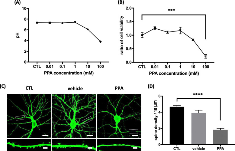
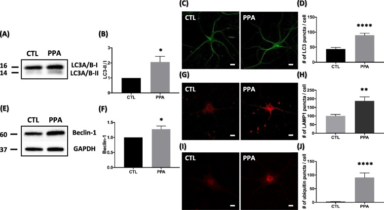
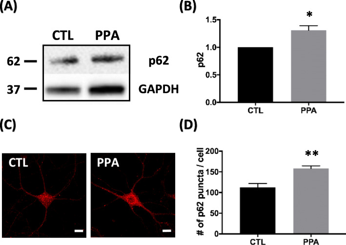
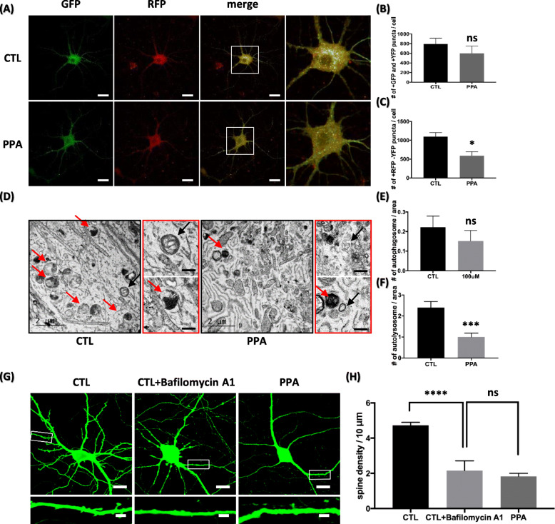
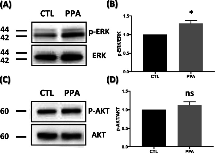
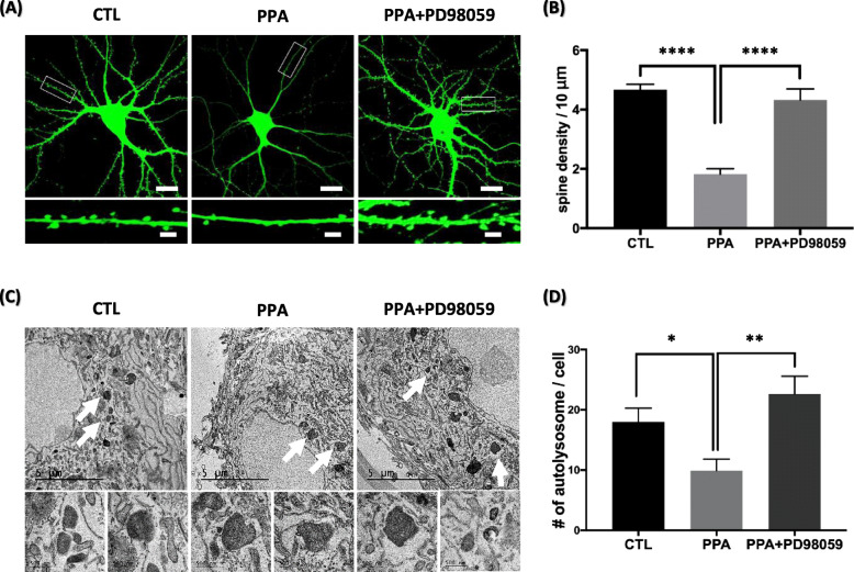
Propionic acid (PPA) is a short-chain fatty acid that is an important mediator of cellular metabolism. It is also a by-product of human gut enterobacteria and a common food preservative. A recent study found that rats administered with PPA showed autistic-like behaviors like restricted interest, impaired social behavior, and impaired reversal in a T-maze task. This study aimed to identify a link between PPA and autism phenotypes facilitated by signaling mechanisms in hippocampal neurons. Findings indicated autism-like pathogenesis associated with reduced dendritic spines in PPA-treated hippocampal neurons. To uncover the mechanisms underlying this loss, we evaluated autophagic flux, a functional readout of autophagy, using relevant biomedical markers. Results indicated that autophagic flux is impaired in PPA-treated hippocampal neurons. At a molecular level, the mitogen-activated protein kinase (MAPK)/extracellular signal-regulated kinase (ERK) pathway was activated and autophagic activity was impaired. We also observed that a MAPK inhibitor rescued dendritic spine loss in PPA-treated hippocampal neurons. Taken together, these results suggest a previously unknown link between PPA and autophagy in spine formation regulation in hippocampal neurons via MAPK/ERK signaling. Our results indicate that MAPK/ERK signaling participates in autism pathogenesis by autophagy disruption affecting dendritic spine density. This study may help to elucidate other mechanisms underlying autism and provide a potential strategy for treating ASD-associated pathology.

Keywords: Autophagy; MAPK/ERK signaling; Propionic acid; Short-chain fatty acid; Spine density.


Conflict of interest statement

The authors declare no competing financial interests.

Figures

Fig. 1
Fig. 1
PPA decreases density of dendritic spines in hippocampal neurons (a) pH. b Cell viability. pH was measured in cultured media. Hippocampal neurons were incubated with PPA in concentrations ranging from 0 to 100 mM for 72 h. Viability was measured by CCK-8 assay. Results are expressed as percentage of viable cells and represent mean ± SEM of three samples (n = 3). c Top shows representative pyramidal neurons transfected with PLV-GFP and treated with vehicle (PBS) and PPA (100 μM) for 72 h. Bottom shows high magnification of representative secondary dendrites. Scale bar of top panel is 20 μm; scale bar of bottom panel is 1 μm. d Bar graph showing mean ± SEM for dendritic spine numbers of the representative groups (n = 50). ***p < 0.0005, ****p < 0.0001
Fig. 2
Fig. 2
PPA upregulates biochemical markers of autophagy in hippocampal neurons (a) Representative western blot of LC3 after PPA treatment. b Quantification of LC3-II levels with respect to LC3-I levels (n = 4). c) Representative immunofluorescence images showing LC3 staining in hippocampal neurons and PPA-treated cells. d Quantification of number of LC3 puncta from images in (C) (n = 10). e Representative Western blot showing increased expression of beclin-1 for 72 h after PPA treatment. f Quantification of beclin-1 levels with respect to GAPDH levels (n = 4). g Representative immunofluorescence images showing LAMP1 staining in hippocampal neurons and PPA-treated cells. h Quantification of the number of LAMP1 puncta from images in (G) (n = 12). i Representative Immunofluorescence images showing poly-ubiquitin staining in hippocampal neurons and PPA-treated cells. j Quantification of the number of poly-ubiquitin puncta from the images in (I) (n = 10). Bar graph showing mean ± SEM of the representative groups. Scale bar is 10 μm. *p < 0.05, **p < 0.01, ****p < 0.0001
Fig. 3
Fig. 3
Autophagic degradation impaired by PPA (a) Representative western blot of p62 after PPA treatment. b Quantification of p62 levels with respect to GAPDH (n = 4). c Representative immunofluorescence images showing p62 staining in hippocampal neurons and PPA-treated cells. d Quantification of number of p62 puncta from images in (C) (n = 8). Bar graph showing mean ± SEM of representative groups. Scale bar is 10 μm. *p < 0.05, **p < 0.01
Fig. 4
Fig. 4
PPA dysregulates fusion from autophagosome to autolysosome (a) Representative fluorescent images after transfection with RFP-GFP-LC3B in control hippocampal neurons and PPA-treated cells. Scale bar is 20 μm. b, c Quantification of number of GFP and YFP puncta (autophagosome) and RFP only puncta (autolysosome) from images in (A) (n = 5). d Black box shows representative TEM images in control hippocampal neurons and PPA-treated cells. Red box shows representative TEM high magnification images of autolysosome and autophagosome (black arrow indicates autophagosome and red arrow indicates autolysosome). Black scale bar is 500 μm. e, f Quantification of autophagosome and autolysosome number per area (n = 35). Bar graph showing mean ± SEM of the representative groups. g Top shows representative pyramidal neurons transfected with PLV-GFP and treated with bafilomycin A1 (2 nM) and PPA (100 μM) for 72 h. Bottom shows high magnification of representative secondary dendrites. Scale bar of top panel is 20 μm; scale bar of bottom panel is 1 μm. h Bar graph showing mean ± SEM for dendritic spine numbers of the representative groups (n = 50). *p < 0.05, ***p < 0.0005, ****p < 0.0001
Fig. 5
Fig. 5
PPA activates MAPK/ERK pathway associated with autophagy impairment a Representative western blot of p-ERK and ERK after PPA treatment. b Quantification of p-ERK levels compared to ERK levels (n = 3). c Representative western blot of p-AKT and AKT after PPA treatment. d Quantification of p-AKT levels compared to AKT (n = 3). Bar graph showing mean ± SEM of the representative groups. *p < 0.05
Fig. 6
Fig. 6
PD98059 rescues spine density and autophagy disruption in PPA-treated hippocampal neurons a Top shows representative pyramidal neurons transfected with PLV-GFP and treated with PPA (100 μM) and PD98059 (10 μM) for 72 h. Bottom shows high magnification of representative secondary dendrites. Scale bar of top panel is 20 μm; scale bar of bottom panel is 1 μm. b Bar graph showing mean ± SEM for dendritic spine numbers of the representative groups (n = 50). c Top shows representative TEM images in PPA (100 μM) and PD98059 (10 μM) for 72 h (white arrow is autolysosome). Bottom shows high magnification of representative images of autolysosome. d Quantification of autolysosome number per cell (n = 10). Bar graph showing mean ± SEM of the representative groups. *p < 0.05, **p < 0.01, ****p < 0.0001

References

    1. Johnson CC, Ownby DR. Allergies and asthma: do atopic disorders result from inadequate immune homeostasis arising from infant gut dysbiosis? Expert Rev Clin Immunol. 2016;12(4):379–388. - PMC - PubMed
    1. Nicholson JK, Holmes E, Kinross J, Burcelin R, Gibson G, Jia W, et al. Host-gut microbiota metabolic interactions. Science. 2012;336(6086):1262–1267. - PubMed
    1. Cureau N, AlJahdali N, Vo N, Carbonero F. Epigenetic mechanisms in microbial members of the human microbiota: current knowledge and perspectives. Epigenomics. 2016;8:1259–1273. - PubMed
    1. Woo V, Alenghat T. Host–microbiota interactions: epigenomic regulation. Curr Opin Immunol. 2017;44:52–60. - PMC - PubMed
    1. Bourassa MW, Alim I, Bultman SJ, Ratan RR. Butyrate, neuroepigenetics and the gut microbiome: can a high fiber diet improve brain health? Neurosci Lett. 2016;625:56–63. - PMC - PubMed

Publication types

MeSH terms

LinkOut - more resources