

Zika Virus NS3 Protease Pharmacophore Anchor Model and Drug Discovery
- PMID: 32488021
- PMCID: PMC7265434
- DOI: 10.1038/s41598-020-65489-w
Zika Virus NS3 Protease Pharmacophore Anchor Model and Drug Discovery
Abstract
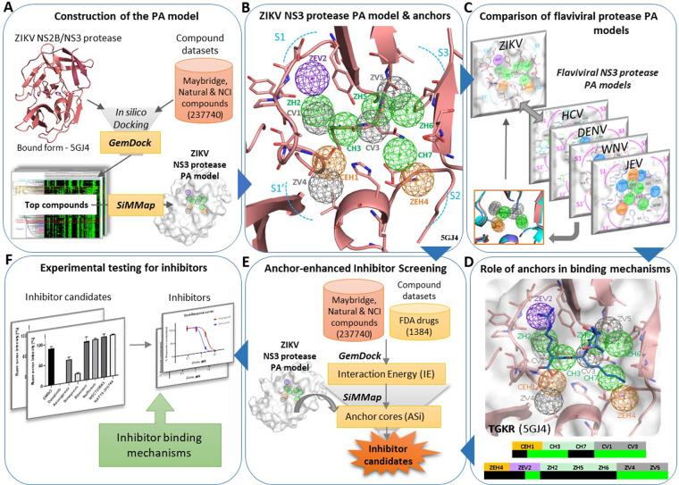
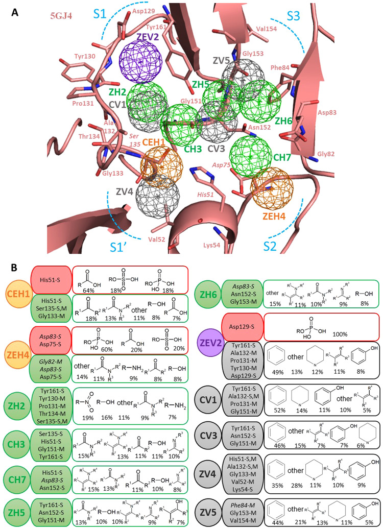
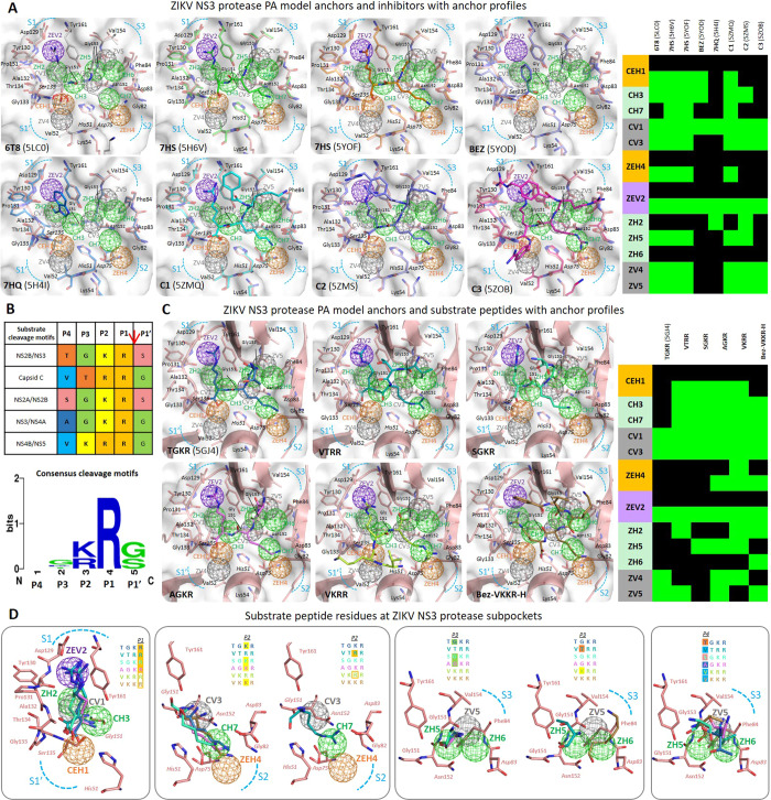
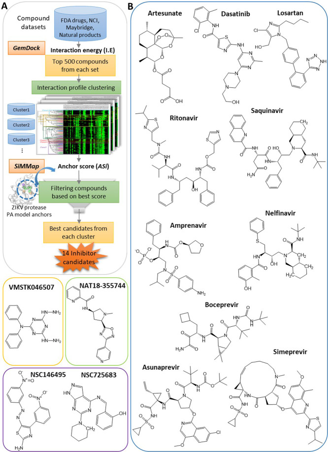
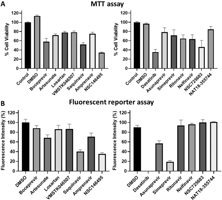
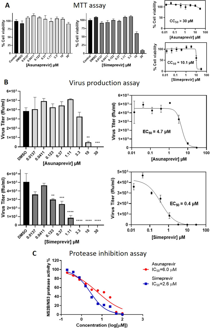
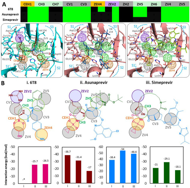
Zika virus (ZIKV) of the flaviviridae family, is the cause of emerging infections characterized by fever, Guillain-Barré syndrome (GBS) in adults and microcephaly in newborns. There exists an urgent unmet clinical need for anti-ZIKV drugs for the treatment of infected individuals. In the current work, we aimed at the promising virus drug target, ZIKV NS3 protease and constructed a Pharmacophore Anchor (PA) model for the active site. The PA model reveals a total of 12 anchors (E, H, V) mapped across the active site subpockets. We further identified five of these anchors to be critical core anchors (CEH1, CH3, CH7, CV1, CV3) conserved across flaviviral proteases. The ZIKV protease PA model was then applied in anchor-enhanced virtual screening yielding 14 potential antiviral candidates, which were tested by in vitro assays. We discovered FDA drugs Asunaprevir and Simeprevir to have potent anti-ZIKV activities with EC50 values 4.7 µM and 0.4 µM, inhibiting the viral protease with IC50 values 6.0 µM and 2.6 µM respectively. Additionally, the PA model anchors aided in the exploration of inhibitor binding mechanisms. In conclusion, our PA model serves as a promising guide map for ZIKV protease targeted drug discovery and the identified 'previr' FDA drugs are promising for anti-ZIKV treatments.
Conflict of interest statement
The authors declare no competing interests.
Figures
Similar articles
-
Pharmacophore anchor models of flaviviral NS3 proteases lead to drug repurposing for DENV infection.BMC Bioinformatics. 2017 Dec 28;18(Suppl 16):548. doi: 10.1186/s12859-017-1957-5. BMC Bioinformatics. 2017. PMID: 29297305 Free PMC article.
-
Structure-based discovery of clinically approved drugs as Zika virus NS2B-NS3 protease inhibitors that potently inhibit Zika virus infection in vitro and in vivo.Antiviral Res. 2017 Sep;145:33-43. doi: 10.1016/j.antiviral.2017.07.007. Epub 2017 Jul 14. Antiviral Res. 2017. PMID: 28712942
-
Characterization of the Zika virus two-component NS2B-NS3 protease and structure-assisted identification of allosteric small-molecule antagonists.Antiviral Res. 2017 Jul;143:218-229. doi: 10.1016/j.antiviral.2017.04.015. Epub 2017 Apr 29. Antiviral Res. 2017. PMID: 28461069 Free PMC article.
-
Strategies for Zika drug discovery.Curr Opin Virol. 2019 Apr;35:19-26. doi: 10.1016/j.coviro.2019.01.005. Epub 2019 Mar 7. Curr Opin Virol. 2019. PMID: 30852345 Review.
-
Exploiting the unique features of Zika and Dengue proteases for inhibitor design.Biochimie. 2019 Nov;166:132-141. doi: 10.1016/j.biochi.2019.05.004. Epub 2019 May 9. Biochimie. 2019. PMID: 31077760 Review.
Cited by
-
C-Terminal Extended Hexapeptides as Potent Inhibitors of the NS2B-NS3 Protease of the ZIKA Virus.Front Med (Lausanne). 2022 Jul 6;9:921060. doi: 10.3389/fmed.2022.921060. eCollection 2022. Front Med (Lausanne). 2022. PMID: 35872792 Free PMC article.
-
ZIKV Inhibitors Based on Pyrazolo[3,4-d]pyridazine-7-one Core: Rational Design, In Vitro Evaluation, and Theoretical Studies.ACS Omega. 2023 Dec 14;8(51):48994-49008. doi: 10.1021/acsomega.3c06612. eCollection 2023 Dec 26. ACS Omega. 2023. PMID: 38162759 Free PMC article.
-
A high-throughput cell-based screening method for Zika virus protease inhibitor discovery.SLAS Discov. 2024 Jul;29(5):100164. doi: 10.1016/j.slasd.2024.100164. Epub 2024 May 24. SLAS Discov. 2024. PMID: 38796112 Free PMC article.
-
Structure-Based Virtual Screening: Identification of a Novel NS2B-NS3 Protease Inhibitor with Potent Antiviral Activity against Zika and Dengue Viruses.Microorganisms. 2021 Mar 6;9(3):545. doi: 10.3390/microorganisms9030545. Microorganisms. 2021. PMID: 33800763 Free PMC article.
-
Employing Machine Learning-Based QSAR for Targeting Zika Virus NS3 Protease: Molecular Insights and Inhibitor Discovery.Pharmaceuticals (Basel). 2024 Aug 15;17(8):1067. doi: 10.3390/ph17081067. Pharmaceuticals (Basel). 2024. PMID: 39204173 Free PMC article.
References
Publication types
MeSH terms
Substances
LinkOut - more resources
Full Text Sources
Medical
