. 2020 Jun 2;11(1):2765.
doi: 10.1038/s41467-020-16533-w.

AGO-bound mature miRNAs are oligouridylated by TUTs and subsequently degraded by DIS3L2

Affiliations

AGO-bound mature miRNAs are oligouridylated by TUTs and subsequently degraded by DIS3L2

Acong Yang et al. Nat Commun. .

Abstract

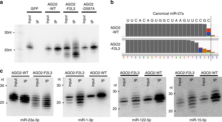
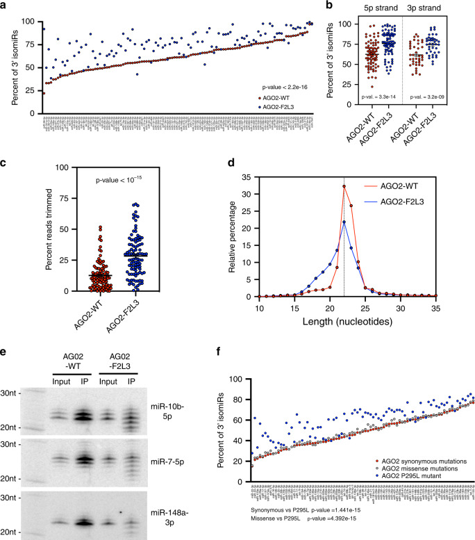
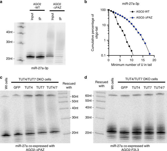
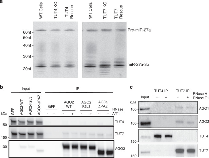
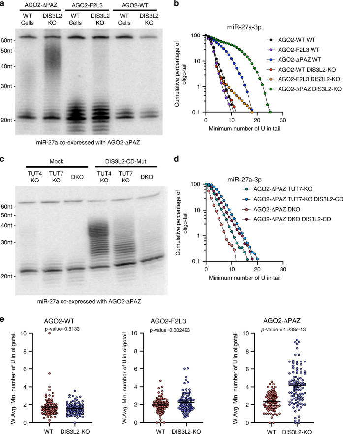
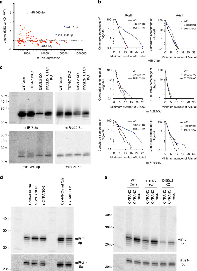
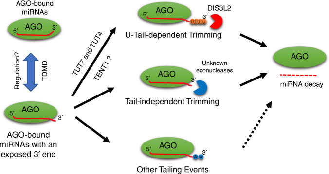
MicroRNAs (miRNAs) associated with Argonaute proteins (AGOs) regulate gene expression in mammals. miRNA 3' ends are subject to frequent sequence modifications, which have been proposed to affect miRNA stability. However, the underlying mechanism is not well understood. Here, by genetic and biochemical studies as well as deep sequencing analyses, we find that AGO mutations disrupting miRNA 3' binding are sufficient to trigger extensive miRNA 3' modifications in HEK293T cells and in cancer patients. Comparing these modifications in TUT4, TUT7 and DIS3L2 knockout cells, we find that TUT7 is more robust than TUT4 in oligouridylating mature miRNAs, which in turn leads to their degradation by the DIS3L2 exonuclease. Our findings indicate a decay machinery removing AGO-associated miRNAs with an exposed 3' end. A set of endogenous miRNAs including miR-7, miR-222 and miR-769 are targeted by this machinery presumably due to target-directed miRNA degradation.


Conflict of interest statement

The authors declare no competing interests.

Figures

Fig. 1
Fig. 1. Disrupting the AGO PAZ domain promotes miRNA 3′ modification.
a Co-expression of pri-miR-27a and FLAG-AGO2 constructs: wild-type (WT), PAZ mutant (F2L3), and slicer-dead (D597A). Detection of miR-27a-3p in input and FLAG-immunoprecipitate (IP) by Northern blot. b Sequence composition of miR-27a-3p reads bound to AGO2-WT and AGO2-F2L3 on IGV (integrative genomics viewer). c Co-expression of FLAG-AGO2 constructs and miRNAs with preferential loading of 3p and 5p arms (miR-23a-3p, miR-1-3p, miR-122-5p, and miR-15-5p). Detection of mature miRNA in input and FLAG-immunoprecipitate (IP) by Northern blot. Source data are provided as a Source Data file.
Fig. 2
Fig. 2. Endogenous miRNAs with exposed 3′ ends undergo modification.
a Plot of percentage of isomiRs (y-axis) for the endogenous miRNA with highest expression (x-axis, N = 116 miRNAs) bound to AGO2-WT and AGO2-F2L3. Wilcoxon’s test for paired values was used to calculate the p value. b Plot of percentage of isomiRs. Unpaired Student’s t test was used for comparing endogenous miRNAs associated with AGO2-WT from miRNAs associated with AGO2-F2L3 (mean ± SEM, N5p-arm = 76 miRNAs, N3p-arm = 40 miRNAs). c Plot of percentage of trimmed reads in endogenous miRNA (mean ± SEM, N = 100 miRNAs). d Histogram of the length distribution in endogenous miRNA bound to AGO2-WT and AGO2-F2L3. e Detection of endogenous mature miRNA in input and FLAG-immunoprecipitate (IP) by Northern blot. f Plot of percentage of isomiRs for the endogenous miRNA with highest expression (N = 85) in TCGA samples with synonymous (51 patients), missense (81 patients), and P295L mutation on AGO2. Wilcoxon’s test for paired values (two-sided) were used to calculate the p values. Source data are provided as a Source Data file.
Fig. 3
Fig. 3. TUT4 and TUT7 oligouridylate the 3′ end of mature miRNAs.
a Co-expression of pri-miR-27a and FLAG-AGO2 constructs: wild-type (WT) and PAZ domain deletion (ΔPAZ). Detection of miR-27a-3p in input and FLAG-immunoprecipitate (IP) by Northern blot. b Nucleotide composition of miR-27a-3p oligo-tail. Horizontal axis indicates the absolute number of non-templated U nucleotides in tail, and the vertical axis indicates the cumulative percentage from the longest to the shortest oligo-tails. c, d Co-expression of pri-miR-27a and AGO2-ΔPAZ (c) or AGO2-F2L3 (d) in wild-type HEK293T or TUT4/7 DKO, and TUT4/7 DKO cells rescued with either GFP, TUT4, TUT7, or TUT4 and TUT7. Detection of miR-27a-3p by Northern blot. Source data are provided as a Source Data file.
Fig. 4
Fig. 4. TUT7 associates with Argonaute proteins.
a Co-expression of pri-miR-27a and AGO2-ΔPAZ in wild-type, TUT4 KO and TUT7 KO cells with or without rescue. Detection of miR-27a-3p by Northern blot. b HEK293T cells were transfected with GFP, AGO2-WT, AGO2-F2L3, or AGO2-ΔPAZ. FLAG-tagged GFP or AGO2 was immunoprecipitated (IP) in the presence or absence of RNasesA/T1. Detection of TUT4, TUT7 and AGO2 by Western blot in input and FLAG-immunoprecipitate. c Endogenous TUT4 or TUT7 was immunoprecipitated by specific antibodies from HEK293T cells in the presence or absence of RNases. Detection of AGO1, AGO2, TUT4, and TUT7 by Western blot in input and immunoprecipitate. Source data are provided as a Source Data file.
Fig. 5
Fig. 5. DIS3L2 degrades oligouridylated mature miRNAs.
a Co-expression of pri-miR-27a and FLAG-AGO2 constructs (WT, F2L3, and ΔPAZ) in HEK293T wild-type and DIS3L2 KO background. Detection of miR-27a-3p in input and FLAG-immunoprecipitate (IP) by Northern blot. b Composition of miR-27a-3p oligo-tail, number of U nucleotides in tail (x-axis) and cumulative percentage (y-axis), comparing AGO2 constructs (WT, F2L3, and ΔPAZ) in wild-type and DIS3L2 KO backgrounds. c Co-expression of pri-miR-27a, AGO2-ΔPAZ, and mock or DIS3L2 catalytic-dead mutant (DIS3L2-CD-mut) in TUT4 and TUT7 single and double KO cells. d Composition of miR-27a-3p oligo-tail, number of U nucleotides in tail (x-axis) and cumulative percentage (y-axis), comparing DIS3L2-CD-mut U-tail protection in TUT7 KO and TUT4/7 DKO cells. e Weighted average number of oligo-U-tail nucleotides added to endogenous miRNA loaded into AGO2 (WT, F2L3, and ΔPAZ) in wild-type and DIS3L2 KO backgrounds (mean ± SEM, N = 100 miRNAs). Wilcoxon’s test for paired values (two-sided) were used to calculate the p values. Source data are provided as a Source Data file.
Fig. 6
Fig. 6. TUT-DIS3L2 is implicated in but not required for TDMD.
a Scatter plot of U-score (change in average number of oligo-U between DIS3L2 KO and wild-type cells, y-axis) and expression of endogenous miRNA (in counts per million, x-axis). Highlighted in blue, miR-7-5p, miR-222-3p, miR-769-5p, and miR-21-5p (control miRNA). b Comparing the composition of oligo-tail (number of U or A in the tail, x-axis) and cumulative percentage (y-axis) of miR-7-5p, miR-222-3p, and miR-769-5p, in wild-type, TUT4/7 DKO and DIS3L2 KO cells. c Detection of tailing of endogenous miR-7-5p, miR-222-3p, and miR-769-5p in wild-type, TUT4/7 DKO, DIS3L2 KO, and TUT4/7/DIS3L2 Triple KO (TKO) by Northern blot. miR-21-5p was also detected as a control. d Northern blot detecting endogenous miR-7-5p and miR-21-5p (control miRNA) in HEK293T cells upon knockdown or overexpression of CYRANO by siRNA (two independent sequences) or plasmids with miR-7 binding site (CYRANO) or mutated site (CYRANO-mut). e Northern blot detecting endogenous miR-7-5p and miR-21-5p (control miRNA) in wild-type, TUT4/7 DKO and DIS3L2 KO cells transfected with plasmids expressing CYRANO or CYRANO-mut. Source data are provided as a Source Data file.
Fig. 7
Fig. 7. Proposed model of mature miRNA degradation via 3′ modifications.
The 3′ ends of miRNAs are protected by the AGO PAZ domain and become accessible upon specific regulations such as TDMD. Exposed miRNA 3′ ends are subjected to 3′ modifications, including (1) oligouridylation which leads to subsequent degradation by DIS3L2, (2) tail-independent trimming which results in decay, and (3) other tailing events such as adenylation which may or may not destabilize miRNAs.

References

    1. Bartel DP. Metazoan microRNAs. Cell. 2018;173:20–51. - PMC - PubMed
    1. Sheu-Gruttadauria J, MacRae IJ. Structural foundations of RNA silencing by argonaute. J. Mol. Biol. 2017;429:2619–2639. - PMC - PubMed
    1. Gu S, Kay MA. How do miRNAs mediate translational repression? Silence. 2010;1:11. - PMC - PubMed
    1. Friedman RC, Farh KK-H, Burge CB, Bartel DP. Most mammalian mRNAs are conserved targets of microRNAs. Genome Res. 2009;19:92–105. - PMC - PubMed
    1. Lin S, Gregory RI. MicroRNA biogenesis pathways in cancer. Nat. Rev. Cancer. 2015;15:321–333. - PMC - PubMed

Publication types

MeSH terms