. 2020 Jun 2;10(1):8932.
doi: 10.1038/s41598-020-65745-z.

Vagus nerve stimulation (VNS)-induced layer-specific modulation of evoked responses in the sensory cortex of rats

Affiliations

Vagus nerve stimulation (VNS)-induced layer-specific modulation of evoked responses in the sensory cortex of rats

Hirokazu Takahashi et al. Sci Rep. .

Abstract

Neuromodulation achieved by vagus nerve stimulation (VNS) induces various neuropsychiatric effects whose underlying mechanisms of action remain poorly understood. Innervation of neuromodulators and a microcircuit structure in the cerebral cortex informed the hypothesis that VNS exerts layer-specific modulation in the sensory cortex and alters the balance between feedforward and feedback pathways. To test this hypothesis, we characterized laminar profiles of auditory-evoked potentials (AEPs) in the primary auditory cortex (A1) of anesthetized rats with an array of microelectrodes and investigated the effects of VNS on AEPs and stimulus specific adaptation (SSA). VNS predominantly increased the amplitudes of AEPs in superficial layers, but this effect diminished with depth. In addition, VNS exerted a stronger modulation of the neural responses to repeated stimuli than to deviant stimuli, resulting in decreased SSA across all layers of the A1. These results may provide new insights that the VNS-induced neuropsychiatric effects may be attributable to a sensory gain mechanism: VNS strengthens the ascending input in the sensory cortex and creates an imbalance in the strength of activities between superficial and deep cortical layers, where the feedfoward and feedback pathways predominantly originate, respectively.


Conflict of interest statement

The authors declare no competing interests.

Figures

Figure 1
Figure 1
Experimental procedure. The experiments consisted of two main conditions: pre- and post-VNS. The pre-VNS sessions completed prior to VNS and always preceded the post-VNS sessions to avoid confounding the effect of VNS. During post-VNS sessions, 30-s period of VNS was made with an interval of 300 s (5 min). AEPs were characterized in the click sequence, tone sequence, oddball paradigm, etc., each test block of which was designed to be shorter than 5 min. Each oddball paradigm consisted of 2 blocks; in the second block (oddball X′), the tones frequencies of standard and deviant stimuli were inverted from those in the first block (oddball X). Tone sequences of an arbitrary tone burst (Tone Z), whose frequency was close to CF at the test shank, were used to characterize CSD. Prior to the main experiments, we characterized FRA and identified the CF at each of the recording sites in the auditory cortex and thalamus. VNS, vagus nerve stimulation; AEP, auditory evoked potential; FRA, frequency response area; CF, characteristic frequency; CSD, current source density.
Figure 2
Figure 2
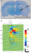
Electrophysiological experiments. (a) Recording with microelectrode array in the A1 and MGB. A coronal histological section of a representative test animal. (b) Laminar recording and current source density analysis in the A1. Upon recording the AEPs across A1 layers (black traces on the image), the CSD analysis was performed to locate the test sites in L1, L2/3, L4, L5 and L6. A1, primary auditory cortex; MGB, thalamus; AEP, auditory evoked potential; CSD, current source density; L, layer.
Figure 3
Figure 3
Click sequences. (a) AEPs across A1 and in MGB layers for the pre- and post-VNS conditions. The present AEPs were grand-averages across test animals. (b) Pre- vs. post-VNS AEP amplitudes. (c) Layer-specific increase of AEP amplitude following VNS. Outline markers with error bars indicate the means with standard deviations. AEP, auditory evoked potential; A1, primary auditory cortex; MGB, thalamus; VNS, vagus nerve stimulation. * indicates statistical significance (p < 0.05).
Figure 4
Figure 4
Oddball paradigm. (a) AEPs across layers in A1. Standard (Std)- and deviant (Dev)-evoked AEPs in either the pre- or post-VNS condition are presented. The present AEPs were grand averages across test animals. (b) Pre- vs. Post-VNS AEP amplitudes: (i) Std; (ii) Dev. (c) Layer-specific increase of AEP amplitude following VNS. Outline markers with error bars indicate the means with standard deviations. (d) SI across layers under pre- and post-VNS conditions. (e) VNS effects on Std- and Dev-evoked AEPs across layers. The proportional increases of AEPs following VNS were quantified. VNS-induced increases of standard-evoked AEPs were larger than those of deviant evoked AEPs, resulting in the decrease of SSA. AEP, auditory evoked potential; A1, primary auditory cortex; VNS, vagus nerve stimulation; SI, stimulus specific adaptation index; SSA, stimulus specific adaptation. * indicates statistical significance (p < 0.05).

Similar articles

Cited by

References

    1. Theodore WH, Fisher RS. Brain stimulation for epilepsy. Lancet Neurol. 2004;3:111–118. - PubMed
    1. Dario JE, Edward FC, Kurtis IA. Vagus nerve stimulation for epilepsy: a meta-analysis of efficacy and predictors of response. J. Neurosurg. JNS. 2011;115:1248–1255. - PubMed
    1. Rush AJ, et al. Vagus nerve stimulation (VNS) for treatment-resistant depressions: A multicenter study. Biol. Psychiat. 2000;47:276–286. - PubMed
    1. Wani A, Trevino K, Marnell P, Husain MM. Advances in brain stimulation for depression. Ann. Clin. psychiatry: Off. J. Am. Acad. Clin. Psychiatrists. 2013;25:217–224. - PubMed
    1. Groves DA, Brown VJ. Vagal nerve stimulation: a review of its applications and potential mechanisms that mediate its clinical effects. Neurosci. Biobehav. Rev. 2005;29:493–500. - PubMed

Publication types