. 2020 Jun 2;11(6):414.
doi: 10.1038/s41419-020-2612-z.

Dendritic cells-derived interferon-λ1 ameliorated inflammatory bone destruction through inhibiting osteoclastogenesis

Affiliations

Dendritic cells-derived interferon-λ1 ameliorated inflammatory bone destruction through inhibiting osteoclastogenesis

Yueqi Chen et al. Cell Death Dis. .

Abstract

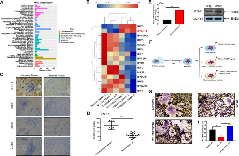
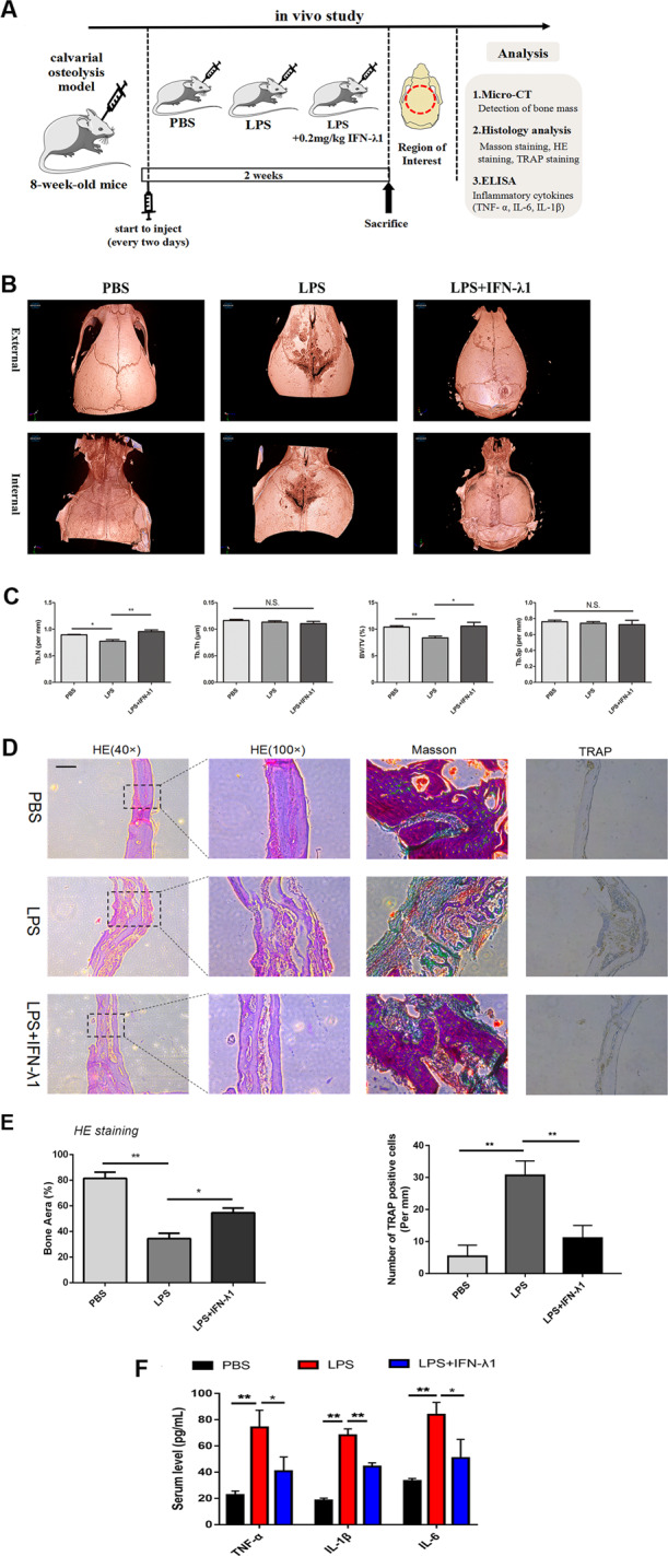
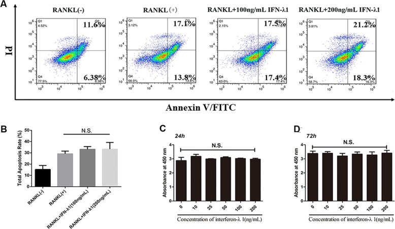
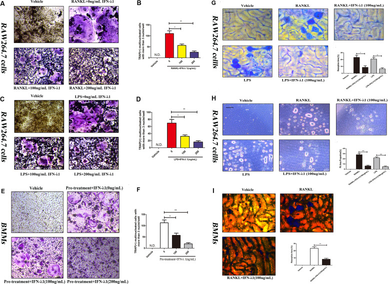
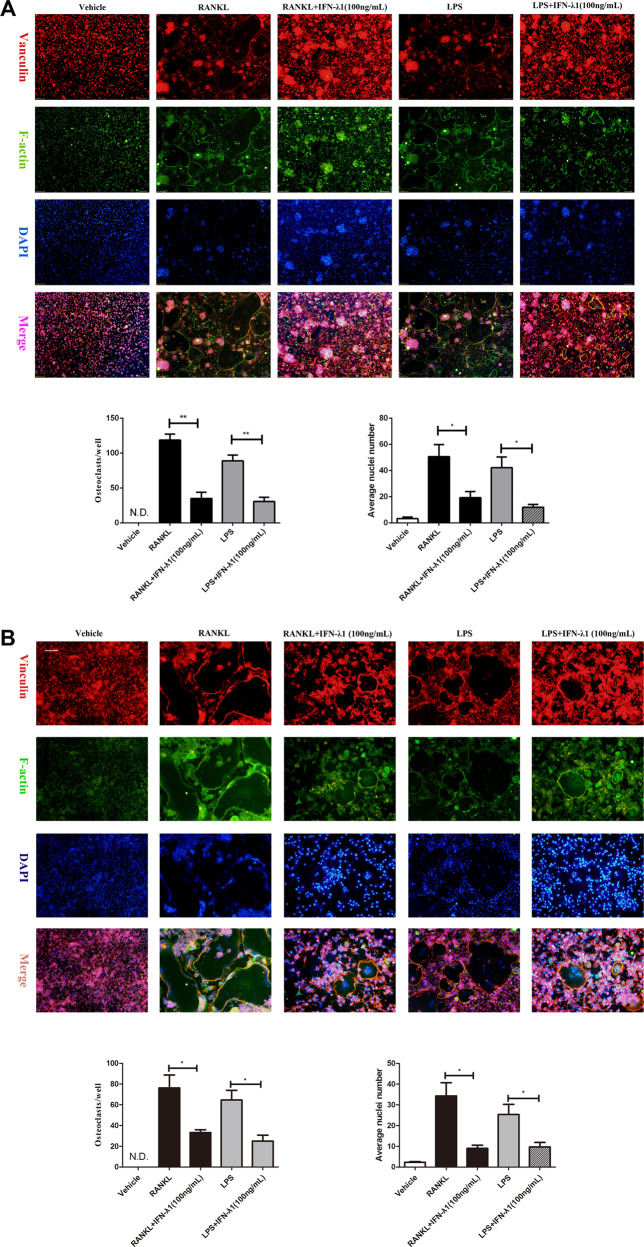
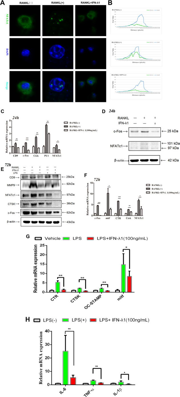
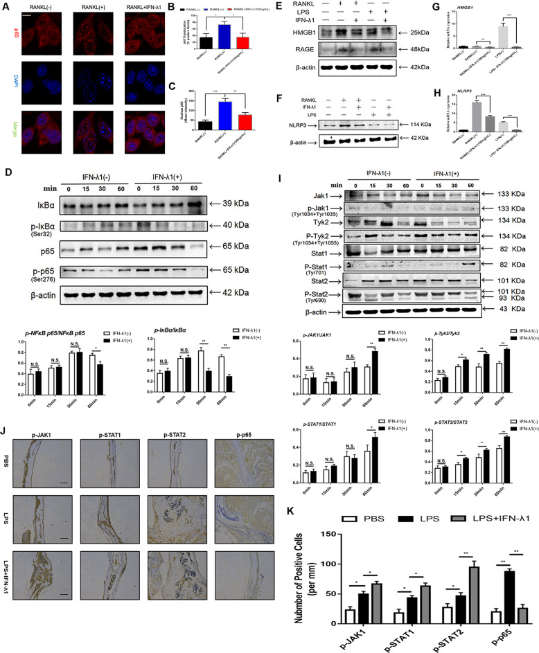
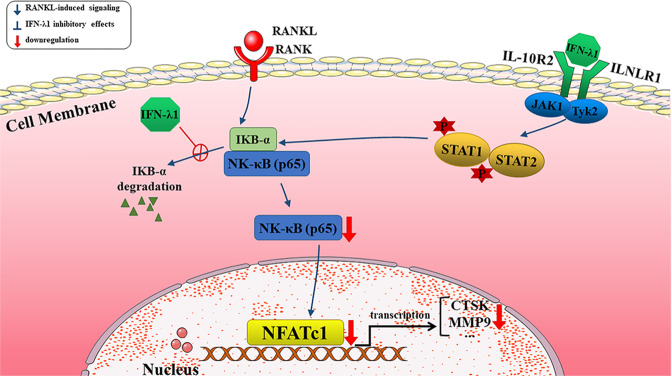
Bone infection contributing to inflammatory osteolysis is common in orthopedic surgery. The dynamic balance between bone formation and bone resorption is destroyed due to excessive osteoclast fusion and differentiation, which results in severe bone matrix loss. Many therapeutic approaches that restrain osteoclast formation and function act as efficient ways to prevent inflammatory bone erosion. We have demonstrated for the first time that dendritic cells-derived interferon-λ1 (IFN-λ1) inhibited inflammatory bone destruction in vivo and explored its underlying mechanisms on osteoclast formation in vitro. We found that IFN-λ1 was highly expressed in infectious bone tissue compared with that of non-infectious bone tissue. Additionally, dendritic cells marker genes such as CD80, CD86, and CD1a were higher expressed in infectious bone tissue than that of non-infectious bone tissue. Dendritic cells that were pretreated with LPS showed high expression of IFN-λ1. Moreover, conditioned medium of LPS-pretreated dendritic cells significantly inhibited osteoclast differentiation, as determined by TRAP staining assay. This suppressive effect was reversed by adding an IFN-λ1 monoclonal antibody. It was also investigated whether exogenous IFN-λ1 restrained osteoclastogenesis, bone resorption, F-actin ring formation, osteoclast-specific gene expression, release of pro-inflammatory cytokines, and translocation of p65 and NFATc1 by preventing the NF-κB signaling pathway and NLRP3 inflammasome formation, as well as by inducing the JAK-STAT signaling pathways in vitro. In vivo study indicated that IFN-λ1 prevents lipopolysaccharide (LPS)-induced inflammatory bone destruction by inhibiting excessive osteoclast fusion and bone resorption activity. In conclusion, our findings confirmed that dendritic cells-derived IFN-λ1 could attenuate osteoclast formation and bone resorptive activity in vitro and in vivo. These novel findings pave the way for the use of exogenous IFN-λ1 as a potential therapeutic treatment for excessive osteoclast-related diseases, such as inflammatory osteolysis, by regulating osteoclastogenesis to maintain the dynamic balance between bone formation and bone resorption.


Conflict of interest statement

The authors declare that they have no conflict of interest.

Figures

Fig. 1
Fig. 1. IFN-λ1 was higher expressed from dendritic cells during inflammatory statement.
a The genes expression in immune system were analyzed by KEGG pathway enrichment analysis between chronic osteomyelitis-infected samples and non-infectious fractures samples. b Profiling of differentially expressed interferon-related genes between chronic osteomyelitis-infected samples and non-infectious fractures samples. c Representative images of immunohistochemical staining with monoclonal antibodies against IFN-λ1, CD80, CD86, and CD1a. d ELISA for IFN-λ1 concentrations from the serum of chronic osteomyelitis patients and non-infectious fracture patients. e Relative expression of IFN-λ1 on mRNA and protein level between LPS (100 ng/ml) pretreatment with dendritic cells and vehicle for 72 h. β-actin and GAPDH were used as an internal control. f Schematic representation of the experimental design of the establishment the co-culture between osteoclast and LPS-induced dendritic cells. g Representative images of TRAP staining of RAW264.7 cells treated with different groups. h Quantification of multinucleated TRAP-positive osteoclast number per well. The data in the figures represent the averages ± SD. Scale bars = 200 μm. Significant differences are indicated as *p < 0.05 or **p < 0.01 paired using Student’s t-test unless otherwise specified.
Fig. 2
Fig. 2. Exogenous IFN-λ1 inhibited LPS-induced inflammatory osteolysis.
a Schematic representation of the design of in vivo experiments. b Representative 3D μCT images of reconstructed mouse calvarial (from internal to external/from external to internal). c Quantification of trabecular number (Tb. N), trabecular separation (Tb. Sp), trabecular thickness (Tb.Th), and trabecular bone volume fraction (BV/TV). d Representative images of cranial sections stained with H&E, Masson, and TRAP from each group. Scale bars=200μm. e Quantification of percentage of bone area in the border zone of the cranial H&E staining by using Image J software and the analysis of TRAP-positive cells in each group. f ELISA to detect the concentration of IL-1β, IL-6, and TNF-α in serum. The data in the figures represent the averages±SD. N.S. represented as no significant difference. Significant differences are indicated as *p<0.05 or **p<0.01 paired using Student’s t-test unless otherwise specified.
Fig. 3
Fig. 3. IFN-λ1 could not make cytotoxicity during osteoclastogenesis.
a Flow cytometry analysis of the apoptosis rate of RAW264.7 cells treated with RANKL (100 ng/ml) and M-CSF (50 ng/ml) for 72 h with various doses of IFN-λ1. b Quantitative analysis of total apoptosis rate during osteoclastogenesis. c, d CCK-8 was performed in triplicate to analyze the cell viability of BMMs treated with varying doses of IFN-λ1 for 24 and 72 h with or without RANKL (100 ng/ ml) and M-CSF (50 ng/ml). The data in the figures represent the averages ± SD. N.S. represented as no significant difference. Significant differences are indicated as *p < 0.05 or **p < 0.01 paired using Student’s t-test unless otherwise specified.
Fig. 4
Fig. 4. IFN-λ1 could inhibit osteoclast differentiation and bone resorption activity, respectively.
a, c, e Representative TRAP stain images of RAW264.7 cells and BMMs treated with RANKL or LPS-induced osteoclastogenesis. b, d, f Quantification of osteoclasts number per well. g RAW264.7 cells were plated on the bone slices and were cultured with RANKL or LPS for 6 days in the presence or absence of 100 ng/ml IFN-λ1. Scale bar = 200 μm. Quantification of the bone resorption area on the bone slices. h RAW264.7 cells were plated on the Osteo Assay Surface and were cultured with RANKL or LPS for 6 days in the presence or absence of 100 ng/ml IFN-λ1. Scale bar = 200 μm. Quantification of the bone resorption area on the bone slices. i BMMs were plated on the bone slices and were cultured with RANKL for 6 days in the presence or absence of 100 ng/ml IFN-λ1. Scale bar = 200 μm. Quantification of the bone resorption area on the bone slices. The data in the figures represent the averages ± SD. N.S. represented as no significant difference. Significant differences are indicated as *p < 0.05 or **p < 0.01 paired using Student’s t-test unless otherwise specified.
Fig. 5
Fig. 5. IFN-λ1 suppressed RANKL or LPS-induced osteoclast fusion significantly.
a Representative images of FAK staining of RAW264.7 cells treated with RANKL and M-CSF alone or together with the indicated concentrations of IFN-λ1 treatment. F-actin using tetramethylrhodamine-conjugated phalloidin (red), focal contacts using anti-vinculin mAb, and nuclear counterstaining using DAPI (blue). Scale bar = 200 μm. Quantitative analysis of osteoclasts (nucleiå 3) and average osteoclast nuclei number in each field. b Representative images of FAK staining of RAW264.7 cells treated with RANKL and M-CSF alone or together with the indicated concentrations of IFN-λ1 treatment. F-actin using tetramethylrhodamine-conjugated phalloidin (red), focal contacts using anti-vinculin mAb, and nuclear counterstaining using DAPI (blue). Scale bar = 200 μm. Quantitative analysis of osteoclasts (nuclei > 3) and average osteoclast nuclei number in each field. The data in the figures represent the averages ± SD. Significant differences are indicated as *p < 0.05 or **p < 0.01 paired using Student’s t-test unless otherwise specified.
Fig. 6
Fig. 6. IFN-λ1 inhibited the nuclei translocation of NFATc1 and the expression of osteoclast-specific genes.
a RAW264.7 cells were seeded in 96-well plates and treated with IFN-λ1 (100 ng/ml) for 24 h, followed by stimulation with 100 ng/ml RANKL and 50 ng/ml M-CSF. The intracellular location of the NFATc1 was observed by immunofluorescence staining using confocal microscopy. Scale bar = 800 μm. b The gray values of the Green and Blue staining were measured using the Image J software, and the mean values were plotted using excel. c Relative mRNA expression of CD9, c-Fos, Ctsk, PU.1, and NFATc1 during treatment with RANKL in the presence or absence of IFN-λ1 (100 ng/ml) for 24 h. d Relative expression of c-Fos and NFATc1 during treatment with RANKL in the presence or absence of IFN-λ1 (100 ng/ml) for 24 h in protein level. β-actin was used as an internal control. e Relative expression of CD9, MMP-9, CTSK, c-Fos, and NFATc1 during treatment with RANKL or LPS in the presence or absence of IFN-λ1 (100 ng/ml) for 72 h in protein level. β-actin was used as an internal control. f Relative mRNA expression of mitf, c-Fos, Ctsk, CTR, and NFATc1 during treatment with RANKL in the presence or absence of IFN-λ1 (100 ng/ml) for 72 h. g Relative mRNA expression of mitf, Ctsk, CTR, and OC-STAMP during treatment with LPS in the presence or absence of IFN-λ1 (100 ng/ml) for 72 h. h Relative mRNA expression of IL-1β, IL-6, and TNF-α during LPS-induced osteoclastogenesis in the presence or absence of IFN-λ1 (100 ng/ml). The data in the figures represent the averages ± SD. Significant differences are indicated as *p < 0.05 or **p < 0.01 paired using Student’s t-test unless otherwise specified.
Fig. 7
Fig. 7. IFN-λ1 inhibited the classical NF-κB signal pathway, the formation of NLRP3 inflammasome and activated JAK-Stat signal pathway.
a RAW264.7 cells were seeded in 96-well plates and treated with IFN-λ1 (100 ng/ml) for 60 min, followed by stimulation with 100 ng/ml RANKL and 50 ng/ml M-CSF. The intracellular location of the NF-κB p65 was observed by immunofluorescence staining using confocal microscopy. Scale bar = 800 μm. b Quantitative analysis of the percentage of positive cells (NF-ĸB p65 translocation from cytosol to nuclear) in all cells. c Quantitative analysis of the mean intensity of NF-ĸB p65 in the cells nuclear. d RAW264.7 cells were stimulated with RANKL with or without IFN-λ1 (100 ng/ml) for the 0–60 min. The cell lysates were analyzed using western blotting for p-NFκB p65, NF-κB p65, p-IκBα, and IκBα. β-actin was used as an internal control. e, f The expression of HMGB1, RAGE, and NLRP3 during osteoclastogenesis in the presence or absence of IFN-λ1 (100 ng/ml) on protein level. β-actin was used as an internal control. g, h Relative expression of HMGB1 and NLRP3 during osteoclastogenesis in the presence or absence of IFN-λ1 (100 ng/ml) on mRNA level. i RAW264.7 cells were stimulated with RANKL with or without IFN-λ1 (100 ng/ml) for the 0–60 min. The cell lysates were analyzed using western blotting for p-Jak1, Jak1, p-Tyk2, Tyk2, p-Stat1, Stat1, p-Stat2, and Stat2. β-actin was used as an internal control. j Representative images of immunohistochemical staining with monoclonal antibodies against p-p65, p-JAK1, p-STAT1, and p-STAT2. Scale bar = 200 μm. k Quantitative analysis of p-p65, p-JAK1, p-STAT1, and p-STAT2-positive cells per mm in each field. The data in the figures represent the averages ± SD. Significant differences are indicated as *p < 0.05 or **p < 0.01 paired using Student’s t-test unless otherwise specified.
Fig. 8
Fig. 8
Schematic diagrams showing the potential mechanism in the protective effects of dendritic cells derived IFN-λ1 on inflammatory bone destruction through inhibiting osteoclastogenesis.

Similar articles

Cited by

References

    1. Waldvogel FA, et al. Osteomyelitis: a review of clinical features, therapeutic considerations and unusual aspects. N. Engl. J. Med. 1970;282:198e206. - PubMed
    1. van Staa TP, et al. Clinical assessment of the long-term risk of fracture in patients with rheumatoid arthritis. Arthritis Rheum. 2006;54:3104–3112. - PubMed
    1. Gravallese EM, et al. Identification of cell types responsible for bone resorption in rheumatoid arthritis and juvenile rheumatoid arthritis. Am. J. Pathol. 1998;152:943–951. - PMC - PubMed
    1. Lew DP, Waldvogel FA. Osteomyelitis. Lancet. 2004;364:369e79. - PubMed
    1. Cassat JE, et al. A secreted bacterial protease tailors the Staphylococcus aureus virulence repertoire to modulate bone remodeling during osteomyelitis. Cell Host Microbe. 2013;13:759–772. - PMC - PubMed

Publication types

MeSH terms