. 2020 Jun 2;11(6):415.
doi: 10.1038/s41419-020-2626-6.

TNF-alpha-induced microglia activation requires miR-342: impact on NF-kB signaling and neurotoxicity

Affiliations

TNF-alpha-induced microglia activation requires miR-342: impact on NF-kB signaling and neurotoxicity

João Paulo Brás et al. Cell Death Dis. .

Abstract

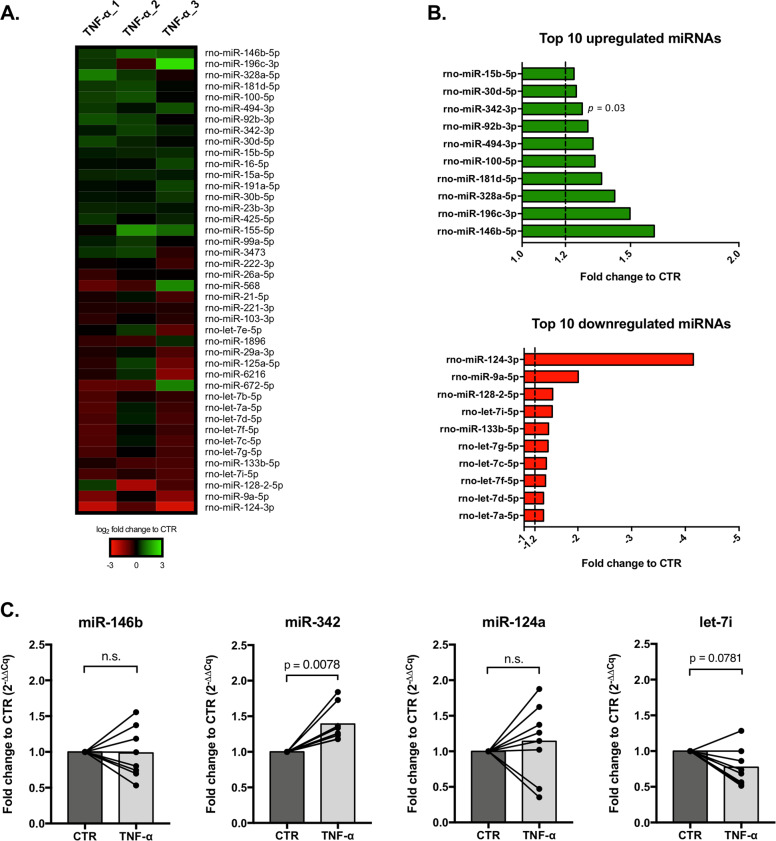
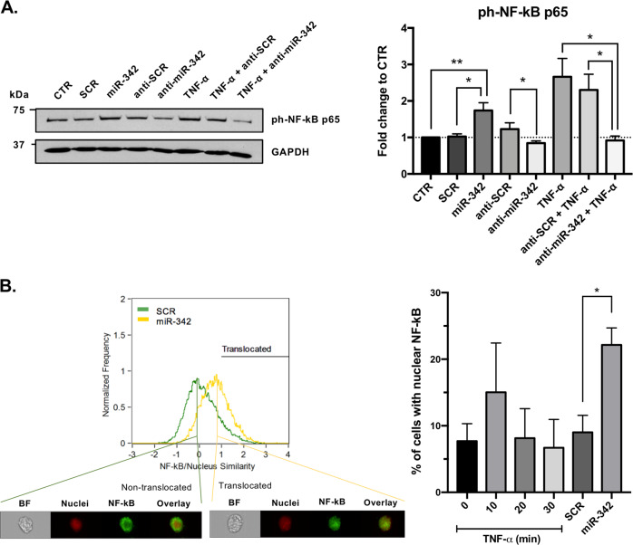
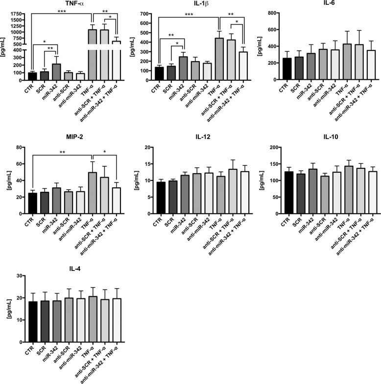
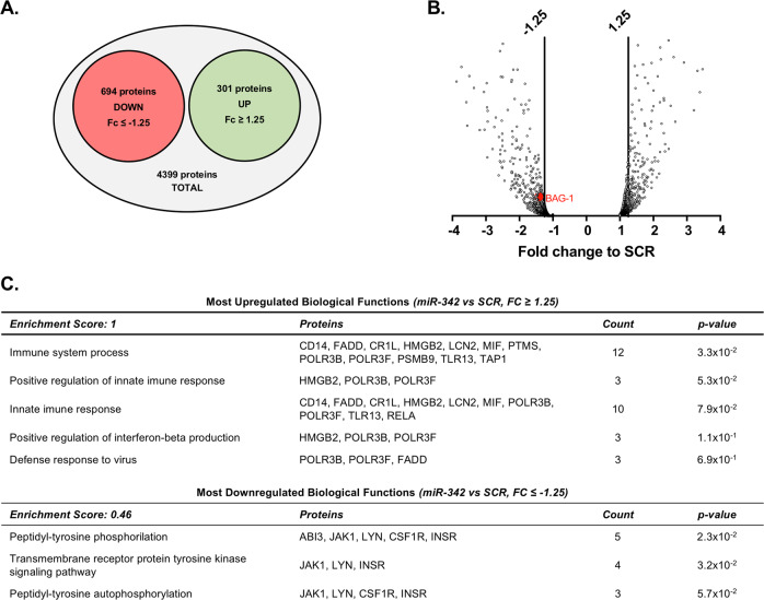
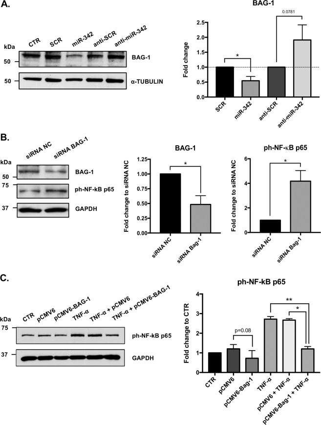
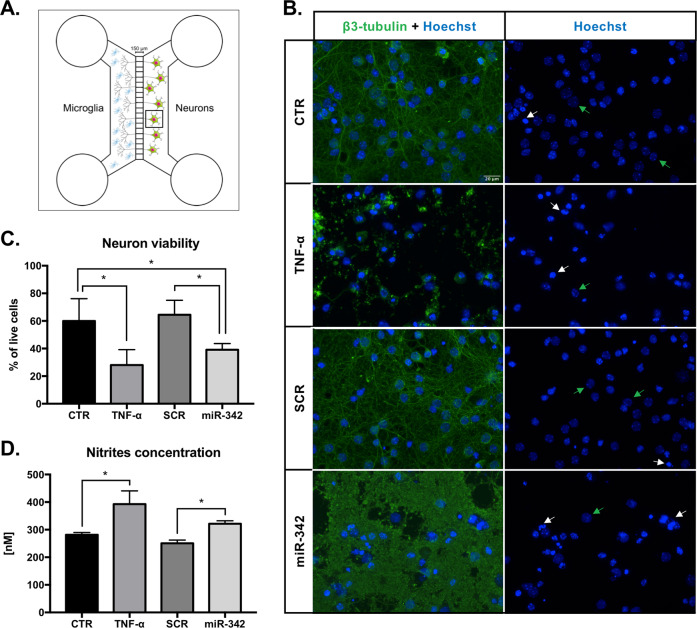
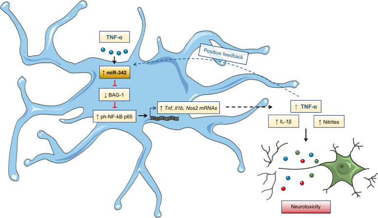
Growing evidences suggest that sustained neuroinflammation, caused by microglia overactivation, is implicated in the development and aggravation of several neurological and psychiatric disorders. In some pathological conditions, microglia produce increased levels of cytotoxic and inflammatory mediators, such as tumor necrosis factor alpha (TNF-α), which can reactivate microglia in a positive feedback mechanism. However, specific molecular mediators that can be effectively targeted to control TNF-α-mediated microglia overactivation, are yet to be uncovered. In this context, we aim to identify novel TNF-α-mediated micro(mi)RNAs and to dissect their roles in microglia activation, as well as to explore their impact on the cellular communication with neurons. A miRNA microarray, followed by RT-qPCR validation, was performed on TNF-α-stimulated primary rat microglia. Gain- and loss-of-function in vitro assays and proteomic analysis were used to dissect the role of miR-342 in microglia activation. Co-cultures of microglia with hippocampal neurons, using a microfluidic system, were performed to understand the impact on neurotoxicity. Stimulation of primary rat microglia with TNF-α led to an upregulation of Nos2, Tnf, and Il1b mRNAs. In addition, ph-NF-kB p65 levels were also increased. miRNA microarray analysis followed by RT-qPCR validation revealed that TNF-α stimulation induced the upregulation of miR-342. Interestingly, miR-342 overexpression in N9 microglia was sufficient to activate the NF-kB pathway by inhibiting BAG-1, leading to increased secretion of TNF-α and IL-1β. Conversely, miR-342 inhibition led to a strong decrease in the levels of these cytokines after TNF-α activation. In fact, both TNF-α-stimulated and miR-342-overexpressing microglia drastically affected neuron viability. Remarkably, increased levels of nitrites were detected in the supernatants of these co-cultures. Globally, our findings show that miR-342 is a crucial mediator of TNF-α-mediated microglia activation and a potential target to tackle microglia-driven neuroinflammation.


Conflict of interest statement

The authors declare that they have no conflict of interest.

Figures

Fig. 1
Fig. 1. TNF-α-induced overexpression of pro-inflammatory genes, through activation of NF-kB pathway in microglia.
Rat microglia were obtained from mixed glial cultures. Isolated microglia were re-seeded in six-well plates, allowed to adhere for 48 h and then stimulated for 6 h with LPS (100 ng/mL) or TNF-α (20 ng/mL). a Gene expression profile of activated microglia evaluated by RT-qPCR. Results were normalized with Gapdh and are expressed in fold change to CTR (mean ± SD, n = 5). b NF-kB p65 phosphorylation levels evaluation by western blot after microglia activation with LPS or TNF-α (mean ± SD, n = 5). GAPDH was used as normalizer. Statistical significance: **p < 0.01, *p < 0.05, ns–non significant; Friedman test followed by Dunn’s multiple comparisons test.
Fig. 2
Fig. 2. miR-342 is overexpressed in TNF-α-stimulated microglia.
a Heat map of miRNA microarray expression results in TNF-α activated microglia (n = 3). Only miRNAs with −0.2 ≤ log2 FC to CTR ≥ 0.2 and a detection signal ≥20 are represented. b Ten most upregulated (green) or downregulated (red) miRNAs in TNF-α stimulated microglia (mean FC to CTR, n = 3). c Fold change to control based on the relative expression of the selected miRNAs, evaluated by RT-qPCR (mean, n = 8). U6 SnRNA was used as reference. Cq: quantification cycle. Statistical significance: p < 0.05; Wilcoxon matched-pairs test.
Fig. 3
Fig. 3. miR-342 regulates TNF-α-mediated microglia activation through NF-kB.
a ph-NF-kB p65 expression evaluation by western blot after TNF-α stimulation and/or mirVana miRNA mimic/inhibitor mmu-miR-342–3p or mirVana miRNA mimic/inhibitor negative control (SCR) transfection. Results were normalized with GAPDH and compared with non-stimulated N9 microglia (mean ± SD, n = 3–6). b Representative plot of the similarity coefficient between NF-kB and nuclei staining’s in cells transfected with mirVana miRNA mimic negative control (SCR, green) or miRNA mimic mmu-miR-342–3p (miR-342, yellow). The black line (Translocated) corresponds to gated cells with nuclear translocated NF-kB (similarity coefficient >1). Representative images of cells with a similarity coefficient <1 (non-translocated) and with a similarity coefficient >1 (translocated) are shown below. BF brightfield. On the right, graph shows the quantification of the percentage of cells with nuclear translocated NF-κB (translocated gate, similarity coefficient >1) after exposure to TNF-α for the indicated times or transfection with SCR or miR-342. Results are mean ± SD of three independent experiments. *p < 0.05, **p < 0.01; ANOVA followed by Sidak’s multiple comparison test.
Fig. 4
Fig. 4. miR-342 impacts cytokine secretion levels in microglia.
N9 microglia were transfected with mirVana miRNA mimic/inhibitor mmu-miR-342–3p or mirVana miRNA mimic/inhibitor Negative Control (SCR), using Lipofectamine 2000. When indicated, microglia were also stimulated with TNF-α (20 ng/mL) for 6 h. After that, supernatants were collected and stored at −80 °C. Levels of TNF-α, IL-1β, IL-6, MIP-2, IL-12, IL-10, and IL-4 were quantified by ELISA (mean ± SD, n = 5–7). Cytokine concentration was obtained using corresponding standard curve, according to manufacturer’s instructions. Statistical significance: *p < 0.05, **p < 0.01, and ***p < 0.001; Friedman test followed by Dunn’s multiple comparisons test.
Fig. 5
Fig. 5. Protein expression profile of miR-342-transfected microglia.
Protein identification and quantitation was performed by nano LC-MS/MS. Diagram a and volcano plot b represent the most differently expressed proteins within all identified proteins (−1.25 ≤ FC to SCR ≥ 1.25, n = 2). c Most up and downregulated biological functions based on protein expression fold change between miR-342 and SCR. Analysis was performed with DAVID using Functional Annotation Tool. The entire list proteins detected was used as background. “Enrichment score” indicates the biological relevance of the group of proteins involved in the respective set of biological functions, based on the p values of all enriched annotation terms. “Count” indicates the number of dysregulated proteins involved in that specific biological function. Full protein names can be found in Supplementary Table 4.
Fig. 6
Fig. 6. miR-342 induces NF-kB activation by inhibiting BAG-1.
a BAG-1 expression after miR-342 overexpression/inhibition was addressed by western blot. Results were normalized with α-tubulin and compared with the respective controls (mean ± SD, n = 6). To evaluate the involvement of BAG-1 on NF-kB activation, N9 microglia were transfected with a siRNA to silence b or with a plasmid (1ug/mL) to overexpress BAG-1 c. BAG-1 and ph-NF-kB p65 expression levels were evaluated by western blot. Results were normalized with GAPDH and compared with the respective controls (mean ± SD, n = 2–4). Statistical significance: *p < 0.05, Wilcoxon matched-pairs test.
Fig. 7
Fig. 7. miR-342 overexpression in microglia induces neurotoxicity.
a Neurons were cultured in PDL-coated coverslips previously attached to the Axon Investigation System. At day 13 of neuronal culture, transfected, TNF-α, or non-stimulated N9 microglia were added to the respective system, in direct contact with axons for 24 h. b Immufluorescence images of neurons after co-culture with N9 microglia. Left panel shows neurons stained with anti-β3-tubulin (anti-Alexa 488, green) and Hoechst (blue). Right panel shows only Hoechst nuclear staining used for neuron viability evaluation. White arrows highlight the nucleus of dead neurons, whereas green arrows highlight the nucleus of healthy neurons. Scale bar: 20 µm. c Neuron viability was addressed after counting the number of living and dead cells of 10 images per condition (mean ± SD, n = 4). d Co-cultures’ supernatants were collected for nitrite levels quantification using Griess reagent (mean ± SD, n = 4). Statistical significance: *p < 0.05 and **p < 0.01, Friedman test followed by Dunn’s multiple comparisons test.
Fig. 8
Fig. 8. Graphical abstract–TNF-alpha-induced miR-342 promotes microglia activation through NF-kB and induces neurotoxicity.
We found miR-342 to be upregulated in microglia activated with TNF-α. miR-342 promotes NF-kB activation by inhibiting BAG-1, leading to the overexpression of pro-inflammatory mediators, including TNF-α, in a positive feedback loop, possibly perpetuating microglia activation. Importantly, inhibition of miR-342 attenuated TNF-α-driven microglia activation. Moreover, microglia activation by miR-342 led to increased neurotoxicity with high levels of nitrites being detected in co-cultures supernatants.

References

    1. Miller AH, Raison CL. The role of inflammation in depression: from evolutionary imperative to modern treatment target. Nat. Rev. Immunol. 2016;16:22–34. - PMC - PubMed
    1. Yirmiya R, Rimmerman N, Reshef R. Depression as a microglial disease. Trends Neurosci. 2015;38:637–658. - PubMed
    1. Ginhoux F, et al. Fate mapping analysis reveals that adult microglia derive from primitive macrophages. Science. 2010;330:841–845. - PMC - PubMed
    1. Thion MS, Ginhoux F, Garel S. Microglia and early brain development: an intimate journey. Science. 2018;362:185–189. - PubMed
    1. Ransohoff RM, Perry VH. Microglial physiology: unique stimuli, specialized responses. Annu. Rev. Immunol. 2009;27:119–145. - PubMed

Publication types

MeSH terms

LinkOut - more resources