

Icariside II attenuates cerebral ischemia/reperfusion-induced blood-brain barrier dysfunction in rats via regulating the balance of MMP9/TIMP1
- PMID: 32488170
- PMCID: PMC7921596
- DOI: 10.1038/s41401-020-0409-3
Icariside II attenuates cerebral ischemia/reperfusion-induced blood-brain barrier dysfunction in rats via regulating the balance of MMP9/TIMP1
Abstract
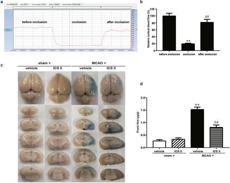
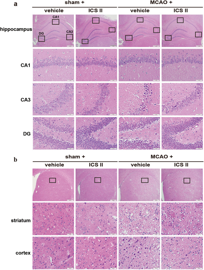
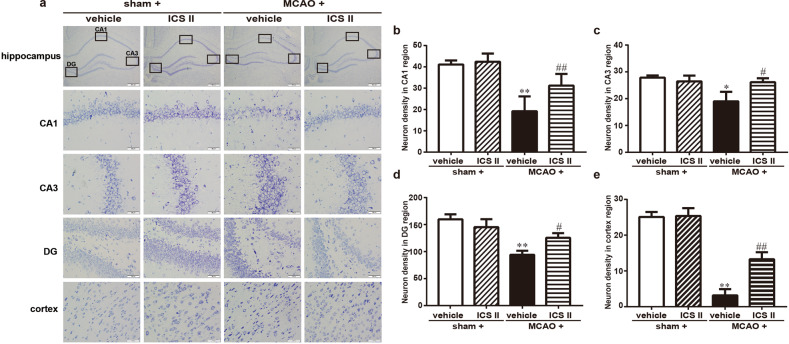
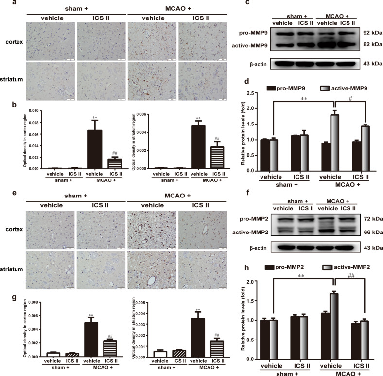
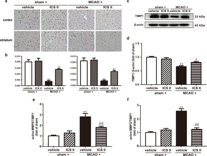
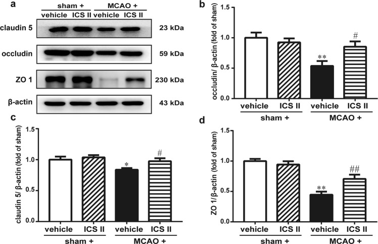
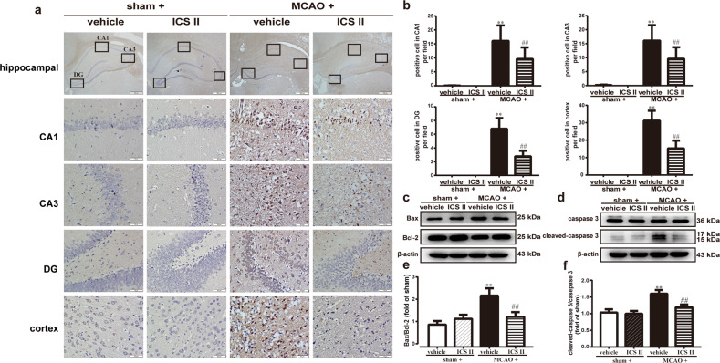
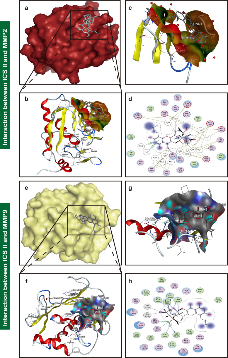
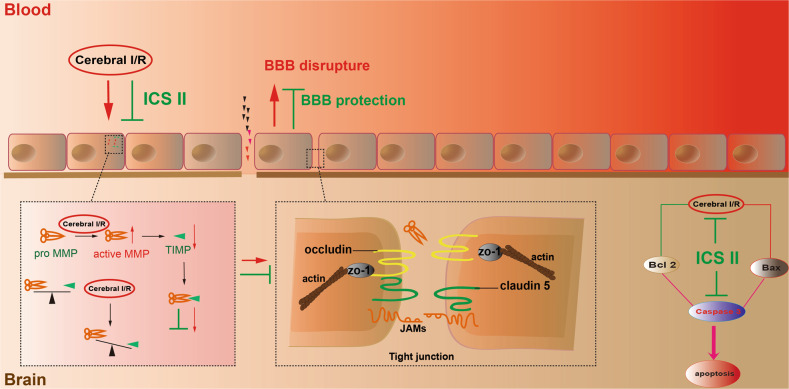
Cerebral ischemia/reperfusion (I/R) results in harmful consequences during ischemic stroke, especially the disruption of the blood-brain barrier (BBB), which leads to severe hemorrhagic transformation through aggravation of edema and brain hemorrhage. Our previous study demonstrated that icariside II (ICS II), which is derived from Herba Epimedii, attenuates cerebral I/R injury by inhibiting the GSK-3β-mediated activation of autophagy both in vitro and in vivo. However, the effect of ICS II on the BBB remains unclear. Thus, in this study, we investigated the regulation of BBB integrity by ICS II after cerebral I/R injury and further explored the underlying mechanism in rats. Cerebral I/R injury was induced by middle cerebral artery occlusion (MCAO), and the treatment groups were administered ICS II at a dose of 16 mg/kg by gavage twice a day for 3 days. The results showed that ICS II effectively prevented BBB disruption, as evidenced by Evans Blue staining. Moreover, ICS II not only significantly reduced the expression of MMP2/9 but also increased TIMP1 and tight junction protein (occludin, claudin 5, and ZO 1) expression. Intriguingly, ICS II may directly bind to both MMP2 and MMP9, as evidenced by molecular docking. In addition, ICS II also inhibited cerebral I/R-induced apoptosis and ameliorated the Bax/Bcl-2 ratio and cleaved-caspase 3 level. Collectively, our findings reveal that ICS II significantly ameliorates I/R-induced BBB disruption and neuronal apoptosis in MCAO rats by regulating the MMP9/TIMP1 balance and inhibiting the caspase 3-dependent apoptosis pathway.
Keywords: MCAO rats; Stroke; apoptosis; blood–brain barrier; cerebral ischemia/reperfusion; icariside II; molecular docking; tight junction.
Conflict of interest statement
The authors declare no competing interests.
Figures
Similar articles
-
Rosmarinic acid attenuates blood-brain barrier dysfunction to improve cerebral ischemia/reperfusion injury in mice.Eur J Pharmacol. 2025 Sep 15;1003:177882. doi: 10.1016/j.ejphar.2025.177882. Epub 2025 Jul 1. Eur J Pharmacol. 2025. PMID: 40609608
-
The protective effect of HET0016 on brain edema and blood-brain barrier dysfunction after cerebral ischemia/reperfusion.Brain Res. 2014 Jan 28;1544:45-53. doi: 10.1016/j.brainres.2013.11.031. Epub 2013 Dec 6. Brain Res. 2014. PMID: 24316243
-
Trilobatin attenuates cerebral ischaemia/reperfusion-induced blood-brain barrier dysfunction by targeting matrix metalloproteinase 9: The legend of a food additive.Br J Pharmacol. 2024 Apr;181(7):1005-1027. doi: 10.1111/bph.16239. Epub 2023 Nov 10. Br J Pharmacol. 2024. PMID: 37723895
-
Brain endothelial cell junctions after cerebral hemorrhage: Changes, mechanisms and therapeutic targets.J Cereb Blood Flow Metab. 2018 Aug;38(8):1255-1275. doi: 10.1177/0271678X18774666. Epub 2018 May 8. J Cereb Blood Flow Metab. 2018. PMID: 29737222 Free PMC article. Review.
-
Therapeutic Potentials of MicroRNA-126 in Cerebral Ischemia.Mol Neurobiol. 2023 Apr;60(4):2062-2069. doi: 10.1007/s12035-022-03197-4. Epub 2023 Jan 4. Mol Neurobiol. 2023. PMID: 36596965 Review.
Cited by
-
Association between TIMP1 polymorphism and female neuromyelitis optica spectrum disorder in Chinese population.Heliyon. 2024 Aug 28;10(17):e37091. doi: 10.1016/j.heliyon.2024.e37091. eCollection 2024 Sep 15. Heliyon. 2024. PMID: 39296182 Free PMC article.
-
Dental Pulp Stem Cell-Derived Conditioned Medium Alleviates Subarachnoid Hemorrhage-Induced Microcirculation Impairment by Promoting M2 Microglia Polarization and Reducing Astrocyte Swelling.Transl Stroke Res. 2023 Oct;14(5):688-703. doi: 10.1007/s12975-022-01083-8. Epub 2022 Oct 1. Transl Stroke Res. 2023. PMID: 36181630 Free PMC article.
-
Treatment with β-sitosterol ameliorates the effects of cerebral ischemia/reperfusion injury by suppressing cholesterol overload, endoplasmic reticulum stress, and apoptosis.Neural Regen Res. 2024 Mar;19(3):642-649. doi: 10.4103/1673-5374.380904. Neural Regen Res. 2024. PMID: 37721296 Free PMC article.
-
Sec62 promotes gastric cancer metastasis through mediating UPR-induced autophagy activation.Cell Mol Life Sci. 2022 Feb 15;79(2):133. doi: 10.1007/s00018-022-04143-2. Cell Mol Life Sci. 2022. PMID: 35165763 Free PMC article.
-
Targeting the PANoptosis signaling pathway for myocardial protection: therapeutic potential of Xian Ling Gu Bao capsule.Front Pharmacol. 2024 May 10;15:1391511. doi: 10.3389/fphar.2024.1391511. eCollection 2024. Front Pharmacol. 2024. PMID: 38799163 Free PMC article.
References
MeSH terms
Substances
LinkOut - more resources
Full Text Sources
Research Materials
Miscellaneous
