. 2020 Oct;17(4):1825-1835.
doi: 10.1007/s13311-020-00854-1.

Pharmacological Inhibition of Soluble Epoxide Hydrolase as a New Therapy for Alzheimer's Disease

Affiliations

Pharmacological Inhibition of Soluble Epoxide Hydrolase as a New Therapy for Alzheimer's Disease

Christian Griñán-Ferré et al. Neurotherapeutics. 2020 Oct.

Abstract

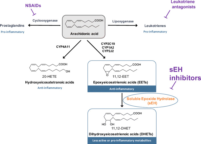
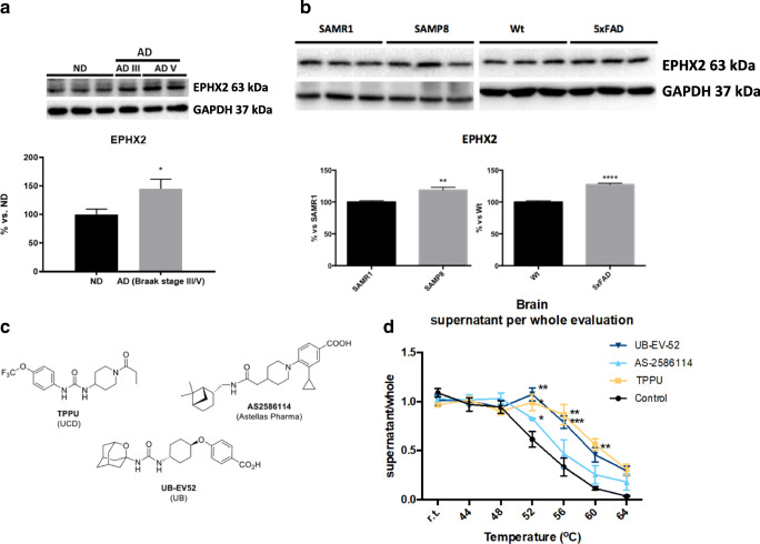
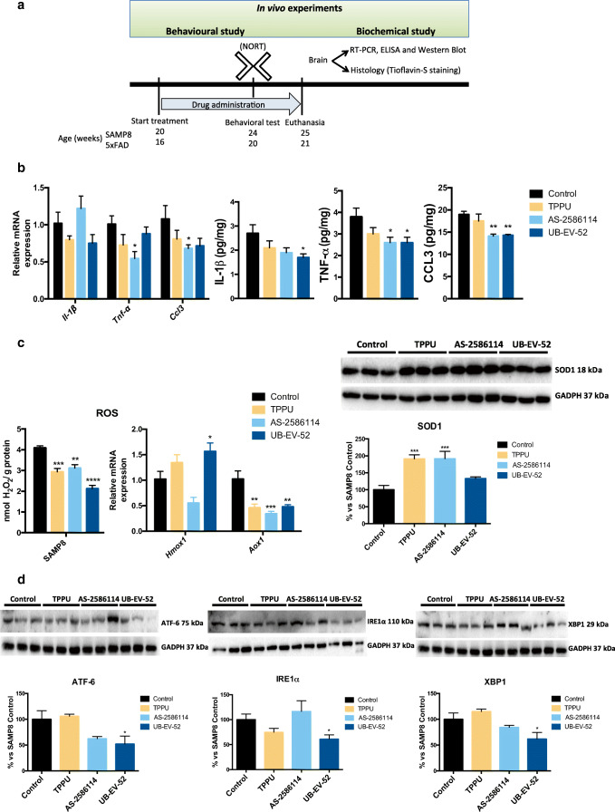
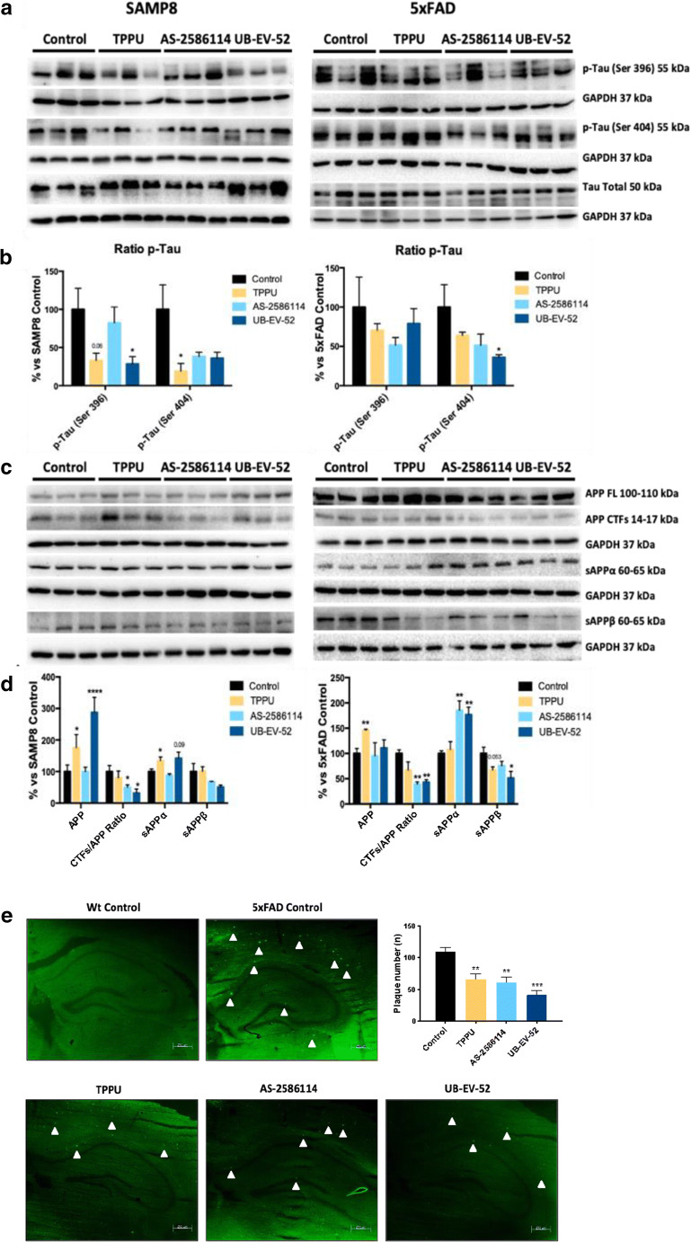
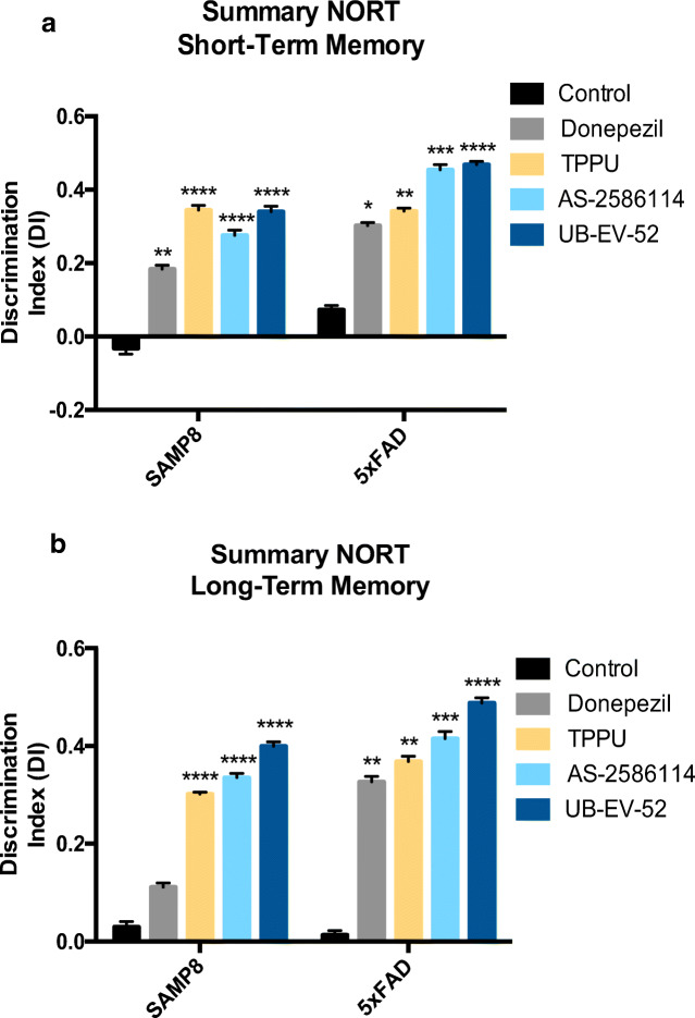
The inhibition of the enzyme soluble epoxide hydrolase (sEH) has demonstrated clinical therapeutic effects in several peripheral inflammatory-related diseases, with 3 compounds in clinical trials. However, the role of this enzyme in the neuroinflammation process has been largely neglected. Herein, we disclose the pharmacological validation of sEH as a novel target for the treatment of Alzheimer's disease (AD). Evaluation of cognitive impairment and pathological hallmarks were used in 2 models of age-related cognitive decline and AD using 3 structurally different and potent sEH inhibitors as chemical probes. sEH is upregulated in brains from AD patients. Our findings supported the beneficial effects of central sEH inhibition, regarding reducing cognitive impairment, neuroinflammation, tau hyperphosphorylation pathology, and the number of amyloid plaques. This study suggests that inhibition of inflammation in the brain by targeting sEH is a relevant therapeutic strategy for AD.

Keywords: Druggability; Inflammation; Soluble epoxide hydrolase; Target engagement; Tau; β-amyloid.


Figures

Fig. 1
Fig. 1
The arachidonic acid cascade. The arachidonic acid (AA) cascade is a group of metabolic pathways in which AA and other polyunsaturated fatty acids are the central molecules. Metabolism via the cyclooxygenase (COX) and lipoxygenase (LOX) pathways gives rise to largely proinflammatory and proalgesic metabolites. Both pathways have been pharmaceutically targeted. CYP enzymes either hydroxylate or epoxidize AA leading to hydroxyeicosatetranoic acids (HETEs) or epoxyeicosatrienoic acids (EETs), respectively. The latter compounds, which are endowed with potent anti-inflammatory properties, are rapidly subjected to hydrolysis to their corresponding diols by the soluble epoxide hydrolase (sEH) enzyme. Inhibitors of sEH block this degradation and stabilize EET levels in vivo [14]. They also reduce the corresponding diols which have some inflammatory properties. Major CYPs that oxidize AA are listed in the figure, but many others make a contribution
Fig. 2
Fig. 2
Soluble epoxide inhibition and its relevance in AD. a Immunoblot of sEH (EPHX2) of human brains from AD patients (Braak stage III-V). Groups were compared by Student t test (n = 4-7). *p < 0.05 vs. non-demented. b Immunoblot of sEH (EPHX2) in the hippocampus of SAMP8 mice (groups were compared by Student t test, n = 12-14, **p < 0.01 vs. SAMR1) and 5xFAD mice (groups were compared by Student t test, n = 12-14, ****p < 0.0001 vs. Wt). c Chemical structure of the sEH inhibitors employed. d CETSA experiments to monitor brain target engagement. Groups were compared by Student t test or 2-way ANOVA and post hoc Dunnett’s, n = 3 per group, *p < 0.05, **p < 0.01, and ***p < 0.001 vs. control
Fig. 3
Fig. 3
Role of sEH inhibitors in neurodegenerative biomarkers. a Scheme of experimental procedures in in vivo experiments. b Gene expression of neuroinflammatory markers (Il-1β, Tnf-α, and Ccl3) and protein levels of proinflammatory cytokines IL-1β, TNF-α, and CCL3 in the hippocampus of SAMP8 mice after treatment with sEH inhibitors. c Oxidative stress measured by hydrogen peroxide concentration in homogenates of the hippocampus. Representative gene expression of Hmox1 and Aox1 and representative Western blot and quantification of protein levels for (antioxidant enzyme) SOD1 in the hippocampus of SAMP8 mice after treatment with sEH inhibitors. d Representative Western blot and quantification of protein levels for ER stress markers ATF-6, IRE1α, and XBP1 in the hippocampus of SAMP8 mice after treatment with sEH inhibitors. Gene expression levels were determined by real-time PCR, cytokine protein levels by ELISA, and SOD1 by immunoblotting. Results are expressed as a mean ± SEM and were significantly different from the control group. Groups were compared by Student t test or by 1-way ANOVA and post hoc Dunnett’s, n = 4-6 per group, (*p < 0.05, **p < 0.01, ***p < 0.001, and ****p < 0.0001) vs. control. See partial correlations between selected variables in Fig. S2 and Table S7
Fig. 4
Fig. 4
AD hallmarks in both SAMP8 and 5xFAD mice models after treatment with sEH inhibitors. a and b Representative Western blot and quantifications for p-Tau Ser396 and p-Tau Ser404. c and d Representative Western blot and quantifications for CTFs/APP ratio, sAPPα, and sAPPβ. e Histological images and quantification of amyloid plaques stained with Thioflavin-S in Wt and 5xFAD. Values in bar graphs are adjusted to 100% for a protein of the control group from each strain. Results are expressed as a mean ± SEM and were significantly different from the control group. Groups were compared by Student t test or by 1-way ANOVA and post hoc Dunnett’s, n = 12 per group (*significant at p < 0.05, **significant at p < 0.01, ***significant at p < 0.001, and ****significant at p < 0.0001). See partial correlations between selected variables in Fig. S2, Fig. S3, Table S7, and Table S8
Fig. 5
Fig. 5
Characterization of the effect of sEH inhibitors and donepezil on cognitive status in both SAMP8 and 5xFAD mice models. a Short-term memory evaluation after 2 h acquisition trial by discrimination index and b long-term memory evaluation after 24 h acquisition trial by discrimination index after exposure to novel objects. Results are expressed as a mean ± SEM and were significantly different from the control group. Groups were compared by Student t test or by 1-way ANOVA and post hoc Dunnett’s, n = 12 per group (*significant at p < 0.05, **significant at p < 0.01, ***significant at p < 0.001, and ****significant at p < 0.0001). See partial correlations between selected variables in Fig. S3, Fig. S4, Table S7, and Table S8

References

    1. Krstic D, Knuesel I. Deciphering the mechanism underlying late-onset Alzheimer disease. Nat. Rev. Neurol. 2012;9:25–34. - PubMed
    1. Calsolaro V, Edison P. Neuroinflammation in Alzheimer’s disease: current evidence and future directions. Alzheimer’s Dementia. 2016;12:719–732. - PubMed
    1. Fu W-Y, Wang X, Ip NY. Targeting neuroinflammation as a therapeutic strategy for Alzheimer’s disease: mechanisms, drug candidates, and new opportunities. ACS Chem. Neurosci. 2019;10:872–879. - PubMed
    1. Kodamullil AT, Zekri F, Sood M, et al. Tracing investment in drug development for Alzheimer disease. Nat. Rev. Drug Discovery. 2017;16:819. - PubMed
    1. Tönnies E, Trushina E. Oxidative stress, synaptic dysfunction, and Alzheimer’s disease. J. Alzheimer’s Dis. 2017;57:1105–1121. - PMC - PubMed

Publication types

MeSH terms

Substances

LinkOut - more resources