

Functions of lactate in the brain of rat with intracerebral hemorrhage evaluated with MRI/MRS and in vitro approaches
- PMID: 32488963
- PMCID: PMC7539841
- DOI: 10.1111/cns.13399
Functions of lactate in the brain of rat with intracerebral hemorrhage evaluated with MRI/MRS and in vitro approaches
Abstract
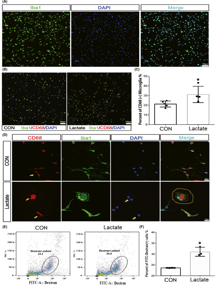
Introduction: Lactate accumulation in the brain is caused by the anaerobic metabolism induced by ischemic damages, which always accompanies intracerebral hemorrhages (ICH). Our former findings showed that microglia's movement was always directly toward hemorrhagic center with the highest lactate concentration, and penumbra area has the largest density of compactly arrayed microglia. However, the relationship between microglia and lactate concentration has not been well documented.
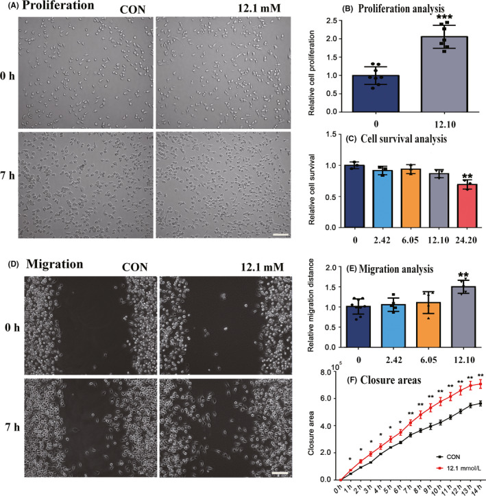
Methods: Cerebral hemorrhage model was successfully achieved by injecting collagenase VII (causing stabile localized bleeding) in CPu (striatum) of SD rats. Emodin was used as a potential therapeutic for ICH. The function of the lactate was examined with in vitro culture studies. Then, the effect of lactate on the proliferation, cell survival, migration, and phagocytosis property of microglia was investigated by in vitro culture studies.
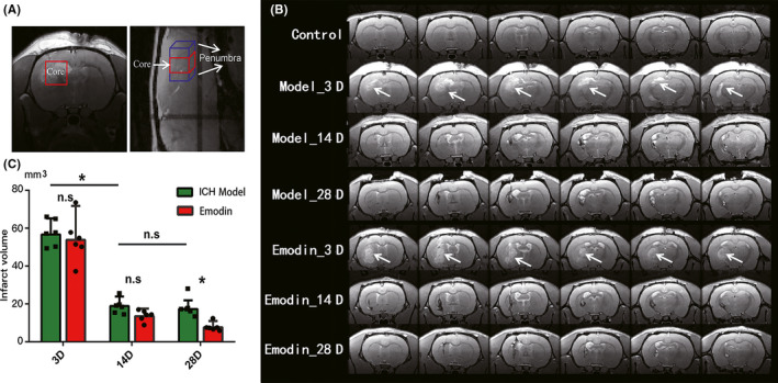
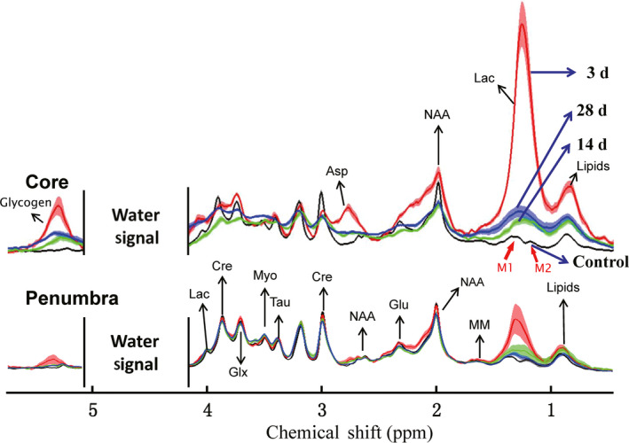
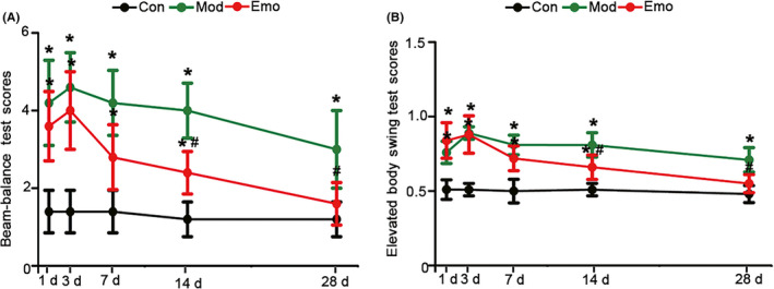
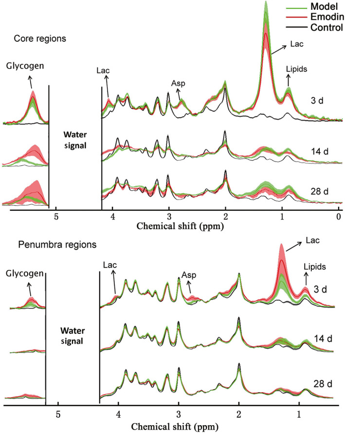
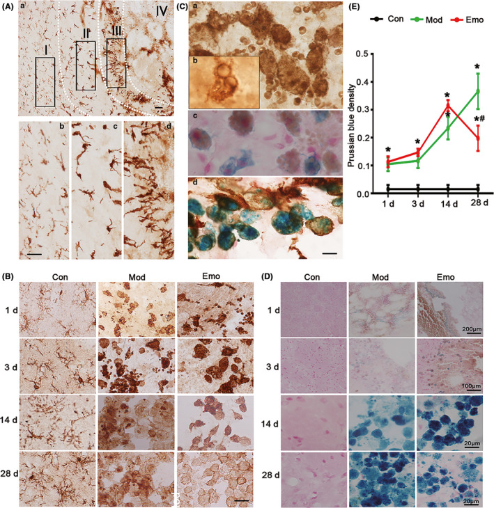
Results: Lactate accumulation was observed with in vivo MRS method, and its concentration was monitored during the recovery of ICH and treatment of emodin. Lactate concentration significantly increased in the core and penumbra regions of hemorrhagic foci, and it decreased after the treatment of emodin. The in vitro culture study was verified that lactate was beneficial for the proliferation, cell survival, migration, and phagocytosis property of the microglia.
Conclusion: Results from in vitro verification study, investigations from the recovery of ICH, and treatment of emodin verify that lactate plays an important role during the recovery of ICH. This could provide a novel therapeutic approach for ICH.
Keywords: emodin; hemorrhages; in vivo MRS; lactate; microglia.
© 2020 The Authors. CNS Neuroscience & Therapeutics published by John Wiley & Sons Ltd.
Conflict of interest statement
The authors declare that there is no conflict of interest.
Figures
Similar articles
-
Protective effects of Da-cheng-qi decoction in rats with intracerebral hemorrhage.Phytomedicine. 2021 Sep;90:153630. doi: 10.1016/j.phymed.2021.153630. Epub 2021 Jun 17. Phytomedicine. 2021. PMID: 34217968
-
Lactate potentiates angiogenesis and neurogenesis in experimental intracerebral hemorrhage.Exp Mol Med. 2018 Jul 6;50(7):1-12. doi: 10.1038/s12276-018-0113-2. Exp Mol Med. 2018. PMID: 29980670 Free PMC article.
-
Exploring Metabolic Aberrations after Intracerebral Hemorrhage In Vivo with Deuterium Metabolic Spectroscopy Imaging.Anal Chem. 2024 Oct 1;96(39):15563-15571. doi: 10.1021/acs.analchem.4c01999. Epub 2024 Sep 18. Anal Chem. 2024. PMID: 39295127
-
Sesamin suppresses activation of microglia and p44/42 MAPK pathway, which confers neuroprotection in rat intracerebral hemorrhage.Neuroscience. 2013 Mar 1;232:45-52. doi: 10.1016/j.neuroscience.2012.11.057. Epub 2012 Dec 8. Neuroscience. 2013. PMID: 23228810
-
High Morphologic Plasticity of Microglia/Macrophages Following Experimental Intracerebral Hemorrhage in Rats.Int J Mol Sci. 2016 Jul 21;17(7):1181. doi: 10.3390/ijms17071181. Int J Mol Sci. 2016. PMID: 27455236 Free PMC article.
Cited by
-
NLRP3 inflammasome and gut microbiota-brain axis: A new perspective on white matter injury after intracerebral hemorrhage.Neural Regen Res. 2026 Jan 1;21(1):62-80. doi: 10.4103/NRR.NRR-D-24-00917. Epub 2025 Jan 29. Neural Regen Res. 2026. PMID: 39885662 Free PMC article.
-
Metabolic regulation of microglial phagocytosis: Implications for Alzheimer's disease therapeutics.Transl Neurodegener. 2023 Oct 31;12(1):48. doi: 10.1186/s40035-023-00382-w. Transl Neurodegener. 2023. PMID: 37908010 Free PMC article. Review.
-
Metabolism navigates neural cell fate in development, aging and neurodegeneration.Dis Model Mech. 2021 Aug 1;14(8):dmm048993. doi: 10.1242/dmm.048993. Epub 2021 Aug 4. Dis Model Mech. 2021. PMID: 34345916 Free PMC article. Review.
-
B-Mode Ultrasound, a Reliable Tool for Monitoring Experimental Intracerebral Hemorrhage.Front Neurol. 2021 Dec 23;12:771402. doi: 10.3389/fneur.2021.771402. eCollection 2021. Front Neurol. 2021. PMID: 35002926 Free PMC article.
-
The roles of lactate and the interplay with m6A modification in diseases.Cell Biol Toxicol. 2024 Dec 2;40(1):107. doi: 10.1007/s10565-024-09951-9. Cell Biol Toxicol. 2024. PMID: 39617813 Free PMC article. Review.
References
-
- Feigin VL, Lawes CM, Bennett DA, Barker‐Collo SL, Parag V. Worldwide stroke incidence and early case fatality reported in 56 population‐based studies: a systematic review. Lancet Neurol. 2009;8(4):355‐369. - PubMed
-
- Kuramatsu JB, Huttner HB, Schwab S. Advances in the management of intracerebral hemorrhage. J Neural Transm. 2013;120(Suppl 1):S35‐S41. - PubMed
-
- Magistretti PJ, Allaman I. A cellular perspective on brain energy metabolism and functional imaging. Neuron. 2015;86(4):883‐901. - PubMed
Publication types
MeSH terms
Substances
LinkOut - more resources
Full Text Sources
