. 2020 Aug 25;142(8):758-775.
doi: 10.1161/CIRCULATIONAHA.119.044340. Epub 2020 Jun 3.

Loss of Protease-Activated Receptor 4 Prevents Inflammation Resolution and Predisposes the Heart to Cardiac Rupture After Myocardial Infarction

Affiliations

Loss of Protease-Activated Receptor 4 Prevents Inflammation Resolution and Predisposes the Heart to Cardiac Rupture After Myocardial Infarction

Mikhail A Kolpakov et al. Circulation. .

Abstract

Background: Cardiac rupture is a major lethal complication of acute myocardial infarction (MI). Despite significant advances in reperfusion strategies, mortality from cardiac rupture remains high. Studies suggest that cardiac rupture can be accelerated by thrombolytic therapy, but the relevance of this risk factor remains controversial.

Methods: We analyzed protease-activated receptor 4 (Par4) expression in mouse hearts with MI and investigated the effects of Par4 deletion on cardiac remodeling and function after MI by echocardiography, quantitative immunohistochemistry, and flow cytometry.

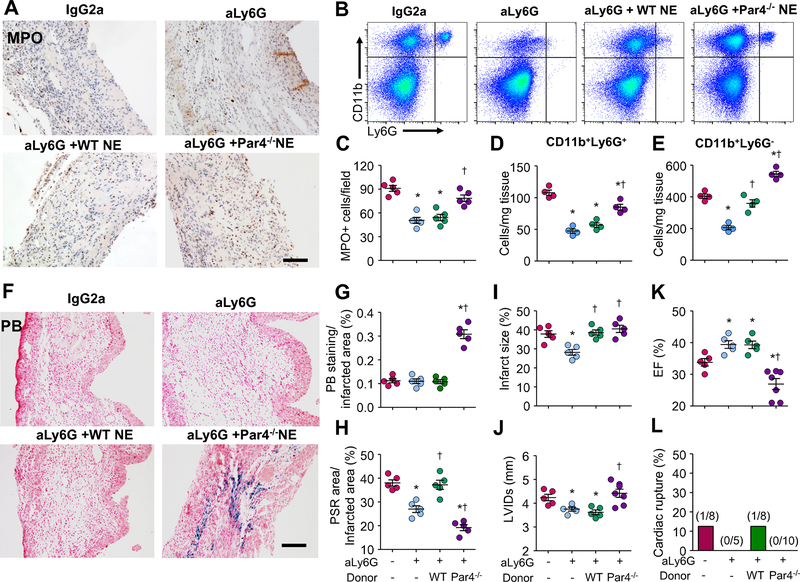
Results: Par4 mRNA and protein levels were increased in mouse hearts after MI and in isolated cardiomyocytes in response to hypertrophic and inflammatory stimuli. Par4-deficient mice showed less myocyte apoptosis, reduced infarct size, and improved functional recovery after acute MI relative to wild-type (WT). Conversely, Par4-/- mice showed impaired cardiac function, greater rates of myocardial rupture, and increased mortality after chronic MI relative to WT. Pathological evaluation of hearts from Par4-/- mice demonstrated a greater infarct expansion, increased cardiac hemorrhage, and delayed neutrophil accumulation, which resulted in impaired post-MI healing compared with WT. Par4 deficiency also attenuated neutrophil apoptosis in vitro and after MI in vivo and impaired inflammation resolution in infarcted myocardium. Transfer of Par4-/- neutrophils, but not of Par4-/- platelets, in WT recipient mice delayed inflammation resolution, increased cardiac hemorrhage, and enhanced cardiac dysfunction. In parallel, adoptive transfer of WT neutrophils into Par4-/- mice restored inflammation resolution, reduced cardiac rupture incidence, and improved cardiac function after MI.

Conclusions: These findings reveal essential roles of Par4 in neutrophil apoptosis and inflammation resolution during myocardial healing and point to Par4 inhibition as a potential therapy that should be limited to the acute phases of ischemic insult and avoided for long-term treatment after MI.

Keywords: heart rupture; inflammation; myocardial infarction; neutrophils; protease-activated receptor 4.


Figures

Figure 1:
Figure 1:. Par4 expression is upregulated after MI in mice.
Mice were subjected to permanent left anterior descending artery ligation to induce ischemia. A, Representative Par4 immunostainings of paraffin-embedded sections from mouse hearts subjected to MI for 2, 7, and 28 days. Arrows indicate myocytes (black) or leukocytes (blue). Scale bar, 40 μm. B, Infarcted and border-zone lysates were assessed for immunoblot analysis. Arrows indicate splicing from the same gel exposure. Left, Representative immunoblots with GAPDH taken as a loading control. Right, Fold induction (n=6 each group). C, Expression of Par1, Par2, and Par4 mRNA levels in infarcted border-zone hearts as assessed by real-time quantitative polymerase chain reaction. Data were normalized to hypoxanthine phosphoribosyl-transferase and were expressed relative to levels in sham hearts. *P<0.05 vs shams. D, Isolated neonatal rat cardiomyocytes were untreated or treated with thrombin (Thr; 1 U/mL), epidermal growth factor (EGF; 100 ng/mL), norepinephrine (NE; 10 μmol/L), or tumor necrosis factor-α (TNFα; 100 ng/mL) for 48 hours. Left, Representative immunoblots of partially purified plasma membrane lysates with caveolin-3 (Cav-3) taken as a loading control. Right, Quantification of experiments expressed as mean±SE from 3 separate cultures. *P<0.05 vs control (Ctrl).
Figure 2:
Figure 2:. Pleiotropic effects of Par4 deletion following acute and chronic MI.
Mice were subjected to permanent MI. (A) Heart tissue sections were assessed for apoptosis using TUNEL assay (green), tropomyosin (Tropom, red), and DAPI (4’,6-diamidino-2-phenylindole) (blue) staining. Scale bar: 40 μm. (B) TUNEL-positive myocytes and non-myocytes in the infarct border area expressed as a percentage of total nuclei detected by DAPI staining. (C) Serum levels of cardiac troponin I (cTnI) after MI in wild-type (WT) and Par4−/− mice. (D) Heart weight to body weight (HW/BW) and (E) Left ventricular ejection fraction (EF) assessment in WT and Par4−/− mice following MI. (F) Representative images of Masson’s trichrome staining on transverse heart sections at day 7 post-MI. Arrows indicate necrotic myocytes. Scale bars: 0.5 mm. (G) Area at risk (AAR) and infarct area expressed as a percentage of total left ventricular area (5 section levels per heart). (H) Post-MI mortality of WT and Par4−/− mice showed a decrease in Par4−/− mice survival after MI compared to WT. (I) Percentage of cardiac rupture between Par4−/− and WT mice post-MI. Values are presented as mean ± S.E. *p<0.05 vs. WT shams, †p<0.05 vs. WT MI.
Figure 3:
Figure 3:. Par4 deficiency impairs myocardial healing after chronic MI.
A, Hematoxylin-eosin (HE), Prussian blue (PB), picrosirius red (PSR), and smooth muscle α-actin (SMA) staining on transverse heart sections at day 7 after MI showing residual presence of necrotic myocytes, hemosiderin deposits, and impaired accumulation of collagen (Col I) and SMA-positive fibroblasts in Par4−/− infarct compared with wild-type (WT). Scale bar, 40 μm. B through E, Semiquantitative analysis of necrotic area (B), PB (C), collagen (D), and SMA (E) staining expressed as a percentage of total infarct area. F, Infarcted and border zone lysates were assessed for immunoblot analysis. Top, Representative immunoblots with GAPDH taken as a loading control. Arrows indicate splicing from the same gel exposure. Bottom, Fold induction. G, Top, Gelatin zymography of extracts from 7 days sham and MI hearts showed significantly enhanced matrix metalloprotease (MMP) 9 activity in Par4−/− compared with WT infarcts. Arrows indicate splicing from the same gel exposure. Bottom, Quantification of cleaved MMP2 and MMP9 accumulation levels. *P<0.05 vs WT sham; †P<0.05 vs WT MI.
Figure 4:
Figure 4:. Par4 deficiency delays infiltration of neutrophils and macrophages after chronic MI.
(A) Heart sections were stained with anti-myeloperoxidase (MPO) or anti-Mac3, to visualize infiltrating inflammatory cells in the infarcted border region at day 7 post-MI. Scale bar: 40 μm. (B and C) The total number of MPO-positive neutrophils (B) and Mac3-positive macrophages (C) infiltrated into the infarct was quantified on 3 serial sections per heart. (D) Single cell suspensions isolated from hearts of shams or mice subjected to MI for 7 days were stained with anti-CD11b, -Ly-6G, -F4/80, and -CD11c antibodies and analyzed by FACS. Neutrophils were identified as CD11b+Ly-6G+ and mononuclear phagocytes were identified as CD11b+Ly-6G. (E-G) Quantification of the number of neutrophils (E), all mononuclear phagocytes (F) or macrophages/dendritic cells (G) relative to the leukocyte-enriched gate or mononuclear phagocyte population, respectively, in mice undergoing MI for 7 days (n=4–5 per time point). (H) ELISA quantification of lipoxin A4 in hearts showing increased lipoxin A4 levels at day 2 post-MI in WT infarct, whereas this increase was delayed in Par4−/− hearts and was only observed at day 7 post-MI. (I and J) Flow cytometric analysis showing increased numbers of CD206+F4/80+CD11b+Ly6G M2-like and CD86+F4/80+CD11b+Ly6G M1-like macrophages in Par4−/− hearts compared to WT at day 7 post-MI (n = 4 in each group). (K) Real time-qPCR analysis of hearts from WT and Par4−/− mice at day 7 post-MI (n=6 per group) showed higher expression of M1 and M2 inflammatory markers compared to WT following MI. Data were normalized to HPRT and were expressed relative to levels in WT sham hearts. Data represent mean ± S.E. *p<0.05 vs. WT shams. †p<0.05 vs. WT MI.
Figure 5:
Figure 5:. Par4 deficiency in platelets does not impair cardiac healing and adverse cardiac remodeling post-MI.
(A and B) Representative aggregation (top) and dense granule secretion (bottom) tracings of washed platelets from Par4−/− or WT controls (A) or mice subjected to MI for 2 days (B) treated with Par4-agonist peptide (AYPGKF) or collagen, respectively, for 4 minutes. (C and D) Quantification of extent of aggregation (C) and dense granule secretion (D) of at least 3 independent experiments from panel B. (E-M) WT mice were treated with IgG or anti-platelet antibodies (aPLT) and then intravenously received platelets from WT or Par4−/− mice prior to MI. (E) Prussian blue (PB), myeloperoxidase (MPO) and picro-sirius red (PSR) staining on transverse heart sections at day 7 post-MI. Scale bar: 40 μm. The area of hemosiderin deposition (F), MPO-positive neutrophils (G) and collagen deposition (H) in the infarct were quantified on 3 serial sections per heart. (I) Infarcted area was measured using Masson’s trichrome staining on transverse heart sections showing no effect of Par4−/− platelet transfer on infarct size compared to WT platelets. (J-L) Echocardiographic measurement of left ventricular (LV) internal diameter in systole (LVIDs) (J) and diastole (LVIDd) (K) and LV ejection fraction (EF) (L). (M) The percentage of cardiac rupture (M) between the groups were shown. *p<0.05 vs. IgG-treated WT. †p<0.05 vs. a-PLT-treated WT.
Figure 6:
Figure 6:. Par4 stimulation induces neutrophil apoptosis.
(A) Heart sections were stained with anti-Ly6G and TUNEL to visualize apoptotic neutrophils in the infarcted border region at day 2 and 7 post-MI. Scale bar: 40 μm. (B) Representative flow cytometry plots showing apoptotic neutrophils stained with annexin 5 (A5). (C and D) The total number of apoptotic neutrophils in MI and sham was quantified on 3 serial sections per heart (C) and was confirmed by FACS analysis using A5 and Ly6G staining (D). Data represent mean ± S.E. *p<0.05 vs. WT shams. †p<0.05 vs. WT MI. (E-I) Neutrophils were isolated from bone marrow of WT or Par4−/− mice and were untreated or treated with Par1-agonist peptide (AP) (TFLLRN, 300 μM), Par4-AP (AYPGKF, 500 μM) or thrombin (10 U/ml) for 24 h. (E and F) Flow cytometric quantification of neutrophil death as detected by A5 and propidium iodide (PI) staining. Left: Representative flow cytometry plots (E); right: Quantification of A5+ cells which included both PI/A5+ (early apoptotic) and PI+/A5+ (late apoptotic and necrotic) cells (F). (G) Quantification of percent TUNEL-positive neutrophils. (H) Several inflammatory cytokines were detected by Mouse cytokine/Chemokine array from isolated WT and Par4−/− neutrophil lysates and were presented in fold changes (over WT). Data are pooled from 4 independent experiments. (I) Oxidation of dichlorodihydrofluoresceindiacetate to fluorescent dichlorofluorescein (DCF) reflected by fluorescent activity (RFU) was measured in WT and Par4−/− neutrophils treated with Par4-AP, thrombin or phorbol myristate acetate (PMA) for 2 h. Samples were assayed in triplicate and results are representative of 3 independent experiments. Data are mean ± S.E. *p<0.05 vs. WT-untreated control; †p<0.05 vs. WT-treated neutrophils.
Figure 7:
Figure 7:. Adoptive transfer of Par4−/− neutrophils impairs inflammation resolution in WT recipient mice post-MI.
WT mice treated with neutrophil-depleting antibody (Ly6G) or isotype (IgG2a) for 2 days were injected with either WT or Par4−/− neutrophils and then subjected to MI for 7 days. (A and B) Neutrophil accumulation within the infarcted area identified by myeloperxodase (MPO) staining (A) or flow cytometry (B). Scale bar: 40 μm. (C-E) The number of neutrophils per microscope field (C) or per mg of heart tissue were shown (D), along with the number of mononuclear phagocytes (E). Data are pooled from 4 independent experiments (n = 9–12 mice per group). (F) Heart sections stained with Prussian blue (PB) (F) show accumulation of hemosiderin in WT mice receiving Par4−/−, but not WT, neutrophils after MI. Scale bar: 40 μm. (G and H) Semi-quantitative analysis of PB and picro-Sirius red (PSR) staining expressed as percentage of infarct area. (I) Quantitative analysis of infarct size (I) and echocardiographic measurement of left ventricular (LV) internal diameter in systole (LVIDs) (J) and ejection fraction (EF) (K) at day 7 post-MI. (L) The percentage of cardiac rupture between the groups were shown. *p<0.05 vs. IgG2a-treated WT. †p<0.05 vs. aLy6G-treated WT.
Figure 8:
Figure 8:. Adoptive transfer of WT neutrophils protects Par4−/− mice against cardiac rupture post-MI.
Mice were subjected to permanent left anterior descending artery ligation to induce ischemia. Immediately after MI, CM-Dil fluorescent dye-labeled WT or Par4−/− neutrophils (red) were injected intravenously into Par4−/− recipient mice. (A) Heart sections were stained with anti-Ly6G and DAPI to visualize WT neutrophils (green) and nuclei (blue), respectively, in Par4−/− recipient hearts at different time post-MI. Scale bar: 40 μm. (B) Flow-cytometric gating strategy to determine the number of neutrophils and macrophages-positive CM-Dil at day 2 post-MI. (C and D) Semi-quantitative analysis (C) and flow cytometric quantification (D) of exogenous CM-Dil labeled neutrophils/macrophages in the infarcted myocardium. Data are pooled from 4 independent experiments (n=9–10 mice per group). (E) Heart sections stained with myeloperoxidase (MPO), Prussian blue (PB) or picro-sirius red (PSR) at day 7 post-MI show reduced accumulation of neutrophils, hemosiderin deposition or collagen in Par4−/− mice receiving WT, but not Par4−/−, neutrophils. Scale bar: 40 μm. (F-H) Semi-quantitative analysis of PB (F), MPO-positive neutrophils (G) and PSR (H) staining expressed as percentage of total infarct area. Comparison of the percentage of cardiac rupture (I) and post-MI mortality rate (J) between the groups were shown. (K-M) Echocardiographic measurement of left ventricular (LV) internal diameter in diastole (LVIDd) (K) and systole (LVIDs) (L) and LV ejection fraction (EF) (M) at day 7 post-MI. (N) Infarcted area was measured using Masson’s trichrome staining on transverse heart sections. *p<0.05 vs. WT. †p<0.05 vs. Par4−/− mice.

References

    1. Institute NHLaB. Morbidity and mortality: 2012 chart book on cardiovascular and lung diseases. Bethesda, MD: National Institute of Health. 2012
    1. Pasotti M, Prati F, Arbustini E. The pathology of myocardial infarction in the pre- and post-interventional era. Heart. 2006;92:1552–1556 - PMC - PubMed
    1. Becker RC, Gore JM, Lambrew C, Douglas Weaver W, Michael Rubison R, French WJ, Tiefenbrunn AJ, Bowlby LJ, Rogers WJ. A composite view of cardiac rupture in the united states national registry of myocardial infarction. J Am Coll Cardiol. 1996;27:1321–1326 - PubMed
    1. Gao X-M, White DA, Dart AM, Du X-J. Post-infarct cardiac rupture: Recent insights on pathogenesis and therapeutic interventions. Pharmacol Therap. 2012;134:156–179 - PubMed
    1. Frangogiannis NG. Regulation of the inflammatory response in cardiac repair. Circ Res. 2012;110:159–173 - PMC - PubMed

Publication types

MeSH terms

Substances