Review
. 2020 May 13:9:F1000 Faculty Rev-355.
doi: 10.12688/f1000research.21914.1. eCollection 2020.

Recent trends in airway management

Affiliations
Review

Recent trends in airway management

Basem B Abdelmalak et al. F1000Res. .

Abstract

Clinical airway management continues to advance at a fast pace. To help update busy anesthesiologists, this abbreviated review summarizes notable airway management advances over the past few years. We briefly discuss advances in video laryngoscopy, in flexible intubation scopes, in jet ventilation, and in extracorporeal membrane oxygenation (ECMO). We also discuss noninvasive ventilation in the forms of high-flow nasal cannula apneic oxygenation and ventilation and nasal continuous positive airway pressure (CPAP) masks. Emerging concepts related to airway management, including the physiologically difficult airway and lower airway management, new clinical subspecialties and related professional organizations such as Anesthesia for Bronchoscopy, the Society for Head and Neck Anesthesia, and fellowship training programs related to advanced airway management are also reviewed. Finally, we discuss the use of checklists and guidelines to enhance patient safety and the value of large databases in airway management research.

Keywords: Airway Management; Jet ventilation; Lower Airway Management; Videolaryngoscope; the physiologically difficult Airway.


Conflict of interest statement

Competing interests: The authors receive royalties from the sales of two related textbooks that they co-edited: Anesthesia for Otolaryngologic Surgery and Clinical Airway Management: An Illustrated Case-Based Approach published by Cambridge University Press, London, UK.No competing interests were disclosed.No competing interests were disclosed.No competing interests were disclosed.

Figures

Figure 1.
Figure 1.. The GlideScope Core™ airway visualization workstation.
The GlideScope Core™ airway visualization workstation offers a 10 inch high-resolution color touch-screen display with picture-in-picture capability to allow simultaneous laryngoscopy and bronchoscopy. Still images and videos can be captured for later review. An adjustable arm enables one to position the monitor for optimal viewing. Other features include the display of patient SpO 2 and 180 degree image rotation. This image has been reproduced with permission from Verathon Inc.
Figure 2a.
Figure 2a.. Hunsaker catheter.
This figure has been reproduced with permission from Cleveland Clinic Center for Medical Art & Photography © 2020. All rights reserved.
Figure 2b.
Figure 2b.. Subglottic jetting using Hunsaker catheter (see Figure 2a).
Please note that the catheter sideport is attached to the airway pressure monitoring tube and, through the stopcock, it can be connected temporarily to an EtCO 2 monitor showing a capnogram. This figure has been reproduced with permission from Cleveland Clinic Center for Medical Art & Photography © 2020. All rights reserved. Written informed consent was obtained from the patient for the use and publication of this clinical image.
Figure 3.
Figure 3.. Supraglottic jetting.
The jetting needle is attached to the eye piece of the suspension laryngoscopy scope. This figure has been reproduced with permission from Cleveland Clinic Center for Medical Art & Photography © 2020. All rights reserved. Written informed consent was obtained from the patient for the use and publication of this clinical image.
Figure 4.
Figure 4.. The Ventrain® device.
This simple-to-operate manual ventilator is operated using one’s thumb and index finger. It features active expiration based on the Bernoulli Principle, allowing ventilation through small-bore tubes. In addition to inspiratory (positive pressure) and expiratory (negative pressure) modes of operation, an equilibration (safety) mode is available where no significant positive or negative pressure is present at the tip of the attached ventilation catheter. The figure and text have been reproduced with permission from Ventinova Medical, Eindhoven, The Netherlands.
Figure 5.
Figure 5.. Illustration of the Ventrain® used in conjunction with a small-bore airway catheter, such as those used to exchange endotracheal tubes or those used to assist with tracheal extubation.
In addition to providing a means to facilitate reintubation, such airway catheters can also be used with the Ventrain® to maintain ventilation and oxygenation until a definitive airway is established. The figure and text have been reproduced with permission from Ventinova Medical, Eindhoven, The Netherlands.
Figure 6.
Figure 6.. The Tritube®.
The Tritube® is a narrow-bore cuffed endotracheal tube with inner and outer diameters of 2.4 mm and 4.4 mm, respectively. A Murphy eye is featured at the distal end. The ventilation lumen is attached to the Ventrain®, while an inflatable cuff seals the airway. A pressure measurement lumen permits continuous intratracheal pressure measurements. This device may be useful in both elective and emergency airway settings. The figure and text have been reproduced with permission from Ventinova Medical, Eindhoven, The Netherlands.
Figure 7.
Figure 7.. High-flow nasal oxygenation and ventilation technique is used during balloon dilation of subglottic stenosis in an anesthetized and paralyzed patient.
This figure has been reproduced with permission from Cleveland Clinic Center for Medical Art & Photography © 2020. All rights reserved. Written informed consent was obtained from the patient for the use and publication of this clinical image.
Figure 8.
Figure 8.. Illustration of how a supraglottic airway can be used to provide ventilation while also serving as a conduit for a flexible bronchoscope and balloon dilator used to dilate a subglottic stenosis.
This image has been reproduced with permission from Cleveland Clinic Center for Medical Art & Photography © 2020. All rights reserved.
Figure 9.
Figure 9.. Illustration of how a rigid bronchoscope is used to access the upper airway while an old tracheostomy site is used to access a lower portion so as to re-establish the connection between the upper and lower portions after the connection had closed off because of granulation and fibrous tissue resulting from long-term tracheostomy use.
Ventilation was accomplished with positive pressure ventilation with a semi-closed circuit through the rigid bronchoscope side port or through intermittent intubation of the old tracheostomy site, depending on the stage of the procedure. Alternatively, jet ventilation or high-flow oxygen apneic oxygenation and ventilation could have been used. This figure has been reproduced with permission from Cleveland Clinic Center for Medical Art & Photography © 2020. All rights reserved. Written informed consent was obtained from the patient for the use and publication of this clinical image.

References

    1. Parotto M, Cooper R: Recent advances in laryngoscopy in adults. F1000Res. 2019;8. pii: F1000 Faculty Rev-797. 10.12688/f1000research.18544.1 - DOI - PMC - PubMed
    2. F1000 Recommendation

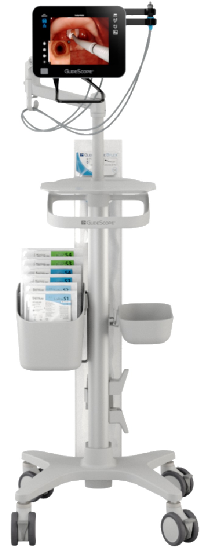
    1. Doyle DJ: The GlideScope Video Laryngoscope: A Narrative Review. TOATJ. 2017;11:48–67. 10.2174/1874321801711010048 - DOI
    1. Schechtman SA, Mathis M, Muller G, et al. : A retrospective analysis of factors associated with difficult endotracheal tube passage with use of the hyper-angled GlideScope blade. Journal of Head & Neck Anesthesia. 2019;3:e14 10.1097/HN9.0000000000000014 - DOI
    2. F1000 Recommendation

    1. Joffe AM, Aziz MF, Posner KL, et al. : Management of Difficult Tracheal Intubation: A Closed Claims Analysis. Anesthesiology. 2019;131(4):818–29. 10.1097/ALN.0000000000002815 - DOI - PMC - PubMed
    1. Mehta AC, Gildea T: Burying Our Heads in the Sand: Cross-Contamination During Bronchoscopy. Chest. 2018;154(5):1001–3. 10.1016/j.chest.2018.07.006 - DOI - PubMed