. 2020 May 9;11(3):547-558.
doi: 10.14336/AD.2019.0715. eCollection 2020 May.

Neuroprotective Effects of Endogenous Secretory Receptor for Advanced Glycation End-products in Brain Ischemia

Affiliations

Neuroprotective Effects of Endogenous Secretory Receptor for Advanced Glycation End-products in Brain Ischemia

Yu Shimizu et al. Aging Dis. .

Abstract

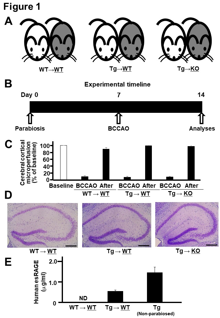
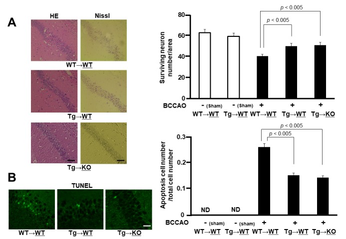
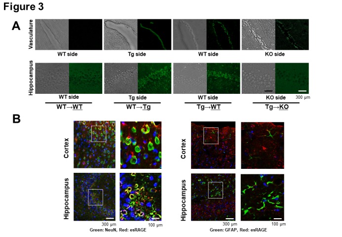
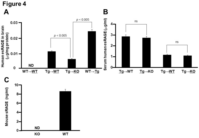
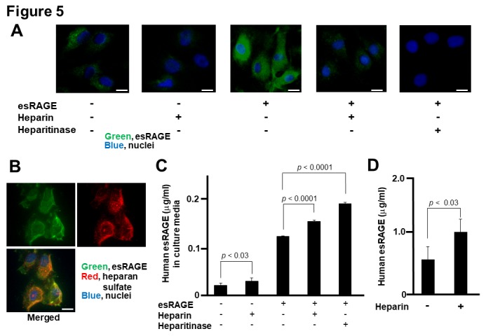
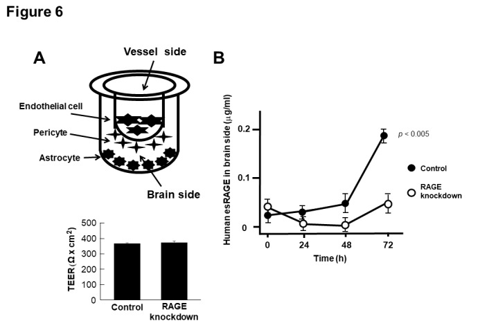
The receptor for advanced glycation end-products (RAGE) is expressed on human brain endothelial cells (HBEC) and is implicated in neuronal cell death after ischemia. We report that endogenous secretory RAGE (esRAGE) is a splicing variant form of RAGE that functions as a decoy against ischemia-induced neuronal cell damage. This study demonstrated that esRAGE was associated with heparan sulphate proteoglycans on HBEC. The parabiotic experiments between human esRAGE overexpressing transgenic (Tg), RAGE knockout (KO), and wild-type (WT) mice revealed a significant neuronal cell damage in the CA1 region of the WT side of parabiotic WT→WT mice, but not of Tg→WT mice, 7 days after bilateral common carotid artery occlusion. Human esRAGE was detected around the CA1 neurons in the WT side of the parabiotic Tg→WT pair, but not in the KO side of the Tg→KO pair. To elucidate the dynamic transfer of esRAGE into the brain, we used the blood-brain barrier (BBB) system (PharmaCo-Cell) with or without RAGE knockdown in endothelial cells. A RAGE-dependent transfer of esRAGE was demonstrated from the vascular to the brain side. These findings suggested that esRAGE is associated with heparan sulphate proteoglycans and is transferred into the brain via BBB to exert its neuroprotective effects in ischemia.

Keywords: Receptor for advanced glycation end-products (RAGE); blood-brain barrier; delayed neuronal cell damage; endogenous secretory RAGE (esRAGE); parabiosis.


Figures

Figure 1.
Figure 1.
Parabiosis and BCCAO. A) Parabiosis was done between wild-type (WT) and WT mice (WT→WT), esRAGE transgenic and WT mice (Tg→WT), and esRAGE transgenic and RAGE knockout (KO) mice (Tg→KO); gray-colored and right-side mice underwent BCCAO and further analyses. B) Experimental timeline. C, Laser-Doppler flowmetry data for evaluating the cerebral cortical microperfusion. Baseline, baseline data (100%); BCCAO, data at 1 min during the occlusion; After, data at 30 min after BCCAO. Values are mean ± SD. D) Hematoxylin-eosin stain of the hippocampus. E) Human esRAGE levels in the sera of WT sides of WT→WT and Tg→WT pairs and non-parabiosed esRAGE Tg mice (n = 4-8). ND, not detected. Values are mean ± SEM.
Figure 2.
Figure 2.
Neuronal cell damage. A) HE and Nissl stains of the hippocampal CA1 region of WT sides of WT→WT and Tg→WT pairs and of KO side of Tg→KO pair after 7 days of BCCAO (left panel). Surviving neuron numbers per area in hippocampal CA1 region were counted in WT sides of WT→WT and Tg→WT pairs and in KO side of Tg→KO pair with or without BCCAO (right panel) (n = 4-8). Values are mean ± SEM. B) TUNEL stain. Green signals indicate apoptotic cells of WT sides of WT→WT and Tg→WT pairs and of KO side of Tg→KO pair after 7 days of BCCAO (left panel). Apoptosis cell numbers per total cell numbers were counted in WT sides of WT→WT and Tg→WT pairs and in KO side of Tg→KO pair with or without BCCAO (right panel) (n = 4-8). Values are mean ± SEM.
Figure 3.
Figure 3.
Immunohistochemical detection of human esRAGE. A) Immunohistochemical study for the detection of human esRAGE (green signals). Hippocampal vasculatures and CA1 regions of WT side of WT→WT and esRAGE Tg (Tg)→WT pairs, the Tg side of the WT→Tg pair, and the RAGE knockout (KO) side of the Tg→KO pair without BCCAO. B) Immunostaining for NeuN (a neuronal marker, green) and human esRAGE (red) (left panel) as well as GFAP (a glial marker, green) and human esRAGE (red) (right panel) in brain cortex and hippocampus of WT sides of Tg→WT pair. Blue signals indicate nuclei [4',6-diamidino-2-phenylindole (DAPI) stain].
Figure 4.
Figure 4.
Quantitative detection of human esRAGE A) Human esRAGE contents in the brain parenchyma of WT side of WT→WT pair, WT side of Tg→WT pair, KO side of Tg→KO pair, and Tg side of WT→Tg pair (n = 3). B) Serum human esRAGE concentrations in Tg side of Tg→WT pair, Tg side of Tg→KO pair, WT side of Tg→WT pair, and KO side of Tg→KO pair (n = 3). C, Serum mouse sRAGE concentrations in KO and WT mice (n = 3). ND, not detected; ns, not significant. Values are mean ± SEM.
Figure 5.
Figure 5.
Association of esRAGE with endothelial cells. A and B) Immunofluorescence studies of human brain endothelial cells (HBEC) in culture. Green, Red and blue signals indicate esRAGE, heparin sulfate and nucleus (DAPI), respectively. Bar, 50 µm. C) Human esRAGE levels in culture media of HBEC (n = 4). esRAGE, 1 µg/ml; Heparin, 0.1 IU/ml; heparitinase, 1 mU/ml. Values are mean ± SEM. C) Serum levels of human esRAGE in the esRAGE Tg mice with or without heparin injection (n = 4-8). Values are mean ± SEM.
Figure 6.
Figure 6.
Transfer of esRAGE through BBB. A) In vitro (BBB) model system composed of primary cultures of monkey brain capillary endothelial cells coupled with rat pericytes and astrocytes. Recombinant esRAGE (20 µg/ml) was added to the upper (vessel side) chambers of the model and transferred esRAGE level was quantified in the lower (brain side) chambers. Endothelial cells were treated with scrambled (control) or RAGE shRNA vectors (knockdown). The integrity of the in vitro primate BBB was unaffected by RAGE knockdown, assessed by high trans-endothelial electrical resistance (TEER) (n = 5). B) Human esRAGE levels in brain side were quantified (n = 5). Control, scrambled vector-treated; RAGE knockdown, RAGE shRNA vectors-treated. Values are mean ± SEM.

References

    1. Moskowitz MA, Lo EH, Iadecola C, et al. (2010). The science of stroke: mechanisms in search of treatments. Neuron, 29: 181-98. - PMC - PubMed
    1. Prabhakaran S, Ruff I and Bernstein RA (2015). Acute stroke intervention: A systematic review. JAMA, 313: 1451-1462. - PubMed
    1. Pulsinelli WA, Levy DE, Duffy TE, et al. (1982). Regional cerebral blood flow and glucose metabolism following transient forebrain ischemia. Ann Neurol, 11: 499-502. - PubMed
    1. Tajiri S, Oyadomari S, Yano S, et al. (2004). Ischemia-induced neuronal cell death is mediated by the endoplasmic reticulum stress pathway involving CHOP. Cell Death and Differentiation, 11: 403-415. - PubMed
    1. Kirino T (1982). Delayed neuronal death in the gerbil hippocampus following ischemia. Brain Res, 6: 57-69. - PubMed

LinkOut - more resources