

Docosahexaenoic Acid (DHA) Inhibits FADS2 Expression in Astrocytes but Increases Survival of Neurons Co-cultured with DHA-enriched Astrocytes
- PMID: 32489952
- PMCID: PMC7241842
- DOI: 10.22088/IJMCM.BUMS.8.3.232
Docosahexaenoic Acid (DHA) Inhibits FADS2 Expression in Astrocytes but Increases Survival of Neurons Co-cultured with DHA-enriched Astrocytes
Abstract
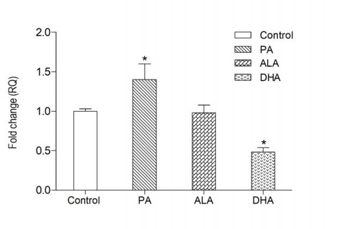
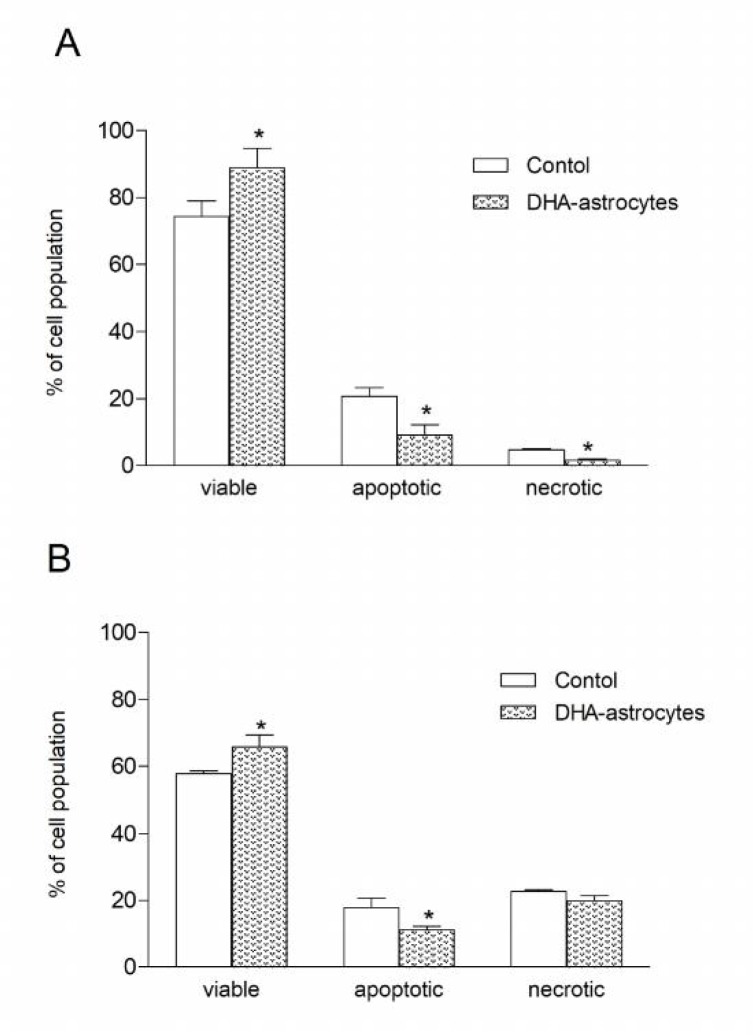
Docosahexaenoic acid (DHA), the most abundant n-3 polyunsaturated fatty acid (n-3PUFA) in the brain, has attracted great importance for a variety of neuronal functions such as signal transduction through plasma membranes, neuronal plasticity, and neuroprotection. Astrocytes that provide structural, functional, and metabolic support for neurons, express ∆6- desaturase encoded by FADS2 gene that can be, next to the plasma DHA pool, additional source of DHA in the brain. Furthermore, the genetic variations of FADS gene cluster has been found in children with developmental disorders, and are associated with cognitive functions. Since, the regulation of DHA biosynthesis in astrocytes remains poorly studied the aim of this study was to determine the effect of palmitic acid (PA), α-linolenic acid (ALA) or docosahexaenoic acid (DHA), on the transcription of FADS2 gene in astrocytes and survival of neurons challenged with oxidative compounds after co-culture with astrocytes exposed to DHA. The lipid profile in cell membranes after incubation with fatty acids was determined by gas chromatography, and FADS2 expression was analyzed using real-time PCR. The viability of neurons cocultured with PUFA-enriched astrocytes was investigated by flow cytometry after staining cells with annexin V-FITC and PI. The results showed that DHA suppressed (P <0.01), PA stimulated (P <0.01), while ALA did not change the FADS2 gene expression after 24 h incubation of astrocytes with fatty acids. Although FADS2 mRNA was down-regulated by DHA, its level in astrocytic membranes significantly increased (P <0.01). Astrocytes with DHA-enriched membrane phospholipids markedly enhanced neuronal resistance to cytotoxic compounds and neuronal survival. These results suggest that beneficial effects of supplementation with n-3 PUFA in Alzheimer disease and in psychiatric disorders is caused, in part, by increased efficacy of DHA-enriched astrocytes to protect neurons under adverse conditions in the brain.
Keywords: Docosahexaenoic acid; FADS2; astrocytes; neuroprotection.
Figures
References
-
- Gharami K, Das M, Das S. Essential role of docosahexaenoic acid towards development of a smarter brain. Neurochem Int. 2015;89:51–62. - PubMed
-
- Wysoczanski T, Sokola-Wysoczanska E, Pekala J, et al. Omega-3 Fatty Acids and their Role in Central Nervous System - A Review. Curr Med Chem. 2016;23:816–31. - PubMed
LinkOut - more resources
Full Text Sources
Research Materials
Miscellaneous