

Lateral Meniscal Oblique Radial Tears Are Common With ACL Injury: A Classification System Based on Arthroscopic Tear Patterns in 600 Consecutive Patients
- PMID: 32490027
- PMCID: PMC7238316
- DOI: 10.1177/2325967120921737
Lateral Meniscal Oblique Radial Tears Are Common With ACL Injury: A Classification System Based on Arthroscopic Tear Patterns in 600 Consecutive Patients
Abstract
Background: Meniscal root tears and ramp lesions have been rigorously characterized in recent literature. However, one of the most common lateral meniscal injuries identified with an acute anterior cruciate ligament (ACL) disruption, a posterior horn lateral meniscal oblique radial tear (LMORT), has not been thoroughly described.
Purpose: To determine the incidence of all meniscal tears and, more specifically, the incidence of posterior horn LMORTs in a multicenter cohort of consecutive, acute ACL reconstructions. Additionally, the authors aimed to develop a new classification system to help guide treatment of posterior horn LMORTs.
Study design: Cross-sectional study; Level of evidence, 3.
Methods: A multicenter retrospective cohort design was used to analyze 200 consecutive cases of acute ACL reconstruction from each of 3 different surgeons, for a total of 600 patients. The operative notes and intraoperative photos were analyzed to determine the incidence and laterality of all meniscal tears. A classification system based on tear characterization was then used to categorize tear patterns into similar groups.
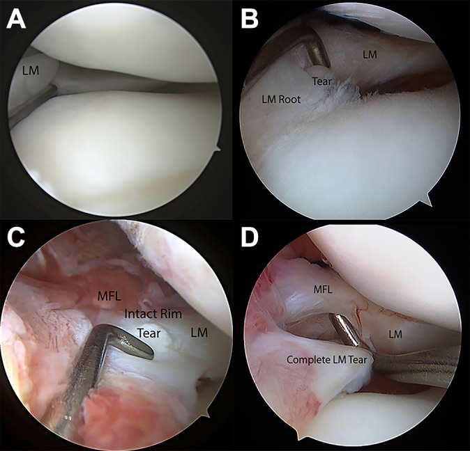
Results: A total of 396 (66%) of the 600 patients with acute ACL disruption had concomitant meniscal tears. Specifically, 187 (31%) had a lateral meniscal injury, 89 (15%) had a medial meniscal injury, and 122 (20%) had both medial and lateral meniscal injuries. The most common lateral meniscal tear was an LMORT; 71 (18%) patients with meniscal tears had a posterior horn LMORT. Overall, the incidence of ACL injury with a concomitant posterior horn LMORT was 12%. A classification was developed, which included type 1 tear (partial thickness <10 mm from the root attachment), type 2 tear (complete radial oblique tear that extended <10 mm from root), type 3 tear (incomplete LMORT that extended >10 mm from root), and type 4 tear (complete LMORT >10 mm from root).
Conclusion: In 600 consecutive acute ACL reconstructions, the incidence of concomitant ACL injury with meniscal injury was 66%, and posterior horn LMORTs represented a large proportion of all meniscal tears (12%). A classification scheme was developed for posterior horn LMORTs to aid reporting and clinical decision making for these common tears.
Keywords: LMORT; lateral meniscus; meniscal tear; oblique meniscal tear; oblique radial tear.
© The Author(s) 2020.
Conflict of interest statement
One or more of the authors has declared the following potential conflict of interest or source of interest: A.J.K. has received research support from Aesculap/B.Braun, Ceterix, Exactech, Gemini Medical, and Histogenics; consulting fees from Arthrex, JRF Ortho, and Vericel; speaking fees from Arthrex; and royalties from Arthrex; he is a board or committee member for the Musculoskeletal Transplant Foundation and has stock/stock options in Responsive Arthroscopy. M.D.L. has a family member with the following disclosures: consulting fees from Arthrex, Linvatec, Ossur, and Smith & Nephew; research support and speaking fees from Smith & Nephew; grant support from the National Institute of Arthritis and Musculoskeletal and Skin Diseases for the Musculoskeletal Research Training Program (T32AR56950); and royalties from Arthrex, Ossur, Smith & Nephew, and Thieme. M.J.S. has received research support from Arthrex and Stryker and consulting fees and royalties from Arthrex. P.A.S. has received research support, consulting fees, and royalties from Arthrex; educational support from Elite Orthopedics; and speaking fees from Arthrex and Alpha Orthopedic Systems and has stock/stock options in Spinal Simplicity. AOSSM checks author disclosures against the Open Payments Database (OPD). AOSSM has not conducted an independent investigation on the OPD and disclaims any liability or responsibility relating thereto.
Figures

References
-
- Ahn JH, Lee YS, Yoo JC, Chang MJ, Park SJ, Pae YR. Results of arthroscopic all-inside repair for lateral meniscus root tear in patients undergoing concomitant anterior cruciate ligament reconstruction. Arthroscopy. 2010;26(1):67–75. - PubMed
-
- Allaire R, Muriuki M, Gilbertson L, Harner CD. Biomechanical consequences of a tear of the posterior root of the medial meniscus: similar to total meniscectomy. J Bone Joint Surg Am. 2008;90(9):1922–1931. - PubMed
-
- Beldame J, Wajfisz A, Lespagnol F, Hulet C, Seil R. Lateral meniscus lesions on unstable knee. Orthop Traumatol Surg Res. 2009;95(8) (suppl 1):S65–S69. - PubMed
-
- Bhatia S, LaPrade CM, Ellman MB, LaPrade RF. Meniscal root tears: significance, diagnosis, and treatment. Am J Sports Med. 2014;42(12):3016–3030. - PubMed
LinkOut - more resources
Full Text Sources
