ASS1 Overexpression: A Hallmark of Sonic Hedgehog Hepatocellular Adenomas; Recommendations for Clinical Practice
- PMID: 32490318
- PMCID: PMC7262286
- DOI: 10.1002/hep4.1514
ASS1 Overexpression: A Hallmark of Sonic Hedgehog Hepatocellular Adenomas; Recommendations for Clinical Practice
Abstract
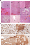
Until recently, 10% of hepatocellular adenomas (HCAs) remained unclassified (UHCA). Among the UHCAs, the sonic hedgehog HCA (shHCA) was defined by focal deletions that fuse the promoter of Inhibin beta E chain with GLI1. Prostaglandin D2 synthase was proposed as immunomarker. In parallel, our previous work using proteomic analysis showed that most UHCAs constitute a homogeneous subtype associated with overexpression of argininosuccinate synthase (ASS1). To clarify the use of ASS1 in the HCA classification and avoid misinterpretations of the immunohistochemical staining, the aims of this work were to study (1) the link between shHCA and ASS1 overexpression and (2) the clinical relevance of ASS1 overexpression for diagnosis. Molecular, proteomic, and immunohistochemical analyses were performed in UHCA cases of the Bordeaux series. The clinico-pathological features, including ASS1 immunohistochemical labeling, were analyzed on a large international series of 67 cases. ASS1 overexpression and the shHCA subgroup were superimposed in 15 cases studied by molecular analysis, establishing ASS1 overexpression as a hallmark of shHCA. Moreover, the ASS1 immunomarker was better than prostaglandin D2 synthase and only found positive in 7 of 22 shHCAs. Of the 67 UHCA cases, 58 (85.3%) overexpressed ASS1, four cases were ASS1 negative, and in five cases ASS1 was noncontributory. Proteomic analysis performed in the case of doubtful interpretation of ASS1 overexpression, especially on biopsies, can be a support to interpret such cases. ASS1 overexpression is a specific hallmark of shHCA known to be at high risk of bleeding. Therefore, ASS1 is an additional tool for HCA classification and clinical diagnosis.
© 2020 The Authors. Hepatology Communications published by Wiley Periodicals, Inc., on behalf of the American Association for the Study of Liver Diseases.
Figures


References
-
- Edmondson HA, Henderson B, Benton B. Liver‐cell adenomas associated with use of oral contraceptives. N Engl J Med 1976;294:470‐472. - PubMed
-
- Baum JK, Bookstein JJ, Holtz F, Klein EW. Possible association between benign hepatomas and oral contraceptives. Lancet 1973;2:926‐929. - PubMed
-
- Nault JC, Bioulac‐Sage P, Zucman‐Rossi J. Hepatocellular benign tumors—from molecular classification to personalized clinical care. Gastroenterology 2013;144:888‐902. - PubMed
-
- Bioulac‐Sage P, Sempoux C, Balabaud C. Hepatocellular adenoma: classification, variants and clinical relevance. Semin Diagn Pathol 2017;34:112‐125. - PubMed
-
- Sempoux C, Paradis V, Komuta M, Wee A, Calderaro J, Balabaud C, et al. Hepatocellular nodules expressing markers of hepatocellular adenomas in Budd‐Chiari syndrome and other rare hepatic vascular disorders. J Hepatol 2015;63:1173‐1180. - PubMed
LinkOut - more resources
Full Text Sources
Other Literature Sources
Miscellaneous

