

Utility of chest CT in diagnosis of COVID-19 pneumonia
- PMID: 32490829
- PMCID: PMC7490028
- DOI: 10.5152/dir.2020.20144
Utility of chest CT in diagnosis of COVID-19 pneumonia
Abstract
Purpose: We aimed to explore the imaging findings of computed tomography (CT) in diagnosing coronavirus disease 2019 (COVID-19) and its clinical value for further evaluation of suspected cases.
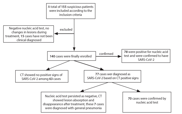
Methods: Files of 155 patients visiting the fever clinics at our hospital and affiliated hospitals from January 20th to February 9th, 2020 were searched. Among them, 140 cases (including 82 males and 58 females) were included as suspected COVID-19 cases based on clinical and epidemiological history; the CT image features of 70 cases with suggestive findings on CT, confirmed by positive nucleic acid test were analyzed and evaluated. The sensitivity and specificity of CT in diagnosing COVID-19 were evaluated in patients with epidemiological history.
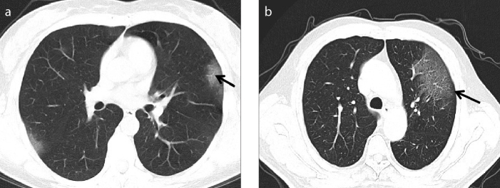
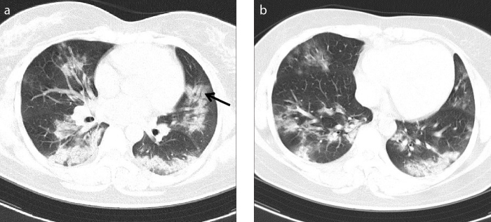
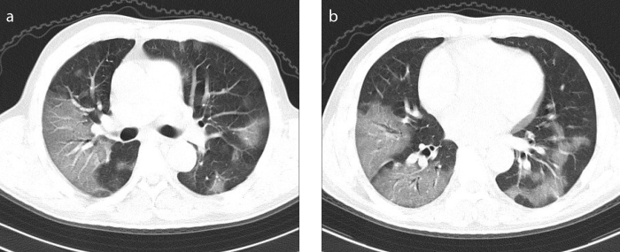
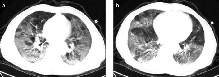
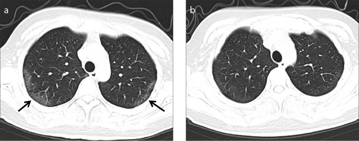
Results: Of the 70 patients, 84.3% showed bilateral lung involvement on CT; 27 cases (38.6%) showed ground-glass opacity (GGO), which was mostly distributed in the subpleural area (55.7%), and this sign was mainly observed in early COVID-19 patients. In addition, 41 cases (58.6%) manifested GGO combined with focal consolidation opacity, 2 (2.8%) had flake-like consolidation opacity, with involvements of the periphery of lung field and the central zone (44.3%), and this sign was mostly observed in severe or critical patients. Concomitant signs such as pleural effusion and mediastinal lymph node enlargement were rare. Among patients with epidemiological history, the sensitivity of CT in diagnosing COVID-19 was 89.7% (70/78), and the specificity was 88.7% (55/62).
Conclusion: CT shows high sensitivity and specificity in diagnosing COVID-19. CT is an important examination method in evaluation of suspected cases and assessment of disease severity.
Conflict of interest statement
The authors declared no conflicts of interest.
Figures
Similar articles
-
[Clinical features and high resolution CT imaging evolution of coronavirus disease 2019].Zhonghua Jie He He Hu Xi Za Zhi. 2020 Jun 12;43(6):509-515. doi: 10.3760/cma.j.cn112147-20200214-00094. Zhonghua Jie He He Hu Xi Za Zhi. 2020. PMID: 32486557 Chinese.
-
CT imaging features of COVID-19 pneumonia: initial experience from Turkey.Diagn Interv Radiol. 2020 Jul;26(4):308-314. doi: 10.5152/dir.2020.20307. Diagn Interv Radiol. 2020. PMID: 32558645 Free PMC article.
-
Thoracic imaging tests for the diagnosis of COVID-19.Cochrane Database Syst Rev. 2020 Sep 30;9:CD013639. doi: 10.1002/14651858.CD013639.pub2. Cochrane Database Syst Rev. 2020. Update in: Cochrane Database Syst Rev. 2020 Nov 26;11:CD013639. doi: 10.1002/14651858.CD013639.pub3. PMID: 32997361 Updated.
-
Imaging features of the initial chest thin-section CT scans from 110 patients after admission with suspected or confirmed diagnosis of COVID-19.BMC Med Imaging. 2020 Jun 15;20(1):64. doi: 10.1186/s12880-020-00464-5. BMC Med Imaging. 2020. PMID: 32539692 Free PMC article.
-
Similarities and Differences of Early Pulmonary CT Features of Pneumonia Caused by SARS-CoV-2, SARS-CoV and MERS-CoV: Comparison Based on a Systemic Review.Chin Med Sci J. 2020 Sep 30;35(3):254-261. doi: 10.24920/003727. Chin Med Sci J. 2020. PMID: 32972503 Free PMC article.
Cited by
-
Novel COVID-19 Diagnosis Delivery App Using Computed Tomography Images Analyzed with Saliency-Preprocessing and Deep Learning.Tomography. 2022 Jun 20;8(3):1618-1630. doi: 10.3390/tomography8030134. Tomography. 2022. PMID: 35736882 Free PMC article.
-
Association of diaphragm thickness and density measured on chest CT with disease severity in COVID-19 patients.Am J Emerg Med. 2022 Nov;61:29-33. doi: 10.1016/j.ajem.2022.08.029. Epub 2022 Aug 18. Am J Emerg Med. 2022. PMID: 36029668 Free PMC article.
-
[Korean Clinical Imaging Guidelines for Justification of Diagnostic Imaging Study for COVID-19].Taehan Yongsang Uihakhoe Chi. 2022 Mar;83(2):265-283. doi: 10.3348/jksr.2021.0117. Epub 2022 Jan 6. Taehan Yongsang Uihakhoe Chi. 2022. PMID: 36237918 Free PMC article. Review. Korean.
-
Radiologic diagnosis of patients with COVID-19.Radiologia (Engl Ed). 2021 Jan-Feb;63(1):56-73. doi: 10.1016/j.rx.2020.11.001. Epub 2020 Nov 24. Radiologia (Engl Ed). 2021. PMID: 33339622 Free PMC article.
-
Diagnostic Performance of Antigen Rapid Diagnostic Tests, Chest Computed Tomography, and Lung Point-of-Care-Ultrasonography for SARS-CoV-2 Compared with RT-PCR Testing: A Systematic Review and Network Meta-Analysis.Diagnostics (Basel). 2022 May 24;12(6):1302. doi: 10.3390/diagnostics12061302. Diagnostics (Basel). 2022. PMID: 35741112 Free PMC article. Review.
References
-
- National Health Commission. [Accessed: Feb 19, 2020];Novel Coronavirus Pneumonia Diagnosis and Treatment Scheme (trial, 6th edition) Available at: http://www.nhc.gov.cn/yzygj/s7653p/202002/8334a8326dd94d329df351d7da8aef....
-
- American College of Radiology (ACR) ACR Recommendations for the use of chest radiography and computed tomography (CT) for suspected COVID-19 Infection. Mar 11, 2020. [Accessed Mar 13, 2020]. Available at: www.acr.org/Advocacy-and-Economics/ACR-Position-Statements/Recommendatio....
MeSH terms
LinkOut - more resources
Full Text Sources
Medical
