. 2020 Jun 3;14(6):e0008364.
doi: 10.1371/journal.pntd.0008364. eCollection 2020 Jun.

Characterization and applications of a Crimean-Congo hemorrhagic fever virus nucleoprotein-specific Affimer: Inhibitory effects in viral replication and development of colorimetric diagnostic tests

Affiliations

Characterization and applications of a Crimean-Congo hemorrhagic fever virus nucleoprotein-specific Affimer: Inhibitory effects in viral replication and development of colorimetric diagnostic tests

Beatriz Álvarez-Rodríguez et al. PLoS Negl Trop Dis. .

Abstract

Crimean-Congo hemorrhagic fever orthonairovirus (CCHFV) is one of the most widespread medically important arboviruses, causing human infections that result in mortality rates of up to 60%. We describe the selection of a high-affinity small protein (Affimer-NP) that binds specifically to the nucleoprotein (NP) of CCHFV. We demonstrate the interference of Affimer-NP in the RNA-binding function of CCHFV NP using fluorescence anisotropy, and its inhibitory effects on CCHFV gene expression in mammalian cells using a mini-genome system. Solution of the crystallographic structure of the complex formed by these two molecules at 2.84 Å resolution revealed the structural basis for this interference, with the Affimer-NP binding site positioned at the critical NP oligomerization interface. Finally, we validate the in vitro application of Affimer-NP for the development of enzyme-linked immunosorbent and lateral flow assays, presenting the first published point-of-care format test able to detect recombinant CCHFV NP in spiked human and animal sera.


Conflict of interest statement

The authors have declared that no competing interests exist.

Figures

Fig 1
Fig 1. Specificity and affinity of Affimer-NP.
(A) Western blot analysis of a pull-down experiment using Affimers as capture molecules and CCHFV NP as target. (B) Western blot analysis of a pull-down experiment using either HAZV or CCHFV NP as target. (C, D) SPR analysis of binding between Affimers and ten-fold dilutions of CCHFV (C) or HAZV NP (D).
Fig 2
Fig 2. CD analyses of Affimer-NP, CCHFV NP and Affimer-NP/CCHFV NP complex.
(A, B, C) Curves representing ellipticity across the far UV spectra of CCHFV NP (A), Affimer-NP (B) or a 1:1 mixture of both (C) at 20ºC-90ºC. Crystal structure of Affimer scaffold (PDB:4n6u) is represented in orange and CCHFV NP (PDB:4akl) in blue. (D, E, F) Curves representing folded/unfolded fractions and melting temperatures (Tm) of Affimer-NP (D), CCHFV NP (E) and their mixture (F).
Fig 3
Fig 3. Inhibitory effects of Affimer-NP.
(A, B) FA analysis of RNA-binding interference of Affimer-NP using 27mer (A) or 48mer (B) RNA molecules. Top-right graphs represent negative control experiments using Affimer-Myo. (C) IncuCyte pictures taken 72h post-co-transfection of CCHFV mini-genome components plus 750 ng of Affimer-NP-RFP, Affimer-YS-RFP or no Affimer (C-). GFP signal corresponds to CCHFV-specific gene expression and RFP signal corresponds to Affimers-RFP. Scale bar = 300 μm. (D) GFP reporter signal 72h post-co-transfection of CCHFV mini-genome components with 750 ng of Affimer-NP, Affimer-YS or no Affimer (C-). (E) GFP and RFP signal 72h post-co-transfection of CCHFV mini-genome components with different amounts of Affimer-NP-RFP. Data are presented as mean ±SD (n = 3). Statistical analysis was performed using a paired t-test (ns = non-significant, *** = p<0.001).
Fig 4
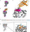
Fig 4. Tertiary structure of Affimer-NP/CCHFV NP complex.
(A) Crystal structure of CCHFV NP (PDB:4akl). Residues involved in RNA-binding, monomer-monomer interactions, or both are highlighted in blue, pink and purple, respectively. (B) Crystal structure of Affimer-NP (represented in orange) bound to CCHFV NP. (C) Close-up of Affimer-NP/CCHFV NP interaction. (D) Alignment of CCHFV NP (residues 329–407, PDB:4akl) and HAZV NP (PDB:4xze). NP residues comprising the Affimer-NP variable loop contacts are highlighted in orange, when conserved in both CCHFV and HAZV NPs, or in cyan if not. (E) Close-up of CCHFV NP residues involved in the interaction with Affimer-NP loops. Non-conserved residues (cyan) are labeled.
Fig 5
Fig 5. Sandwich ELISA for the detection of CCHFV NP.
(A) Affimer-NP was used as capture molecule for CCHFV NP in spiked sera. Anti-CCHFV NP IgGs were used as detection molecule. Anti-rabbit goat polyclonal IgGs labelled with HRP were used to develop the colorimetric output using TMB as substrate. (B, C, D, E, F) ELISA results testing sera from animals (B-E) or human (F) spiked with CCHFV, HAZV, SBV or RVFV NPs.
Fig 6
Fig 6. Development of a LFA for the detection of CCHFV NP using Affimer-NP as detection molecule.
(A, B) Schematic of the LFA. Affimer-NP was used as detection molecule on the membrane of the LFA strip and anti-CCHFV NP polyclonal rabbit IgGs were used as capture antibody for the coating of red latex beads. Control anti-biotin mouse IgGs were used for the control line in the membrane together with biotin-BSA for the coating of blue latex beads. If a CCHFV NP spiked sample (A) is applied to the strip, the CCHFV NP binds to the antibody in the red beads and it is subsequently captured by the Affimer-NP in the membrane, resulting in a red test line. In the case of a negative sample (B), there is no CCHFV NP present and the red latex beads migrate until the end of the strip, resulting in an uncolored test line. In both cases, the BSA-biotin coated blue latex beads bind to the anti-biotin mouse IgG on the membrane, resulting in a blue control line.
Fig 7
Fig 7. Affimer-NP based LFA results and comparison with ELISA.
(A) LFA positive line intensity measured after applying animal sera spiked with CCHFV, HAZV or SBV NP (120 ng NP/strip). (B) Histogram representation of data in (A). (C) LFAs 10 min after application of horse sera samples spiked with CCHFV, HAZV, SBV or RVFV NP. (D) Comparison of performance between ELISA and LFAs using horse sera spiked with CCHFV NP. Primary axes correspond to LFAs and secondary axes to ELISA. Limits of detection are highlighted with an asterisk.

Similar articles

Cited by

References

    1. Vorou R, Pierroutsakos IN, Maltezou HC. Crimean-Congo Hemorrhagic Fever. Curr Opin Infect Dis. 2007;20: 495–500. 10.1097/QCO.0b013e3282a56a0a - DOI - PubMed
    1. Bente DA, Forrester NL, Watts DM, McAuley AJ, Whitehouse CA, Bray M. Crimean-Congo hemorrhagic fever: History, epidemiology, pathogenesis, clinical syndrome and genetic diversity. Antiviral Res. 2013;100: 159–189. 10.1016/j.antiviral.2013.07.006 - DOI - PubMed
    1. Messina JP, Pigott DM, Golding N, Duda KA, Brownstein JS, Weiss DJ, et al. The global distribution of Crimean-Congo hemorrhagic fever. Trans R Soc Trop Med Hyg. 2015;109: 503–513. 10.1093/trstmh/trv050 - DOI - PMC - PubMed
    1. Gargili A, Estrada-Peña A, Spengler JR, Lukashev A, Nuttall PA, Bente DA. The role of ticks in the maintenance and transmission of Crimean-Congo hemorrhagic fever virus: A review of published field and laboratory studies. Antiviral Res. 2017;144: 93–119. 10.1016/j.antiviral.2017.05.010 - DOI - PMC - PubMed
    1. Abudurexiti A, Adkins S, Alioto D, Alkhovsky S V., Avšič-Županc T, Ballinger MJ, et al. Taxonomy of the order Bunyavirales: update 2019. Arch Virol. 2019;164: 1949–1965. 10.1007/s00705-019-04253-6 - DOI - PMC - PubMed

Publication types

MeSH terms