. 2020 Jun 3;15(6):e0233007.
doi: 10.1371/journal.pone.0233007. eCollection 2020.

A missense mutation of ErbB2 produces a novel mouse model of stillbirth associated with a cardiac abnormality but lacking abnormalities of placental structure

Affiliations

A missense mutation of ErbB2 produces a novel mouse model of stillbirth associated with a cardiac abnormality but lacking abnormalities of placental structure

Heba Shawer et al. PLoS One. .

Abstract

Background: In humans, stillbirth describes the death of a fetus before birth after 28 weeks gestation, and accounts for approximately 2.6 million deaths worldwide annually. In high-income countries, up to half of stillbirths have an unknown cause and are described as "unexplained stillbirths"; this lack of understanding impairs efforts to prevent stillbirth. There are also few animal models of stillbirth, but those that have been described usually have significant placental abnormalities. This study describes a novel mutant murine model of fetal death with atrial conduction block due to an ErbB2 missense mutation which is not associated with abnormal placental morphology.

Methods: Phenotypic characterisation and histological analysis of the mutant mouse model was conducted. The mRNA distribution of the early cardiomyocyte marker Nkx2-5 was assessed via in situ hybridisation. Cardiac structure was quantified and cellular morphology evaluated by electron microscopy. Immunostaining was employed to quantify placental structure and cell characteristics on matched heterozygous and homozygous mutant placental samples.

Results: There were no structural abnormalities observed in hearts of mutant embryos. Comparable Nkx2-5 expression was observed in hearts of mutants and controls, suggesting normal cardiac specification. Additionally, there was no significant difference in the weight, placenta dimensions, giant cell characteristics, labyrinth tissue composition, levels of apoptosis, proliferation or vascularisation between placentas of homozygous mutant mice and controls.

Conclusion: Embryonic lethality in the ErbB2 homozygous mutant mouse cannot be attributed to placental pathology. As such, we conclude the ErbB2M802R mutant is a model of stillbirth with a non-placental cause of death. The mechanism of the atrial block resulting from ErbB2 mutation and its role in embryonic death is still unclear. Studying this mutant mouse model could identify candidate genes involved in stillbirth associated with structural or functional cardiac defects.


Conflict of interest statement

The authors have declared that no competing interests exist.

Figures

Fig 1
Fig 1. Morphological and ultrastructural and molecular analysis.
(A) Heterozygous and (B) Homozygous ErbB2M802R mutant embryo yolk sacs showing a difference in vascularisation. (C) Heterozygous and (D) ErbB2M802R homozygous mutant embryos at E12.5 showing haemorrhage in the thoracic cavity in Scale bar (A-D) = 1 mm. (E-H) Haematoxylin and Eosin stained sagittal sections of E12.5 hearts show no structural abnormalities in the ventricles from (E) heterozygous, and (F) homozygous l11Jus8 mutants, or the atria from (G) heterozygous and (H) homozygous l11Jus8 mutants. (E-H) Scale bar = 164 μm. (I-N) Representative ultrastructural features at E11.5 observed in mutant atrial myocardium. (I,J) Images of intact repeating units of sarcomeres (red arrows) in (I) heterozygous and (J) homozygous mutant embryos. (K,L) Swollen mitochondria in homozygous mutants (red arrows). (M,N) Presence of electron lucent membrane bound vesicles in homozygous mutants in between neighbouring myocardium cells or being engulfed by or excreted by myocardium cells (red arrows). (I-N) Scale bars = 1μm. (O-P) Nkx2-5 in situ hybridisations in E10.heterozygous and homozygous l11Jus8 mutants. LA—Left Atrium, LV—Left Ventricle, RA—Right Atrium, RV—Right Ventricle. Scale bar = 1mm.
Fig 2
Fig 2. Morphological features of the heterozygous controls and homozygous mutants.
Representative image of coronal sections and graphical representation of (A,D) atrial myocardium area, (B,E) atrioventricular canal tissue area, and (C,F) ventricular myocardium at E11.5 for wild type (blue bars), heterozygote (red bars) and l11Jus8 (L8) mutant embryos (green bars). (heterozygote n = 9 and homozygous mutant n = 5) (A-C) Scale bars = 1mm. Images for each genotype were taken at equivalent depth through the heart. Dotted lines indicate area measured.
Fig 3
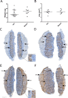
Fig 3. Placenta weight and tissue area in heterozygous and homozygous ErbB2M802R mutants at E12.5.
Quantification of (A) placental weight (B) placental area. Data expressed as median with data points. Example images of (C,E) heterozygous and (D,F) homozygous mutant mouse placentas immunostained with (C,D) MCT4 and (E,F) IL-B4 and captured at x1 magnification. Arrows indicate the transitions at the junctional zone (JZ), labyrinth (Lab), and decidua (Dec). Scale bar = 500μm.
Fig 4
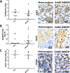
Fig 4. Placenta phenotype in heterozygous and homozygous ErbB2M802R mutants at E12.5.
Quantification of (A) proliferative index (Ki67), (B) apoptotic index (active Caspase 3), (C) vascularisation index (CD34) in placentas from heterozygous (Het) and homozygous (Mutant) embryos. Scale bar 50 μm. A and B are expressed as ratio of stain +ve nuclei/total nuclei, E is expressed as the percentage of CD34+ tissue in the labyrinth zone. Data expressed as median with data points.

References

    1. Blencowe H, Cousens S, Jassir FB, Say L, Chou D, Mathers C, et al. National, regional, and worldwide estimates of stillbirth rates in 2015, with trends from 2000: a systematic analysis. The Lancet Global health. 2016;4(2):e98–e108. 10.1016/S2214-109X(15)00275-2 . - DOI - PubMed
    1. Flenady V, Koopmans L, Middleton P, Froen JF, Smith GC, Gibbons K, et al. Major risk factors for stillbirth in high-income countries: a systematic review and meta-analysis. Lancet. 2011;377(9774):1331–40. Epub 2011/04/19. 10.1016/S0140-6736(10)62233-7 . - DOI - PubMed
    1. Lawn JE, Blencowe H, Waiswa P, Amouzou A, Mathers C, Hogan D, et al. Stillbirths: rates, risk factors, and acceleration towards 2030. Lancet. 2016;387(10018):587–603. 10.1016/S0140-6736(15)00837-5 . - DOI - PubMed
    1. Gardosi J, Kady SM, McGeown P, Francis A, Tonks A. Classification of stillbirth by relevant condition at death (ReCoDe): population based cohort study. Brit Med J. 2005;331(7525):1113–7. 10.1136/bmj.38629.587639.7C ISI:000233417300014. - DOI - PMC - PubMed
    1. Varli IH, Petersson K, Bottinga R, Bremme K, Hofsjo A, Holm M, et al. The Stockholm classification of stillbirth. Acta obstetricia et gynecologica Scandinavica. 2008;87(11):1202–12. 10.1080/00016340802460271 WOS:000261017000017. - DOI - PubMed

Publication types

MeSH terms

Supplementary concepts