. 2020 Jun 16;118(12):3041-3050.
doi: 10.1016/j.bpj.2020.05.014. Epub 2020 May 20.

A Lamin-Associated Chromatin Model for Chromosome Organization

Affiliations

A Lamin-Associated Chromatin Model for Chromosome Organization

Ajoy Maji et al. Biophys J. .

Abstract

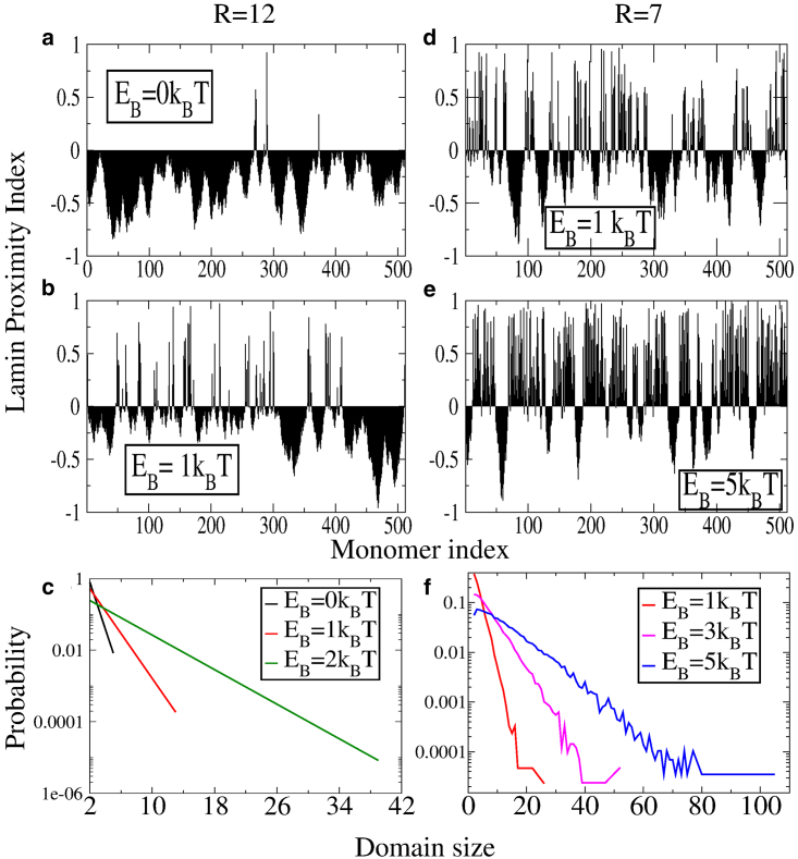
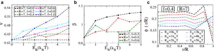
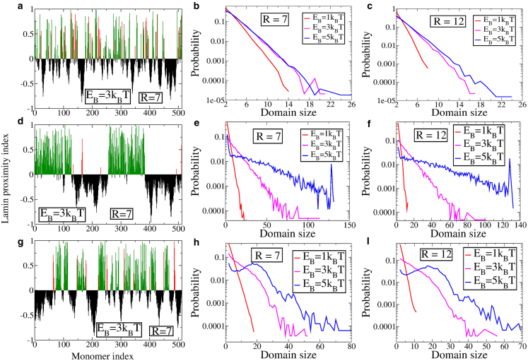
We propose a simple model for chromatin organization based on the interaction of the chromatin fibers with lamin proteins along the nuclear membrane. Lamin proteins are known to be a major factor that influences chromatin organization and hence gene expression in the cells. We provide a quantitative understanding of lamin-associated chromatin organization in a crowded macromolecular environment by systematically varying the heteropolymer segment distribution and the strength of the lamin-chromatin attractive interaction. Our minimal polymer model reproduces the formation of lamin-associated-domains and provides an in silico tool for quantifying domain length distributions for different distributions of heteropolymer segments. We show that a Gaussian distribution of heteropolymer segments, coupled with strong lamin-chromatin interactions, can qualitatively reproduce observed length distributions of lamin-associated-domains. Further, lamin-mediated interaction can enhance the formation of chromosome territories as well as the organization of chromatin into tightly packed heterochromatin and the loosely packed gene-rich euchromatin regions.


Figures

Figure 1
Figure 1
A schematic representation of a bead-spring polymer model with type-A and type-B beads confined within a sphere (see text). To see this figure in color, go online.
Figure 2
Figure 2
(a) Mean-square-distance R2(s) as a function of the separation s for different binding energies and different volume fractions. (b) The contact probability (pc(s)) as a function of the separation s for different binding energies and different volume fractions. (c) The volume fraction within spherical shells as a function of the normalized radial distance is shown. (d) The (normalized) number of monomers within a spherical shell as a function of the normalized radial distance is shown. To see this figure in color, go online.
Figure 3
Figure 3
Lamin proximity indices for R = 12 and binding energies (a) EB = 0 kBT and (b) EB = 1 kBT. Shown are the proximity indices for R = 7 and binding energies (d) EB = 1 kBT and (e) EB = 5 kBT. (c) and (f) Shown are domain size distributions for different binding energies for R = 12 and R = 7, respectively. To see this figure in color, go online.
Figure 4
Figure 4
(a)The mean-square displacement exponent ν as a function of EB for a different fraction of binding monomers f. (b) Shown is the contact probability exponent β as a function of EB for different f. (c) Plots of the radial volume fraction are shown. All results are shown for the random heteropolymer model in a spherical cavity of R = 7. To see this figure in color, go online.
Figure 5
Figure 5
LPI for EB = 3 kBT for a sphere with R = 7 for the three heteropolymer models: (a) random heteropolymer, (d) uniform block copolymer, and (g) Gaussian block copolymer. The red lines indicate type-B monomers that are in proximity to the NL, whereas green lines indicate bond-forming type-A monomers. The corresponding distributions of domain sizes is shown for R = 7 in (b, e, and h) and for R = 12 in (c, f, and i). To see this figure in color, go online.
Figure 6
Figure 6
(a) Contact maps and (c) sample configuration for a system of four polymers, none of which interact with lamin proteins. (b) Shown are contact maps and (d) sample configuration for a system of four polymers of which two interact with lamin proteins, whereas the other two do not. To see this figure in color, go online.

References

    1. Fraser P., Bickmore W. Nuclear organization of the genome and the potential for gene regulation. Nature. 2007;447:413–417. - PubMed
    1. Misteli T. Beyond the sequence: cellular organization of genome function. Cell. 2007;128:787–800. - PubMed
    1. Lanctôt C., Cheutin T., Cremer T. Dynamic genome architecture in the nuclear space: regulation of gene expression in three dimensions. Nat. Rev. Genet. 2007;8:104–115. - PubMed
    1. Horowitz-Scherer R.A., Woodcock C.L. Organization of interphase chromatin. Chromosoma. 2006;115:1–14. - PubMed
    1. Bystricky K., Heun P., Gasser S.M. Long-range compaction and flexibility of interphase chromatin in budding yeast analyzed by high-resolution imaging techniques. Proc. Natl. Acad. Sci. USA. 2004;101:16495–16500. - PMC - PubMed

Publication types

LinkOut - more resources