. 2020 Jun 1;18(6):291.
doi: 10.3390/md18060291.

Absorption and Tissue Distribution of Siphonaxanthin from Green Algae

Affiliations

Absorption and Tissue Distribution of Siphonaxanthin from Green Algae

Zhuosi Li et al. Mar Drugs. .

Abstract

Siphonaxanthin has been known to possess inhibitory effects against obesity, inflammation, and angiogenesis. However, little information on its in vivo bioavailability and biotransformation is available. To assess the bioavailability and metabolism of siphonaxanthin, its absorption and accumulation were evaluated using intestinal Caco-2 cells and Institute of Cancer Research (ICR) mice. Siphonaxanthin was absorbed and exhibited non-uniform accumulation and distribution patterns in tissues of ICR mice. Notably, in addition to siphonaxanthin, three main compounds were detected following dietary administration of siphonaxanthin. Because the compounds showed changes on mass spectra compared with that of siphonaxanthin, they were presumed to be metabolites of siphonaxanthin in ICR mice. Siphonaxanthin mainly accumulated in stomach and small intestine, while putative metabolites of siphonaxanthin mainly accumulated in liver and adipose tissues. Furthermore, siphonaxanthin and its putative metabolites selectively accumulated in white adipose tissue (WAT), especially mesenteric WAT. These results provide useful evidence regarding the in vivo bioactivity of siphonaxanthin. In particular, the results regarding the specific accumulation of siphonaxanthin and its metabolites in WAT have important implications for understanding their anti-obesity effects and regulatory roles in lipid metabolism.

Keywords: dehydro-metabolite; metabolic pathway in vivo; siphonaxanthin; white adipose tissue.


Conflict of interest statement

The authors declare no conflicts of interest.

Figures

Figure 1
Figure 1
Chemical structure of siphonaxanthin (3,19,3’-Trihydroxy-7,8-dihydro-β,ε-caroten-8-one).
Figure 2
Figure 2
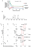
Uptake and metabolite analysis of siphonaxanthin in Caco-2 cells (A) Changes in siphonaxanthin concentration in Caco-2 cells and medium. Values are means ± SEM, n = 3. Data were analyzed by 1-factor ANOVA with repeated measures. “Time effect p < 0.05” indicates that there are differences in siphonaxanthin concentration variables at each time point within every group (cells, medium and cell+medium groups). Mean values without the same letter label indicate that they are significantly different within cells (red), medium (blue), or medium and cells (green), respectively, p < 0.05. (B) Representative HPLC chromatograms of the extract from Caco-2 cells after treatment with siphonaxanthin-containing micelles for 24 h. HPLC analysis was performed as described in the experimental methods. Peaks: 2, 3, and x, unknown metabolites; 4, siphonaxanthin; 4′, cis isomer of siphonaxanthin. Retention time: peak 2 at 25 min, peak 3 at 28 min, peak 4 at 30 min and peak x at 43–48 min. In the Caco-2 cells, we did not detect the peak corresponding to the peak 1 in the Figure 4 (mice data). Thus, in order to compare the corresponding peak at the same retention time compared with the Figure 4, here, the label “1” were not used. The detection wavelength was 450 nm. UV-vis spectra (C) and APCI-MS spectra (D) of peaks 2, 3, 4, and 4’. LC-MS with APCI analysis was performed as described in the experimental methods. HPLC, High-performance liquid chromatography; APCI, Atmospheric pressure chemical ionization; LC, Liquid chromatography; MS, Mass spectrometry.
Figure 3
Figure 3
Body weight (A), food intake (B), and tissue weight (C) of ICR mice. ICR mice were fed a control or siphonaxanthin supplementation diet for 16 days. Body weight and food intake were measured daily. After the 16-day feeding period, the mice were killed and their weight was measured. Values are means ± SEM, n = 4. The difference between the control and siphonaxanthin groups was analyzed using the Student’s t-test. * Different from the control group, p < 0.05. BAT, brown adipose tissue; WAT, white adipose tissue; SPX, siphonaxanthin.
Figure 4
Figure 4
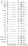
Representative HPLC chromatograms of the extracts from each tissue of mice fed a diet without (A) or with (B) siphonaxanthin for 16 days. HPLC analysis was performed as described in the experimental methods. The detection wavelength was 450 nm. Peaks with the same number in different chromatograms show similar UV-vis and MS spectra, as shown in Figure 5A,B. 1, 2, 3, and 5, unknown metabolites; 4, siphonaxanthin; Retention time: peak 1 at 22 min, peak 2 at 25 min, peak 3 at 28 min, peak 4 at 30 min and peak 5 at 43 min.
Figure 5
Figure 5
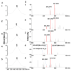
UV-vis spectra (A) and APCI-MS spectra (B) of peaks 1, 2, 3, 4, and 5 are shown in Figure 4. LC-MS (APCI) analysis was performed as described in the experimental methods.

References

    1. Foote C.S., Chang Y.C., Denny R.W. Chemistry of singlet oxygen. XI. Cis-trans isomerization of carotenoids by singlet oxygen and a probable quenching mechanism. J. Am. Chem. Soc. 1970;92:5218–5219. doi: 10.1021/ja00720a037. - DOI - PubMed
    1. Martin H.D., Ruck C., Schmidt M., Sell S., Beutner S., Mayer B., Walsh R. Chemistry of carotenoid oxidation and free radical reactions. Pure Appl. Chem. 1999;71:2253–2262. doi: 10.1351/pac199971122253. - DOI
    1. Fiedor J., Burda K. Potential role of carotenoids as antioxidants in human health and disease. Nutrients. 2014;6:466–488. doi: 10.3390/nu6020466. - DOI - PMC - PubMed
    1. Riccioni G. Marine carotenoids and oxidative stress. Mar. Drugs. 2012;10:116–118. doi: 10.3390/md10010116. - DOI - PMC - PubMed
    1. Gammone M.A., Riccioni G., D’Orazio N. Marine carotenoids against oxidative stress: Effects on human health. Mar. Drugs. 2015;13:6226–6246. doi: 10.3390/md13106226. - DOI - PMC - PubMed