. 2020 Jun 1;20(11):3124.
doi: 10.3390/s20113124.

Aptamer-Conjugated Polydiacetylene Colorimetric Paper Chip for the Detection of Bacillus thuringiensis Spores

Affiliations

Aptamer-Conjugated Polydiacetylene Colorimetric Paper Chip for the Detection of Bacillus thuringiensis Spores

Chaoge Zhou et al. Sensors (Basel). .

Abstract

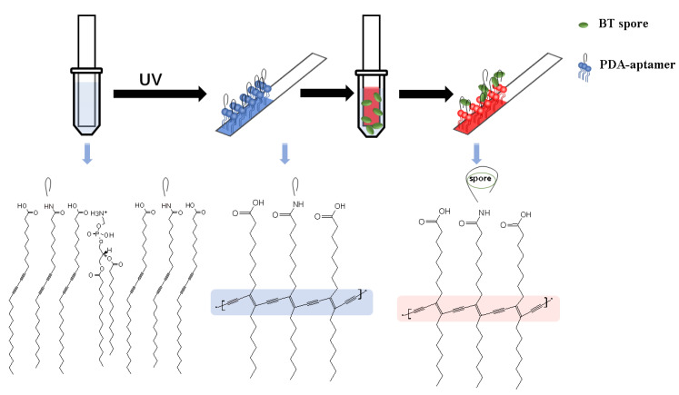
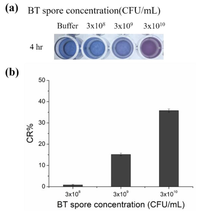
A colorimetric polydiacetylene (PDA) paper strip sensor that can specifically recognize Bacillus thuringiensis (BT) HD-73 spores is described in this work. The target-specific aptamer was combined with PDA, and the aptamer-conjugated PDA vesicles were then coated on polyvinylidene fluoride (PVDF) paper strips by a simple solvent evaporation method. The PDA-aptamer paper strips can be used to detect the target without any pre-treatment. Using the paper strip, the presence of BT spores is directly observable by the naked eye based on the unique blue-to-red color transition of the PDA. Quantitative studies using the paper strip were also carried out by analyzing the color transitions of the PDA. The specificity of this PDA sensor was verified with a high concentration of Escherichia coli, and no discernable change was observed. The observable color change in the paper strip occurs in less than 1 h, and the limit of detection is 3 × 10⁷ CFU/mL, much below the level harmful to humans. The PDA-based paper sensor, developed in this work, does not require a separate power or detection device, making the sensor strip highly transportable and suitable for spore analysis anytime and anywhere. Moreover, this paper sensor platform is easily fabricated, can be adapted to other targets, is highly portable, and is highly specific for the detection of BT spores.

Keywords: Bacillus thuringiensis spores; aptamer; chromatic sensor; paper chip; polydiacetylene (PDA).


Conflict of interest statement

The authors declare no conflict of interest.

Figures

Figure 1
Figure 1
Schematic of the PDA-aptamer paper strip coating, the entire detection process and the key chemical structures. The immobilized PDA-aptamer was polymerized by UV exposure, and it turned blue. After a short incubation period, the BT spores were recognized by the PDA-aptamer, and the reaction between the aptamer and spore induced a color change in the PDA from blue to red.
Figure 2
Figure 2
PDA-aptamer liposomes incubated with different concentrations of spore solutions. (a) Image of PDA-aptamer liposomes after 4 h of incubation in PBS (pH = 7.5, 5 mM) and spore solutions at different concentrations. (b) Color response percentage (CR%) of the PDA-aptamer liposomes after 4 h of incubation in spore solutions of different concentrations. Color response data are presented as the mean ± SD (n = 3).
Figure 3
Figure 3
PDA strip detection of BT spores. (a) Images of the paper strips after incubation in spore solutions of different concentrations for 4 h. The negative and positive control images are from strips dipped in PBS (5 mM, pH = 7.5), and 1 M NaOH, respectively. (b) Red chromatic shift of the strips after 1 h, 2 h, 3 h, and 4 h of incubation in the spore solutions. Red chromatic shift data are presented as the mean ± SD (n = 3).
Figure 4
Figure 4
Testing the specificity of the PDA strips. (a) Images of the paper strips (with aptamer) after incubation in 3 × 10¹¹ CFU/mL BT spore solutions, paper strip (without aptamer) in 3 × 10¹¹ CFU/mL BT spore solution, and paper strip (with aptamer) in 3 × 10¹¹ CFU/mL E. coli for 4 h. The negative and positive control images are strips dipped in PBS (5 mM, pH = 7.5) and 1 M NaOH. (b) Red chromatic shift of the strips after 4 h of incubation. Red chromatic shift data are presented as the mean ± SD (n = 3).

References

    1. Kim W., Kim D., Back S., Lee Y.-S., Abari A.H., Kim J. Removal of Ni2+ and Cd2+ by surface display of polyhistidine on bacillus subtilis spore using CotE anchor protein. Biotechnol. Bioprocess Eng. 2019;24:375–381. doi: 10.1007/s12257-018-0467-2. - DOI
    1. Makino S.-I., Cheun H.I., Watarai M., Uchida I., Takeshi K. Detection of anthrax spores from the air by real-time PCR. Lett. Appl. Microbiol. 2001;33:237–240. doi: 10.1046/j.1472-765x.2001.00989.x. - DOI - PubMed
    1. Kuske C.R., Banton K.L., Adorada D.L., Stark P.C., Hill K.K., Jackson P.J. Small-scale DNA sample preparation method for field PCR detection of microbial cells and spores in soil. Appl. Environ. Microbiol. 1998;64:2463–2472. doi: 10.1128/AEM.64.7.2463-2472.1998. - DOI - PMC - PubMed
    1. Wang D.B., Tian B., Zhang Z.P., Wang X.Y., Fleming J., Bi L.J., Yang R.F., Zhang X.E. Detection of Bacillus anthracis spores by super-paramagnetic lateral-flow immunoassays based on “Road Closure”. Biosens. Bioelectron. 2015;67:608–614. doi: 10.1016/j.bios.2014.09.067. - DOI - PubMed
    1. Ikanovic M., Rudzinski W.E., Bruno J.G., Allman A., Carrillo M.P., Dwarakanath S., Bhahdigadi S., Rao P., Kiel J.L., Andrews C.J. Fluorescence assay based on aptamer-quantum dot binding to Bacillus thuringiensis Spores. J. Fluoresc. 2007;17:193–199. doi: 10.1007/s10895-007-0158-4. - DOI - PubMed

MeSH terms

LinkOut - more resources