. 2020 Jun 1;21(11):3982.
doi: 10.3390/ijms21113982.

AMPK Profiling in Rodent and Human Pancreatic Beta-Cells under Nutrient-Rich Metabolic Stress

Affiliations

AMPK Profiling in Rodent and Human Pancreatic Beta-Cells under Nutrient-Rich Metabolic Stress

Thierry Brun et al. Int J Mol Sci. .

Abstract

Chronic exposure of pancreatic β-cells to elevated nutrient levels impairs their function and potentially induces apoptosis. Like in other cell types, AMPK is activated in β-cells under conditions of nutrient deprivation, while little is known on AMPK responses to metabolic stresses. Here, we first reviewed recent studies on the role of AMPK activation in β-cells. Then, we investigated the expression profile of AMPK pathways in β-cells following metabolic stresses. INS-1E β-cells and human islets were exposed for 3 days to glucose (5.5-25 mM), palmitate or oleate (0.4 mM), and fructose (5.5 mM). Following these treatments, we analyzed transcript levels of INS-1E β-cells by qRT-PCR and of human islets by RNA-Seq; with a special focus on AMPK-associated genes, such as the AMPK catalytic subunits α1 (Prkaa1) and α2 (Prkaa2). AMPKα and pAMPKα were also evaluated at the protein level by immunoblotting. Chronic exposure to the different metabolic stresses, known to alter glucose-stimulated insulin secretion, did not change AMPK expression, either in insulinoma cells or in human islets. Expression profile of the six AMPK subunits was marginally modified by the different diabetogenic conditions. However, the expression of some upstream kinases and downstream AMPK targets, including K-ATP channel subunits, exhibited stress-specific signatures. Interestingly, at the protein level, chronic fructose treatment favored fasting-like phenotype in human islets, as witnessed by AMPK activation. Collectively, previously published and present data indicate that, in the β-cell, AMPK activation might be implicated in the pre-diabetic state, potentially as a protective mechanism.

Keywords: AMPK; ATP; beta-cell; fructose; glucotoxicity; insulin; pancreatic islets.


Conflict of interest statement

The authors declare no conflict of interest.

Figures

Figure 1
Figure 1
Regulation of insulin secretion by the pancreatic β-cell and intracellular/extracellular ATP signaling pathways. Glucose metabolism leads to the production of ATP in mitochondria. The ensuing elevation of the ATP/ADP ratio induces the closure of the K-ATP channels, which promotes the depolarization of the plasma membrane that opens the voltage-sensitive calcium channels. The resulting elevation of cytosolic calcium concentration triggers insulin release (blue arrows). Prolonged treatment with fructose induces a fasting-like phenotype in β-cells, resulting in AMPK activation even in the presence of glucose (black arrows). Fructose exposure activates Panx1 channels, promoting the release of cellular ATP and activation of purinergic P2Y1 receptors on the plasma membrane (red arrows). Chronic fructose exposure potentiates the stimulation of insulin secretion at intermediate glucose concentrations. Therefore, intracellular and extracellular ATP signaling pathways could play a concerted role in the β-cell as potentiators of glucose-induced insulin secretion.
Figure 2
Figure 2
AMPK mRNA levels in rat islets and in INS-1E β-cells under metabolic stress conditions. (AB) Relative expression of the two components of the AMPK catalytic subunits α1 and α2, encoded by the Prkaa1 and Prkaa2 genes, respectively; measured by qRT-PCR and normalized to cyclophilin (n = 6); ** p< 0.01, *** p< 0.005, **** p< 0.001. (CD) Time course of the transcript levels in INS-1E β-cells after low (G5.5), control (G11) and high (G25) glucose exposure (n = 3). (EF) Cells were treated for 3 days with 0.4 mM palmitate (Palm) or oleate (Olea) in the presence of 0.5% BSA (n = 5).
Figure 3
Figure 3
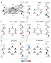
AMPK transcript levels in human islets under metabolic stress conditions. (A) Functional interaction network of human AMPK-associated genes, i.e., AMPK subunits (AMPK box), upstream kinases, and downstream targets. (BF) Effects of culture conditions compared to standard G5.5 medium on transcript levels shown as up-regulated (red), down-regulated (blue), or unchanged (white). Each disk is split into individual changes for the different donors. (B) Genes regulated upon high-glucose conditions (G25). (C–D) Genes regulated upon (C) oleate or (D) palmitate exposure (0.4 mM) in control glucose condition (G5.5). (E–F) Genes regulated upon (E) oleate or (F) palmitate exposure (0.4 mM) in high-glucose conditions (G25). (A) Node connections were established according to the STRING interaction knowledgebase with a confidence score >0.4. Color code reflects the changes in expression in log2 fold changes (log2 FC; quantitative data in Supplementary Table S1) of that particular gene for each individual human donor (described in Table S2). Dashed boxes show, from left to right, the upstream kinases comprising CAMKK (calcium/calmodulin-dependent protein kinase kinase) and STK11 (serine/threonine kinase 11/LKB1); the six subunits of AMPK; the downstream targets RPTOR (regulatory-associated protein of MTOR complex 1), MTOR (mechanistic target of rapamycin kinase), PKM (pyruvate kinase M1/2 isozyme), ACACA (acetyl-CoA carboxylase), ABCC8 (SUR1 subunit), KCNJ11 (KIR6.2 subunit). *adjusted p < 0.05, **adjusted p < 0.01, ***adjusted p < 0.001 between control 5.5 mM glucose and the specific culture condition. Clinical data of donors #1 to #5 are shown in Supplementary Table S2.
Figure 4
Figure 4
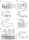
AMPK activation in INS-1E β-cells and in human islets exposed to high glucose and fructose. (A–D) INS-1E β-cells were cultured for (A) 3 days or for (B) 4 days in low (G5.5), standard (G11) and high (G25) glucose supplemented with 5.5 mM fructose (red line). Representative immunoblotting showing levels of pAMPKα, AMPKα (AMPK) and ACTIN in cells right after the culture period. Quantitative analysis of (C) AMPK and (D) pAMPK/AMPK band densities normalized to ACTIN are shown for 3-day-treated cells (n = 5). Results are expressed as protein levels normalized to G11 values. (E-H) Freshly isolated human islets from six donors were treated for 4 days with 5.5 mM fructose (F) in complete CRML-1066 medium. Culture at 5.5 mM glucose (G5.5) served as a control culture condition. (E) Transcript levels of AMPK catalytic subunits α1 (PRKAA1) and α2 (PRKAA2) were quantified in a batch of islets from one representative human islet preparation exposed to different glucose concentrations, normalized to cyclophilin. (F-G) Representative immunoblotting showing levels of pAMPK, AMPK, LKB1 and ACTIN from treated islets isolated from four donors right after culture and after 1 h post-culture of glucose starving at 2.8 mM (G2.8). (H) Quantitative analysis of pAMPK/AMPK and LKB1 band densities normalized to ACTIN is shown (n = 6). Results are expressed as protein levels normalized to G5.5 control values. *p < 0.05 G5.5+F islets versus G5.5 islets.

References

    1. Prentki M., Peyot M.L., Masiello P., Madiraju S.R.M. Nutrient-Induced Metabolic Stress, Adaptation, Detoxification, and Toxicity in the Pancreatic β-Cell. Diabetes. 2020;69:279–290. doi: 10.2337/dbi19-0014. - DOI - PubMed
    1. Kasuga M. Insulin resistance and pancreatic beta cell failure. J. Clin. Investig. 2006;116:1756–1760. doi: 10.1172/JCI29189. - DOI - PMC - PubMed
    1. Rourke J.L., Hu Q., Screaton R.A. AMPK and Friends: Central Regulators of β Cell Biology. Trends Endocrinol. Metab. 2018;29:111–122. doi: 10.1016/j.tem.2017.11.007. - DOI - PubMed
    1. Fu A., Eberhard C.E., Screaton R.A. Role of AMPK in pancreatic β cell function. Mol. Cell. Endocrinol. 2013;366:127–134. doi: 10.1016/j.mce.2012.06.020. - DOI - PubMed
    1. Szkudelski T., Szkudelska K. The relevance of AMP-activated protein kinase in insulin-secreting β cells: A potential target for improving β cell function? J. Physiol. Biochem. 2019;75:423–432. doi: 10.1007/s13105-019-00706-3. - DOI - PMC - PubMed

MeSH terms

LinkOut - more resources