. 2020 Aug 14;127(5):677-692.
doi: 10.1161/CIRCRESAHA.119.316398. Epub 2020 Jun 4.

Long Range Endocrine Delivery of Circulating miR-210 to Endothelium Promotes Pulmonary Hypertension

Affiliations

Long Range Endocrine Delivery of Circulating miR-210 to Endothelium Promotes Pulmonary Hypertension

Jingsi Zhao et al. Circ Res. .

Abstract

Rationale: Unproven theories abound regarding the long-range uptake and endocrine activity of extracellular blood-borne microRNAs into tissue. In pulmonary hypertension (PH), microRNA-210 (miR-210) in pulmonary endothelial cells promotes disease, but its activity as an extracellular molecule is incompletely defined.

Objective: We investigated whether chronic and endogenous endocrine delivery of extracellular miR-210 to pulmonary vascular endothelial cells promotes PH.

Methods and results: Using miR-210 replete (wild-type [WT]) and knockout mice, we tracked blood-borne miR-210 using bone marrow transplantation and parabiosis (conjoining of circulatory systems). With bone marrow transplantation, circulating miR-210 was derived predominantly from bone marrow. Via parabiosis during chronic hypoxia to induce miR-210 production and PH, miR-210 was undetectable in knockout-knockout mice pairs. However, in plasma and lung endothelium, but not smooth muscle or adventitia, miR-210 was observed in knockout mice of WT-knockout pairs. This was accompanied by downregulation of miR-210 targets ISCU (iron-sulfur assembly proteins)1/2 and COX10 (cytochrome c oxidase assembly protein-10), indicating endothelial import of functional miR-210. Via hemodynamic and histological indices, knockout-knockout pairs were protected from PH, whereas knockout mice in WT-knockout pairs developed PH. In particular, pulmonary vascular engraftment of miR-210-positive interstitial lung macrophages was observed in knockout mice of WT-knockout pairs. To address whether engrafted miR-210-positive myeloid or lymphoid cells contribute to paracrine miR-210 delivery, we studied miR-210 knockout mice parabiosed with miR-210 WT; Cx3cr1 knockout mice (deficient in myeloid recruitment) or miR-210 WT; Rag1 knockout mice (deficient in lymphocytes). In both pairs, miR-210 knockout mice still displayed miR-210 delivery and PH, thus demonstrating a pathogenic endocrine delivery of extracellular miR-210.

Conclusions: Endogenous blood-borne transport of miR-210 into pulmonary vascular endothelial cells promotes PH, offering fundamental insight into the systemic physiology of microRNA activity. These results also describe a platform for RNA-mediated crosstalk in PH, providing an impetus for developing blood-based miR-210 technologies for diagnosis and therapy in this disease.

Keywords: endothelium; lymphocytes; macrophage; microRNAs; parabiosis.


Figures

Figure 1.
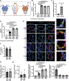
Figure 1.. A majority of circulating, steady-state hypoxia-induced miR-210 is derived from bone marrow cells.
(A) At 12 weeks post-bone marrow transplantation of miR-210 (WT) and miR-210 (KO) mice with WT or KO bone marrow, mice were exposed to chronic hypoxia (10% O2) for 3 weeks to induce miR-210 up-regulation. (B-C) By RT-qPCR, circulating extracellular miR-210 (B, N=5,6,6,6 mice) and intracellular miR-210 (C, N=4,4,5,4 mice) were detected and quantified in plasma (B) and CD31+/CD45− lung endothelial cells (C) of WT mice and KO mice transplanted with WT or KO bone marrow. (One-way ANOVA with post-hoc Bonferroni testing was performed.)
Figure 2.
Figure 2.. Chronic conjoining of the circulatory systems in mice via parabiosis leads to delivery of blood-borne miR-210 to pulmonary arterial endothelial cells in vivo.
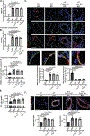
(A) An in vivo parabiosis platform allowed for the conjoining of the circulatory systems of two mice in the context of miR-210 +/+ (WT) and miR-210 −/− (KO) partners. One-week post-surgery, mouse pairs were exposed to chronic hypoxia (10% O2) for 3 weeks to induce endogenous miR-210 up-regulation. (Red arrows denote comparison groups.) (B-D) As assessed by RT-qPCR in plasma (B, N=8,7,9,8 mice) and in CD31+/CD45− lung endothelial cells (D, N=12,11,10,12 mice), while miR-210 expression was negligible in KO-KO pairs, extracellular miR-210 level in KO partners of KO-WT pairs was increased to comparable level as WT-WT pairs whereas intracellular miR-210 in those mice was readily detected but still less than that of WT partners and WT-WT pairs. Extracellular miR-210 in plasma was found to be co-immunoprecipitated specifically in the presence of α-Argonaute 2 (AGO2) as compared with control IgG (C, N=4 mice/group). (E) By in situ staining of miR-210 (red), its target proteins ISCU1/2 (grey, pointed out by red arrows), and the endothelial marker CD31 (green), miR-210 expression in the KO partners of KO-WT pairs was detected specifically in pulmonary arteriolar endothelium (yellow, micrograph inset), accompanied by a reciprocal decrease in ISCU1/2 expression in these miR-210-positive cells, as compared with KO-KO pairs (N=6 mice/group, average of N=10–12 vessels/mice. Scale bar denotes 50 μm.). (F-H) In parallel, there was no significant difference of endothelin-1 content in plasma (F, N=4 mice/group), nitrate and nitrite content in lungs (G, N=5 mice/group) or inflammatory cytokine IL-6 content in plasma (H, N=4 mice/group) between KO partners of KO-WT pairs with KO-KO pair. (Data are presented as mean ± SEM. In x-axes of (B,D,E,F,G,H) bold font denotes parabionts being studied, whereas regular font denotes partner parabionts. Mann Whitney U test for C,F,G,H and one-way ANOVA with post-hoc Bonferroni testing were performed for other panels.)
Figure 3.
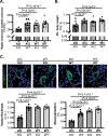
Figure 3.. Delivery of blood-borne miR-210 promotes PH.
(A) By invasive right heart catheterization and measurement of right ventricular systolic pressure (RVSP), while hypoxic KO-KO pairs were protected from PH as compared with WT-WT pairs, the KO partners in KO-WT pairs developed significantly higher RVSP versus KO-KO pairs (N=7,9,6,8 mice). (B) Right ventricular mass/body weight ratio was increased in the KO partners of KO-WT pairs as compared with KO-KO pairs (N=12,8,7,9 mice). (C) In situ staining of α-SMA (green) demonstrated increased pulmonary arteriolar remodeling and muscularization in the KO partners of KO-WT pairs versus KO-KO pairs and comparable to that seen in WT partners and WT-WT pairs (N=6 mice/group, average of 10–12 vessels/mice. Scale bar denotes 50 μm.). (Data are presented as mean ± SEM. In x-axes of (A,B,C), bold font denotes parabionts being studied, whereas regular font denotes partner parabionts. One-way ANOVA with post-hoc Bonferroni testing was performed.)
Figure 4.
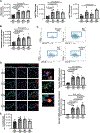
Figure 4.. Hypoxic expansion of miR-210-positive blood monocytes and interstitial lung macrophages.
(A) Employing the same parabiosis system as in Figure 2 and via fluorescence activated cell sorting (FACS) of blood, total blood monocytes (N=6,6,5,6 mice), and subpopulations including Ly6chigh and Ly6clow monocytes (N=5,4,4,4 mice) were increased in hypoxic WT-WT pairs as compared with hypoxic KO-KO pairs. Importantly, KO mice of KO-WT pairs displayed an equivalent level of total blood monocytes and Ly6chigh monocytes, which was significantly higher than KO-KO pairs. (B-C) In parallel, by FACS, expansion of CD64/CD11b-positive interstitial lung macrophages was observed in isolated cells from whole lung of KO mice of KO-WT pairs comparable to that seen in the WT partner and WT-WT pairs but substantially increased compared with KO-KO pairs (quantitation in B (N=6,6,5,6 mice, and representative flow cytometric plots in C. Percentages reflect the frequencies of interstitial macrophages among CD45.2+ Siglec F- Ly-6G- CD11b+ MHC class II- leukocytes in the lung.). (D) Increased presence of miR-210-positive interstitial lung macrophages (yellow, micrograph inset) was also demonstrated by in situ staining of CD68 (red) and miR-210 (green) in the small pulmonary arterioles of KO mice of KO-WT pairs, comparable to WT partners and WT-WT pairs but greater in number than those seen in KO-KO pairs (N=6 mice/group, average of 10–12 vessels/mice. Scale bar denotes 50 μm.). (E) By FACS, no significant difference of lymphocyte presence was observed in lung tissue of KO-KO pairs, KO-WT pairs and WT-WT pairs (N=6,6,4,6 mice). (Data are presented as mean ± SEM. In x-axes of (A,B,D,E), bold font denotes parabionts being studied, whereas regular font denotes partner parabionts. One-way ANOVA with post-hoc Bonferroni testing was performed.)
Figure 5.
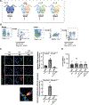
Figure 5.. Transfer of miR-210-positive myeloid cells and lymphocytes was abrogated by Cx3cr1 deficiency and Rag1 deficiency, respectively.
(A) A parabiosis platform similar to that in Figure 2 was used in the context of pairing miR-210 −/− (KO) mice with miR-210 −/− (KO), miR-210 +/+ (WT), miR-210 +/+; Cx3cr1 −/− (WT; Cx3cr1 KO) or miR-210 +/+; Rag1 −/− (WT; Rag1 KO) mice. One week after surgery, the mouse pairs were exposed to chronic hypoxia (10% O2) for three weeks. (Red arrows denote comparison groups) (B) As denoted by frequencies (%) of enriched lymphocytes within the total leukocyte population, lack of mature lymphocytes was confirmed by flow cytometry in blood and lung tissue of (WT; Rag1 KO mice). (C) Decreased presence of miR-210-positive interstitial lung macrophages (yellow, micrograph inset) was demonstrated by in situ staining of CD68 (red) and miR-210 (green) in the small pulmonary arterioles of KO partners of KO-(WT; Cx3cr1 KO) pairs, comparable to those seen in KO-KO pairs (N=6 mice/group, average of 10–12 vessels/mice. Scale bar denotes 50 μm.). (D) In parallel, circulating levels of inflammatory marker IL6 were not altered in plasma of KO partners of KO-WT pairs, KO-(WT; Cx3cr1 KO) pairs and KO-(WT; Rag1 KO) compared to KO-KO pairs, as quantified by ELISA (N=4 mice/group). (Data are presented as mean ± SEM. In x-axes of (C,D), bold font denotes parabionts being studied, whereas regular font denotes partner parabionts. Kruskal Wallis and post-hoc Dunn’s testing for D and one-way ANOVA with post-hoc Bonferroni testing for other panels were performed.)
Figure 6.
Figure 6.. PAEC delivery and pathogenic activity of blood-borne miR-210 depends upon extracellular endocrine, rather than myeloid-based or lymphoid-based paracrine, delivery.
(A-B) Mouse parabiosis pairs as in Figure 5 were exposed to chronic hypoxia (10% O2) for 3 weeks to induce endogenous miR-210 up-regulation. Levels of miR-210 in plasma (A, N=8,7,6,5 mice) and CD31+/CD45− lung endothelial cells (B, N=11,11,10,8 mice) were assessed by RT-qPCR. In KO partners of KO-(WT; Cx3cr1 KO) pairs and KO-(WT; Rag1 KO pairs), they displayed similar extracellular (A) and intracellular (B) miR-210 levels as in KO partners of KO-WT pairs, which was substantially higher than expression in KO-KO pairs. (C) By in situ staining of miR-210 (red), its target proteins ISCU1/2 (grey, pointed out by red arrows), and the endothelial marker CD31 (green), comparable level of miR-210 delivery to pulmonary arteriolar endothelium (yellow, micrograph inset) and corresponding decrease of ISCU1/2 in the KO partners of KO-(WT; Cx3cr1 KO) pairs and of KO-(WT; Rag1 KO) pairs were detected as compared with KO partners of KO-WT pairs (N=6,6,6,5 mice, average of 10–12 vessels/mice. Scale bar denotes 50 μm.). (D-F) In the aspect of pathogenic phenotypes, right ventricular systolic pressure (RVSP) (D, N=7,9,10,6 mice), right ventricular remodeling (E, N=12,8,13,10 mice) and pulmonary arteriolar remodeling and muscularization (F, N=6,6,6,5 mice, average of 10–12 vessels/mice. Scale bar denotes 50 μm.) also showed comparable degree of elevation in KO partners of KO-(WT; Cx3cr1 KO) pairs and of KO-(WT; Rag1 KO) pairs as KO partners of KO-WT pairs, measured by right heart catheterization, RV mass to body weight ratio and in situ staining of α-SMA (white), respectively. (Data are presented as mean ± SEM. In all panels, bold font in x-axes denotes parabionts being studied, whereas regular font denotes partner parabionts. One-way ANOVA with post-hoc Bonferroni testing was performed.)
Figure 7.
Figure 7.. Model of the endocrine delivery and pathogenic actions of blood-borne miR-210 in PH.
Collectively, our findings demonstrate that long range, endocrine transport encompasses the predominant mechanism by which blood-borne miR-210 is delivered into pulmonary arterial endothelial cells with consequent control of PH in vivo.

Comment in

Similar articles

Cited by

References

    1. Negi V, Chan SY. Discerning functional hierarchies of micrornas in pulmonary hypertension. JCI Insight. 2017;2:e91327. - PMC - PubMed
    1. Aliotta JM, Pereira M, Wen S, Dooner MS, Del Tatto M, Papa E, Goldberg LR, Baird GL, Ventetuolo CE, Quesenberry PJ, Klinger JR. Exosomes induce and reverse monocrotaline-induced pulmonary hypertension in mice. Cardiovasc Res. 2016;110:319–330 - PMC - PubMed
    1. Creemers EE, Tijsen AJ, Pinto YM. Circulating micrornas: Novel biomarkers and extracellular communicators in cardiovascular disease? Circ Res. 2012;110:483–495 - PubMed
    1. He Y, Lin J, Kong D, Huang M, Xu C, Kim TK, Etheridge A, Luo Y, Ding Y, Wang K. Current state of circulating micrornas as cancer biomarkers. Clin Chem. 2015;61:1138–1155 - PubMed
    1. Min PK, Chan SY. The biology of circulating micrornas in cardiovascular disease. Eur J Clin Invest. 2015;45:860–874 - PMC - PubMed

Publication types

MeSH terms