

The findings of corneal specular microscopy in patients with type-2 diabetes mellitus
- PMID: 32493325
- PMCID: PMC7271396
- DOI: 10.1186/s12886-020-01488-9
The findings of corneal specular microscopy in patients with type-2 diabetes mellitus
Abstract
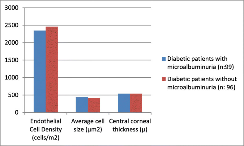
Background: We aimed to compare the morphological characteristics of corneal endothelial cells in type 2 diabetic patients and age-matched healthy subjects by specular microscopy. We also aimed to determine the association of corneal morphological features with the general characteristics and laboratory data of diabetic patients, including disease duration, haemoglobin A1c (HbA1c) levels and urine albumin creatinine ratio.
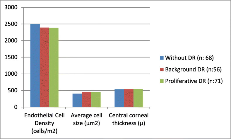
Methods: A total of 195 diabetic patients and 100 healthy controls were enrolled in the study. All participants underwent a complete ophthalmological examination. Corneal endothelial measurements were performed using a noncontact specular microscopy. Laboratory data including serum fasting glucose, haemoglobin A1c levels, creatinine levels, and the urinary albumin-to-creatinine ratio were recorded. Diabetic patients were further subdivided into 3 groups according to the presence and stage of diabetic retinopathy. Specular microscopy findings and central corneal thickness of all patients were compared.
Results: The ECD and hexagonal cell ratio were significantly lower, while the average cell size, CV%, and central corneal thickness were determined to be significantly higher in diabetic patients than in healthy controls (p = 0.001). With the presence and advancement of diabetic retinopathy, the ECD and hexagonal cell ratio decreased, while the average cell size, CV%, and central corneal thickness increased. When correlation analysis was performed between corneal morphological features and laboratory data of diabetic patients, ECD showed a significant negative correlation with diabetes duration (p = 0.028). HbA1c levels, urinary albumin-creatinine ratio (p = 0.041), average cell size and CV showed a positive correlation with these parameters.
Conclusion: In conclusion, keratopathy is an important complication of type 2 diabetes. With an increase in the stage of diabetic retinopathy, alterations in corneal findings also increased. In that respect, we can suggest that keratopathy should be evaluated more cautiously in diabetic patients.
Keywords: Corneal endothelial cells; Diabetes mellitus; Microalbuminuria; Specular microscopy.
Conflict of interest statement
Following our ethical obligations as researchers, all authors report that they have no financial or non-financial conflicts of interest in this research. (Authors: Adem Uğurlu MD, Erel Icel MD, Nurdan Gamze Tasli MD, Hayati Yılmaz MD, Turgay Ucak MD, Yucel Karakurt MD, Emin Murat Akbaş MD).
Figures
References
-
- Molitch ME, Rutledge B, Steffes M, Cleary P. Renal insufficiency in the absence of albuminuria among adults with Type 1 diabetes in the Diabetes Control and Complications Trial (DCCT)/Epidemiology of Diabetes Interventions and Complications (EDIC) Study; ADA Annual Meeting 2006 (Abstract 23-OR).
-
- Kaji Y, Usui T, Oshika T, Matsubara M, Yamashita H, Araie M, Murata T, Ishibashi T, Nagai R, Horiuchi S. Amano S advanced glycation end products in diabetic corneas. Invest Ophthalmol Vis Sci. 2000;41(2):362–368. - PubMed
Publication types
MeSH terms
Substances
LinkOut - more resources
Full Text Sources
Medical
