. 2020 Jun 3;19(1):101.
doi: 10.1186/s12943-020-01221-6.

Circular RNA circSATB2 promotes progression of non-small cell lung cancer cells

Affiliations

Circular RNA circSATB2 promotes progression of non-small cell lung cancer cells

Nan Zhang et al. Mol Cancer. .

Abstract

Background: Lung cancer has high morbidity and mortality worldwide with non-small cell lung cancer (NSCLC) accounting for 85% of the cases. Therapies for lung cancer have relatively poor outcomes and further improvements are required. Circular RNAs have been reported to participate in the occurrence and progression of cancer. Information on the functions and mechanism of circRNAs in lung cancer is limited and needs more exploration.

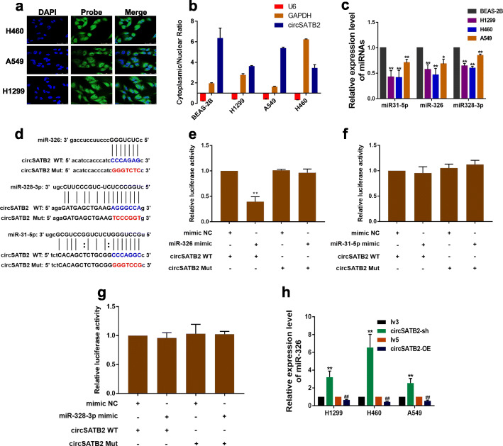
Methods: We detected expression of genes and proteins by qPCR and western blot. Function of circSATB2 was investigated using RNA interference and overexpression assays. Location of circSATB2 was assessed by fluorescence in situ hybridization (FISH). Interaction of circSATB2, miR-326 and FSCN1 was confirmed by dual-luciferase reporter assay.

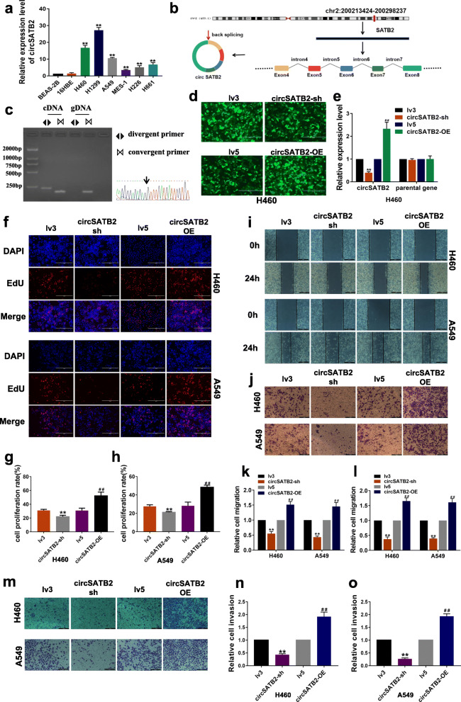
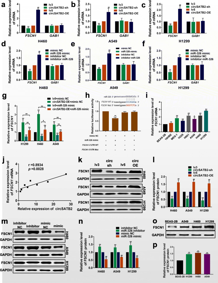
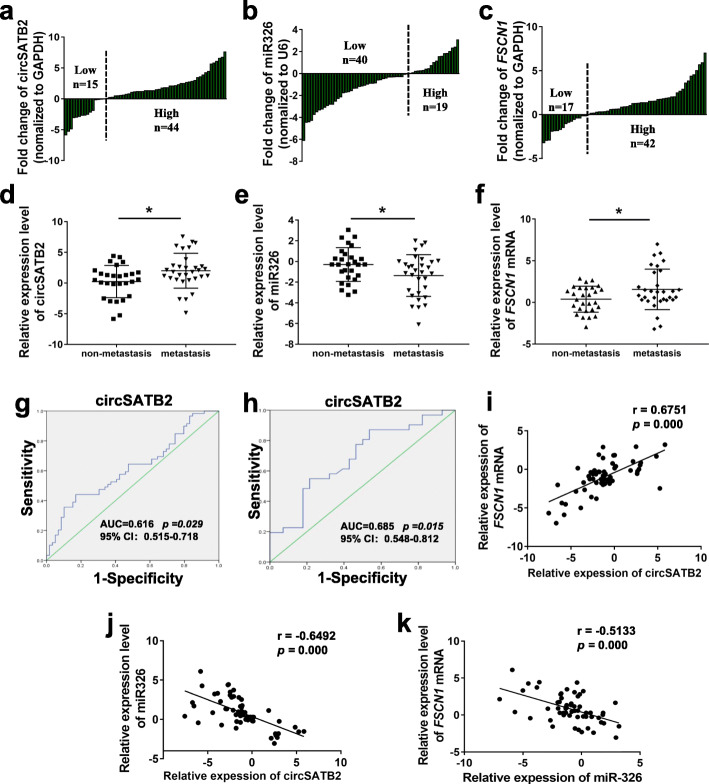
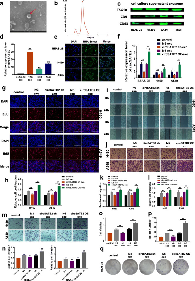
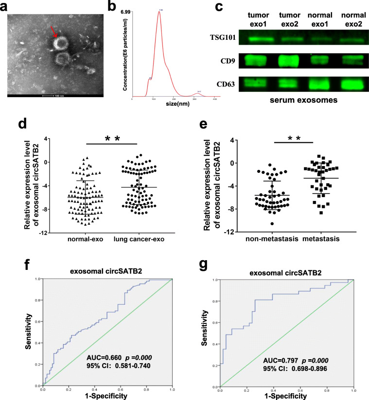
Results: Data from the investigation showed that circSATB2 was highly expressed in NSCLC cells and tissues. circSATB2 positively regulated fascin homolog 1, actin-bundling protein 1 (FSCN1) expression via miR-326 in lung cancer cells. Furthermore, circSATB2 can be transferred by exosomes and promote the proliferation, migration and invasion of NSCLC cells, as well as induce abnormal proliferation in normal human bronchial epithelial cells. Also, circSATB2 was highly expressed in serumal exosomes from lung cancer patients with high sensitivity and specificity for clinical detection and was related to lung cancer metastasis.

Conclusions: circSATB2 participated in the progression of NSCLC and was differentially expressed in lung cancer tissue and serumal exosomes. circSATB2 may be potential biomarker for the diagnosis of NSCLC.

Keywords: Exosome; Lung cancer; Progression; circRNA; miRNA.


Conflict of interest statement

The authors have no conflicts of interest to declare.

Figures

Fig. 1
Fig. 1
circSATB2 promotes proliferation, migration and invasion of NCSLC cells. a circSATB2 expression in normal and lung cancer cells. b Genomic location and splicing mode of circSATB2. c Identification of circSATB2 qPCR amplification products. DNA gel electrophoresis (left) and Sanger sequencing of qPCR product (right). d GFP-tagged lentivirus in cells observed by fluorescence microscope. e Efficiency of stably transfected circSATB2-knockdown and overexpressing H460 cells detected by qPCR. f EdU assay to detect proliferation of circSATB2-knockdown and overexpressing stably transfected H460 and A549 cells. g, h Quantitative analysis of EdU assay. i Wound healing assay to detect migration of circSATB-knockdown and overexpressing stably transfected H460 and A549 cells. j Transwell migration assay detect migration of circSATB-knockdown and overexpressing stably transfected H460 and A549 cells. k Quantitative analysis of wound healing assay. (l) Quantitative analysis of Transwell migration assay. m Transwell invasion assay to detect invasion of circSATB2 knockdown and overexpressing stably transfected H460 and A549 cells. n, o Quantitative analysis of Transwell invasion assay. All experiments were repeated independently three times. Data are presented as means±standard deviation. **P < 0.01 compared with BAES-2B cells or lv3 group; ##P < 0.01 compared with lv5 group
Fig. 2
Fig. 2
circSATB2 directly binds to miR-326 and regulates miR-326 expression in NSCLC cells. a FISH for subcellular localization of circSATB2. 6-FAM labeled the circSATB2 FISH probe, DAPI stained cell nuclei. b Cytoplasmic and nuclear circSATB2 expression detected by qPCR. U6 and GAPDH acted as nuclear and cytoplasmic reference genes, respectively. Relative expression measured by the 2-(cytoplasmic Ct value - nuclear Ct value) method. c miRNA expression in BEAS-2B and NSCLC cells detected by qPCR. d Sequence alignments between circSATB2 and seed sequences of miRNAs. WT, wild-type vector; Mut, mutant sequence vector of circSATB2. eg Dual luciferase reporter gene assay to detect interaction between circSATB2 and miRNAs. Mimic NC, empty vector control; mimic, miRNA mimic. h miR-326 expression in circSATB2-sh and circSATB2-OE stably transfected lung cancer cells detected by qPCR. All experiments repeated independently three times. Data are presented as means±standard deviation. *P < 0.05, **P < 0.01 compared with BEAS-2B cells (3c), mimic NC group (3e-g), and lv3 group (3 h); ##P < 0.01 compared with lv5 group
Fig. 3
Fig. 3
circSATB2 regulates FSCN1 expression via miR-326. acFSCN1 and GAB1 mRNA expression in circSATB2-knockdown and overexpressing stably transfected lung cancer cells detected by qRT-PCR. dfFSCN1 and GAB1 mRNA expression in miR-326-knockdown and overexpressing lung cancer cells detected by qRT-PCR. gFSCN1 mRNA expression in NSCLC cells after co-transfection with circSATB2 and miR-326 overexpression vectors. h Sequence alignments (upper) and interaction between miR-326 and FSCN1 detected by dual luciferase reporter gene assay (under). WT, wild-type vector; Mut, mutant sequence vector of FSCN1; mimic NC, empty vector control; mimic, miR-326 mimic. i, j Correlation between circSATB2 and FSCN1 mRNA expression in NSCLC cells. k, l FSCN1 protein expression in circSATB2-knockdown and overexpressing stably transfected H460, A549 and H1299 cells detected by western blot. m, n FSCN1 protein expression in miR-326-knockdown and overexpressing H460, A549 and H1299 cells detected by western blot. o, p FSCN1 protein expression in BEAS-2B, H460, A549 and H1299 cells detected by western blot. All experiments were independently repeated three times. Data are presented as means±standard deviation. **P < 0.01 compared with BEAS-2B, lv3, or inhibitor NC group; #P < 0.05, ##P < 0.01 compared with lv5 group
Fig. 4
Fig. 4
circSATB2, miR-326 and FSCN1 expression in NSCLC tissues. ac Expression of circSATB2, miR-326 and FSCN1 in lung cancer and paired normal adjacent tissues detected by qPCR. df Expression of circSATB2, miR-326 and FSCN1 in metastatic and non-metastatic lung cancer tissues detected by qPCR. g ROC curve analyses and AUC values for circSATB2 in lung cancer and normal adjacent tissues. h ROC curve analyses and AUC values for circSATB2 in metastatic and non-metastatic lung cancer tissues. ik Correlation between circSATB2, miR-326 and FSCN1 expression in NSCLC tissues. Experimental data are presented as means±standard deviation. *P < 0.05, compared with non-metastatic lung cancer tissues
Fig. 5
Fig. 5
circSATB2 affects cell proliferation, migration and invasion of NSCLC cells and increases proliferation of normal cells via exosomes. a Exosome identification by TEM. b Nanosight-detected size range of cellular exosome diameters. c Western blot detected expression of exosome marker proteins TSG101, CD9 and CD63. d circSATB2 expression in exosomes derived from normal bronchial epithelial cells and NSCLC cells detected by qPCR. e Exosomes from fluorescence-labeled cells were taken up by BEAS-2B and NSCLC cells. f circSATB2 expression after co-culture with circSATB2-OE and circSATB2-sh exosomes detected by qPCR. g, h EdU assay to detect proliferation of H460 and A549 cells after co-culture with circSATB2-sh and circSATB2-OE exosomes. i, k Wound healing assay to detect migration of H460 and A549 cells after co-culture with circSATB2-sh and circSATB2-OE exosomes. j, l Transwell migration assay to detect migration of H460 and A549 cells after co-culture with circSATB2-sh and circSATB2-OE exosomes. m, n Transwell invasion assay to detect invasion of H460 and A549 cells after co-culture with circSATB2-sh and circSATB2-OE exosomes. o CCK-8 assay to detect the cell viability of BEAS-2B cells after co-culture with circSATB2-sh and circSATB2-OE exosomes. p, q Colon formation assay to detect the proliferation of BEAS-2B cells after co-culture with circSATB2-sh and circSATB2-OE exosomes. All experiments were repeated independently three times. Data are presented as means±standard deviation. **P < 0.01 compared with BEAS-2B-exo or lv3-exo group; #P < 0.05, ##P < 0.01 compared with lv5-exo group
Fig. 6
Fig. 6
Exosomal circSATB2 expression in serum from NSCLC patients. a Identification of serum exosomes by TEM. b Size distribution of serumal exosome diameters. c Detection of TSG101, CD9 and CD63 protein expression by western blot. d circSATB2 expression in exosomes in serum from lung cancer patients and from non-cancerous donors detected by qPCR. e circSATB2 expression in exosomes from serum of patients with metastatic and non-metastatic lung cancer detected by qPCR. f ROC curve analyses and AUC values for circSATB2 in exosomes from serum of lung cancer patients and non-cancerous donors. g ROC curve analyses and AUC values for circSATB2 in exosomes from serum of lung cancer patients with metastatic and non-metastatic lung cancer. Experimental data presented as means±standard deviation. **P < 0.01 compared with exosomes from serum of non-cancerous donors or patients with non-metastatic lung cancer

References

    1. Freddie B, Jacques F, Isabelle S, Rebecca L, Lindsey A, Ahmedin J. Global Cancer statistics 2018: GLOBOCAN estimates of incidence and mortality worldwide for 36 cancers in 185 countries. CA Cancer J Clin. 2018;6:394–424. - PubMed
    1. Chen WQ, Zheng RS, Peter D, Zhang SW, Zeng HM, Freddie B, Ahmedin J, Yu XQ, He J. Cancer statistics in China, 2015. CA Cancer J Clin. 2016;66:115–132. - PubMed
    1. Reck M, Popat S, Reinmuth N, De Ruysscher D, Kerr KM, Peters S. Metastatic non-small-cell lung cancer (NSCLC): ESMO clinical practice guidelines for diagnosis, treatment and follow-up. Ann Oncol. 2014;25:27–39. - PubMed
    1. Rebecca L, SiegelKimberly D, Miller AJ. Cancer statistics, 2016. CA Cancer J Clin. 2016;66:7–30.
    1. Qu L, Ding J, Chen C, Wu ZJ, Liu B, Gao Y, Chen W, Liu F, Sun W, Li XF, et al. Exosome-transmitted lncARSR promotes Sunitinib resistance in renal Cancer by acting as a competing endogenous RNA. Cancer Cell. 2016;29:653–668. - PubMed

Publication types

MeSH terms