. 2020 Jun 3;8(1):79.
doi: 10.1186/s40478-020-00954-1.

The lipid phosphatase Synaptojanin 1 undergoes a significant alteration in expression and solubility and is associated with brain lesions in Alzheimer's disease

Collaborators, Affiliations

The lipid phosphatase Synaptojanin 1 undergoes a significant alteration in expression and solubility and is associated with brain lesions in Alzheimer's disease

Kunie Ando et al. Acta Neuropathol Commun. .

Abstract

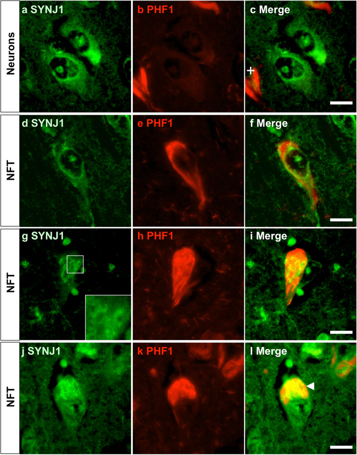
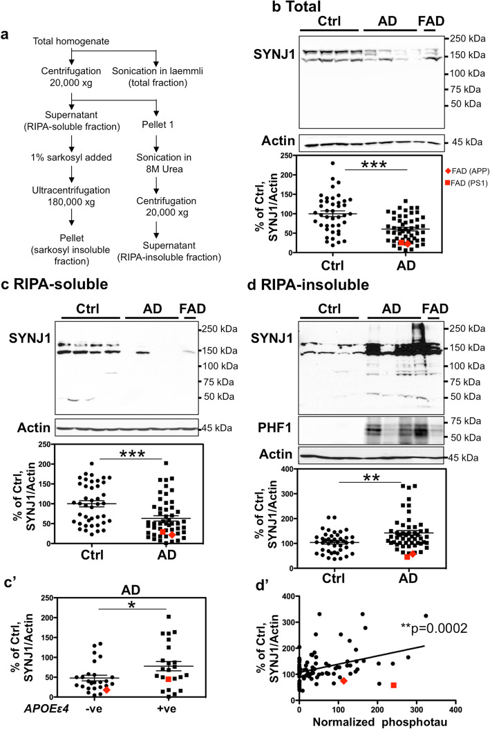
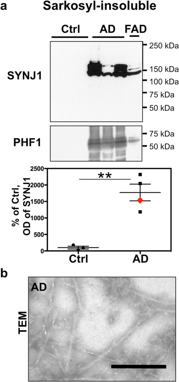
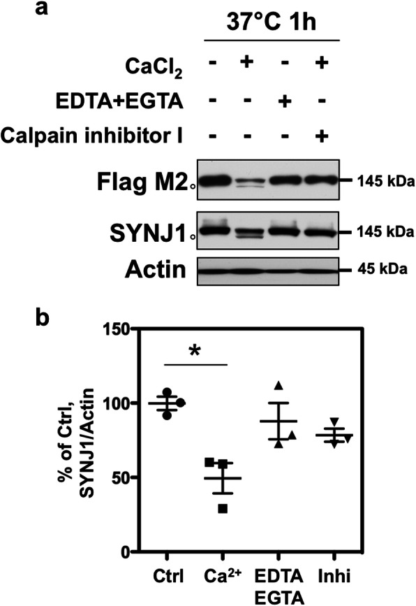
Synaptojanin 1 (SYNJ1) is a brain-enriched lipid phosphatase critically involved in autophagosomal/endosomal trafficking, synaptic vesicle recycling and metabolism of phosphoinositides. Previous studies suggest that SYNJ1 polymorphisms have significant impact on the age of onset of Alzheimer's disease (AD) and that SYNJ1 is involved in amyloid-induced toxicity. Yet SYNJ1 protein level and cellular localization in post-mortem human AD brain tissues have remained elusive. This study aimed to examine whether SYNJ1 localization and expression are altered in post-mortem AD brains. We found that SYNJ1 is accumulated in Hirano bodies, plaque-associated dystrophic neurites and some neurofibrillary tangles (NFTs). SYNJ1 immunoreactivity was higher in neurons and in the senile plaques in AD patients carrying one or two ApolipoproteinE (APOE) ε4 allele(s). In two large cohorts of APOE-genotyped controls and AD patients, SYNJ1 transcripts were significantly increased in AD temporal isocortex compared to control. There was a significant increase in SYNJ1 transcript in APOEε4 carriers compared to non-carriers in AD cohort. SYNJ1 was systematically co-enriched with PHF-tau in the sarkosyl-insoluble fraction of AD brain. In the RIPA-insoluble fraction containing protein aggregates, SYNJ1 proteins were significantly increased and observed as a smear containing full-length and cleaved fragments in AD brains. In vitro cleavage assay showed that SYNJ1 is a substrate of calpain, which is highly activated in AD brains. Our study provides evidence of alterations in SYNJ1 mRNA level and SYNJ1 protein degradation, solubility and localization in AD brains.

Keywords: Alzheimer’s disease; Amyloid β; Hirano bodies; Neurofibrillary tangles; Phosphatidylinositol; SYNJ1; Tau.


Conflict of interest statement

The authors declare that they have no competing interests.

Figures

Fig. 1
Fig. 1
SYNJ1 is accumulated in neurons, Hirano bodies and plaque-associated dystrophic neurites in AD brains. a Immunostaining of SYNJ1 in hippocampal CA1–2 area of a control case carrying APOEε3/3. In control brains, SYNJ1 immunoreactivity was detected in dendrites in the hippocampal pyramidal neurons (a, arrow). b-d Immunostaining of SYNJ1 in hippocampal CA1–2 area of an AD patient carrying APOEε3/3. Neuronal perikarya and a perinuclear rim were immunostained for SYNJ1 (b). Ovoid intraneuronal inclusions similar to Hirano bodies were stained by anti-SYNJ1 antibody (c, arrowhead). Dystrophic neurites surrounding amyloid plaques were also stained for SYNJ1 (d). e-f In AD brains carrying APOEε4 allele(s), the intensity of SYNJ1 staining is stronger than in AD cases without APOEε4 allele (e). SYNJI positive Hirano bodies were also detected (e, arrowhead). A strong SYNJ1 immunoreactivity was observed in plaque-associated dystrophic neurites in CA1–2 area of AD cases carrying APOEε4 alleles (f). g Quantification of neuronal SYNJ1 immunoreactivity by image analysis in three groups: control without APOEε4 allele, AD group without APOEε4 allele and AD group carrying APOEε4 allele(s) (n = 3 for each group). Paraffin section of control cases bearing APOEε4 allele was not available and was not included in the analyses. SYNJ1 immunoreactivity is significantly increased in AD cases carrying APOEε4 alleles compared to AD cases without APOEε4 allele. *p < 0.05 and **p < 0.01 by one way ANOVA with post-hoc Tukey test. h Quantification of SYNJ1 immunoreactivity by image analysis in plaque-associated dystrophic neurites. SYNJ1 immunoreactivity is significantly increased in AD cases carrying APOEε4 alleles compared to AD cases without APOEε4 allele (n = 3 for each group). **p < 0.01 by unpaired t-test. i. In the hippocampus of DSAD brain, a strong immunostaining for SYNJ1 was detected in neuronal perikarya and in dystrophic neurites surrounding amyloid plaques (asterisk). Scale bars 10 μm
Fig. 2
Fig. 2
Detection of SYNJ1 immunoreactivities in intraneuronal granular structures in AD brains. a-l Double immunostaining for SYNJ1 (a, d, g, j, green) and PHF1 (b, e, h, k, red) shows that SYNJ1 immunoreactivity is detected as a perinuclear rim and in the perikarya in non-tangle bearing neurons (a-c) and in tangle bearing neurons (d-l) in the CA1–2 pyramidal neurons of AD hippocampus. SYNJ1-positive intraneuronal granular or donuts-like structures (g, inset) were occasionally detected in tangle bearing neurons and were surrounded and sometimes overlapped with hyperphosphorylated tau in NFTs (l, arrowhead). Representative images of an APOEε3/3 AD case are shown. Scale bars 10 μm
Fig. 3
Fig. 3
Detection of SYNJ1 immunoreactivities in Hirano bodies and in plaque-associated dystrophic neurites in AD brains. a-c A double immunofluorescence labelling for SYNJ1 (a, green) and actin (b, red) confirmed that Hirano bodies were immunostained for SYNJ1 in pyramidal neurons of AD brains (c, merge). While SYNJ1 labelling was stronger in the centre of the Hirano bodies (a), actin labelling was stronger in the periphery of Hirano bodies (b). There were numerous smaller punctiform structures that were actin positive but SYNJ1 negative in AD brains (arrows). d-f A double immunofluorescence labelling for SYNJ1 (d, green) and PHF1 (e, red) does not show association of SYNJ1-positive globular structures and hyperphosphorylated tau in the dystrophic neurites surrounding senile plaques. g-l A double immunofluorescence labelling for SYNJ1 (g, j, green) and Synaptophysin (SY38, h, k, red) shows partial colocalization of SYNJ1 and Synaptophysin (arrowheads) in AD brain (g-i) and in 5XFAD mouse brains at 12 months (j-l). Some plaque-associated dystrophic neurites were yet devoid of SYNJ1 immunoreactivity (l, asterisk). Representative images of an APOEε3/3 AD case (a-i) and 5XFAD (j-l) are shown. Scale bar 10 μm for a-c and 40 μm for d-l
Fig. 4
Fig. 4
Expression of SYNJ1 mRNA in human post-mortem AD brains. a qPCR for SYNJ1 was performed on the mRNA extracted from control (n = 30) and AD (n = 36) T1 isocortex. The level of normalized SYNJ1 mRNA was significantly increased in AD brains. Two FAD cases are shown in red. b No significant difference was observed in the normalised SYNJ1 mRNA levels between non-carriers (n = 15) and carriers (n = 4) of APOEε4 allele(s) in the control cohort. c In AD cohorts, SYNJ1 mRNA level was significantly higher in APOEε4 carriers (n = 21) than in non-carriers (n = 14). FAD cases are shown in red. *p < 0.05, **p < 0.01, by unequal variances t test. d There was a significant correlation between the levels of SYNJ1 mRNA and phosphorylated tau detected by WB using PHF1. (r2 = 0.3713, n = 66, p = 0.0021, by Pearson correlation test)
Fig. 5
Fig. 5
Insoluble SYNJ1 is increased in AD brains and is correlated to tau load. a Summary of the fractionation protocol used to obtain total, RIPA-soluble, RIPA-insoluble and sarkosyl-insoluble fractions. b SYNJ1 was significantly decreased in the total homogenate of AD T1 isocortex. c SYNJ1 was significantly decreased in RIPA-soluble fraction of AD brains. c’ There was a significantly higher level of SYNJ1 protein detected in the AD cases carrying APOEε4 allele(s) compared to the AD cases without APOEε4 allele. d SYNJ1 was increased in RIPA-insoluble fraction of AD brains. Lower MW SYNJ1 positive bands were detected approximately around 100 kDa, 80 kDa and 50 kDa in the AD brain. For b-d, T1 isocortex from control (n = 42) and AD (n = 50) including 2 FAD cases with APP or PSEN1 mutation were analysed. **p < 0.01, ***p < 0.001, by unequal variances t test. d’ There was a significant positive correlation between the levels of SYNJ1 protein and phosphorylated tau detected using PHF1 in the RIPA-insoluble fraction. (r2 = 0.1443, n = 92, p = 0.0002, by Pearson correlation test)
Fig. 6
Fig. 6
SYNJ1 is co-enriched with PHF-tau in sarkosyl-insoluble fraction. a WB for SYNJ1 and PHF1 in sarkosyl-insoluble fraction of control (n = 3) and AD (SAD n = 3, FAD with APP mutation n = 1). SYNJ1 was enriched in sarkosyl-insoluble fractions with PHF-tau. b Representative image of the sarkosyl-insoluble PHF-tau of a sporadic AD case taken by transmission electron microscopy. Scale bar 0.2 μm
Fig. 7
Fig. 7
In vitro cleavage assay for SYNJ1 by calpain. a HEK 293 cells were transiently transfected with Flag-SYNJ1 145 and cultured for 24 h. The cell lysate was incubated at 37 °C for 1 h in the presence or absence of calcium. In the presence of calcium in the lysate, SYNJ1 was significantly decreased by 50% and a 140-kDa band appeared (open circle). The proteolysis was inhibited by adding calcium chelators (EDTA and EGTA) or calpain inhibitor I. b The graph shows the OD of SYNJ1 normalised to actin in each condition of three independent experiments. *p < 0.05 by one-way ANOVA with post-hoc Tukey test

References

    1. Ait Yahya-Graison E, Aubert J, Dauphinot L, Rivals I, Prieur M, Golfier G, et al. Classification of human chromosome 21 gene-expression variations in Down syndrome: impact on disease phenotypes. Am J Hum Genet. 2007;81:475–491. - PMC - PubMed
    1. Ando K, Brion JP, Stygelbout V, Suain V, Authelet M, Dedecker R, et al. Clathrin adaptor CALM/PICALM is associated with neurofibrillary tangles and is cleaved in Alzheimer's brains. Acta Neuropathol. 2013;125:861–878. - PubMed
    1. Ando K, De Decker R, Vergara C, Yilmaz Z, Mansour S, Suain V, Sleegers K, de Fisenne MA, Houben S, Potier MC, Duyckaerts C, Watanabe T, Buée L, Leroy K, Brion JP. Acta Neuropathol. 2020;139(4):773-89. - PubMed
    1. Ando K, Leroy K, Heraud C, Yilmaz Z, Authelet M, Suain V, et al. Accelerated human mutant tau aggregation by knocking out murine tau in a transgenic mouse model. Am J Pathol. 2011;178:803–816. - PMC - PubMed
    1. Ando K, Tomimura K, Sazdovitch V, Suain V, Yilmaz Z, Authelet M, et al. Level of PICALM, a key component of clathrin-mediated endocytosis, is correlated with levels of phosphotau and autophagy-related proteins and is associated with tau inclusions in AD, PSP and pick disease. Neurobiol Dis. 2016;94:32–43. - PubMed

Publication types