. 2020 Jul 30;94(16):e02100-19.
doi: 10.1128/JVI.02100-19. Print 2020 Jul 30.

Ebola Virus Inclusion Body Formation and RNA Synthesis Are Controlled by a Novel Domain of Nucleoprotein Interacting with VP35

Affiliations

Ebola Virus Inclusion Body Formation and RNA Synthesis Are Controlled by a Novel Domain of Nucleoprotein Interacting with VP35

Tsuyoshi Miyake et al. J Virol. .

Abstract

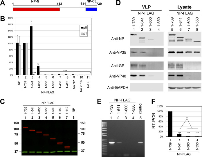
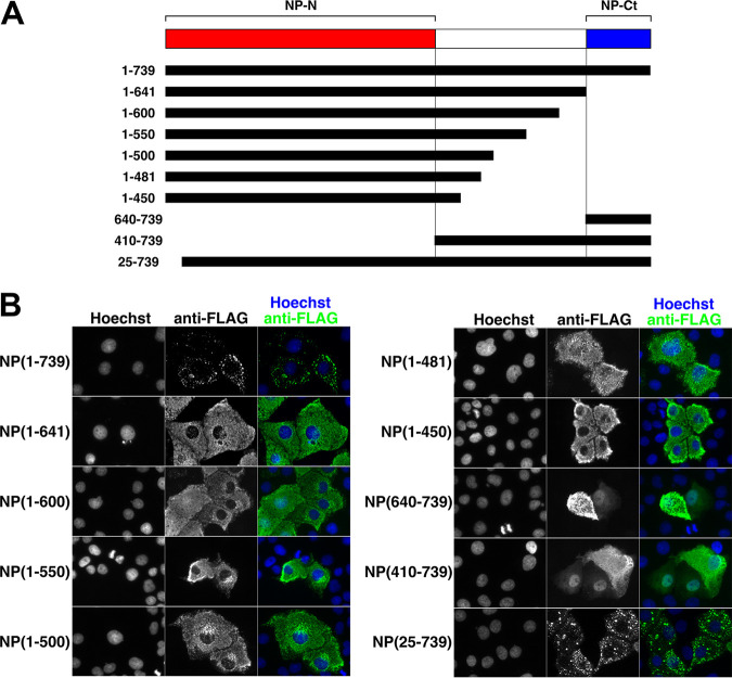
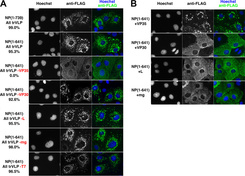
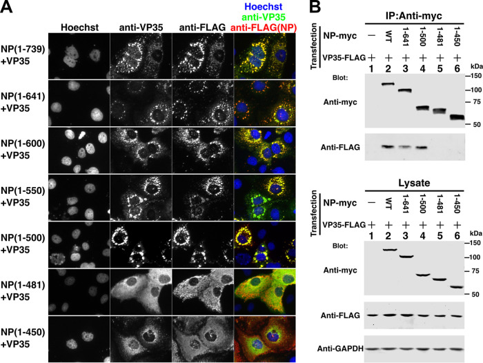
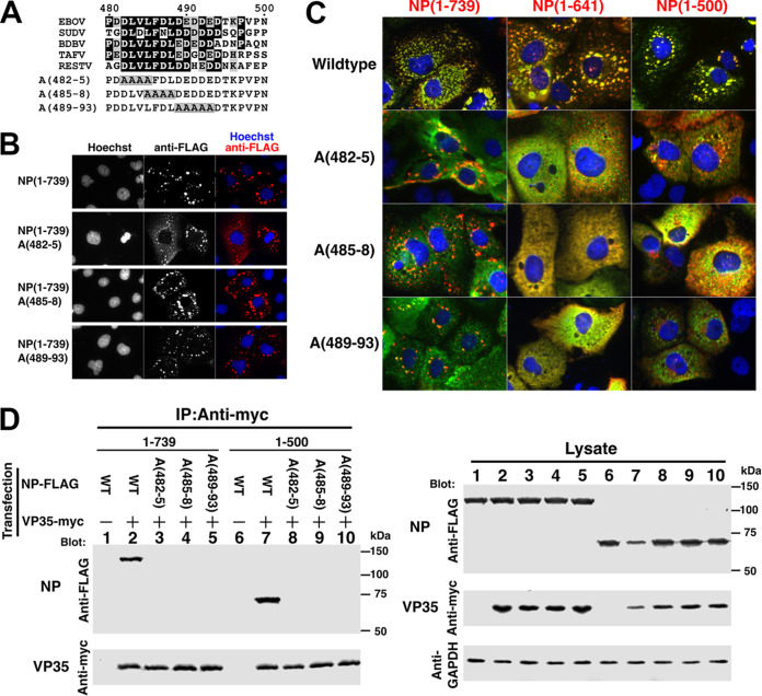
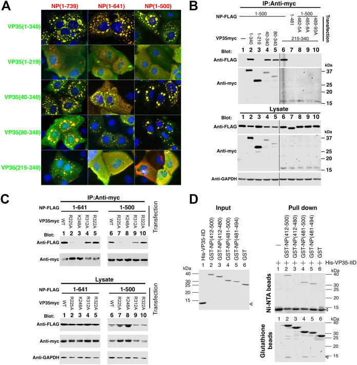
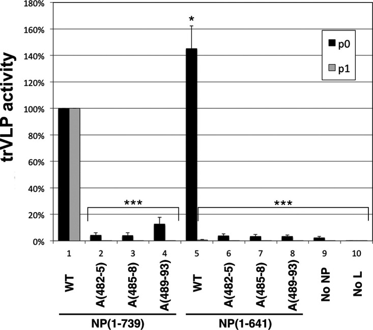
Ebola virus (EBOV) inclusion bodies (IBs) are cytoplasmic sites of nucleocapsid formation and RNA replication, housing key steps in the virus life cycle that warrant further investigation. During infection, IBs display dynamic properties regarding their size and location. The contents of IBs also must transition prior to further viral maturation, assembly, and release, implying additional steps in IB function. Interestingly, the expression of the viral nucleoprotein (NP) alone is sufficient for the generation of IBs, indicating that it plays an important role in IB formation during infection. In addition to NP, other components of the nucleocapsid localize to IBs, including VP35, VP24, VP30, and the RNA polymerase L. We previously defined and solved the crystal structure of the C-terminal domain of NP (NP-Ct), but its role in virus replication remained unclear. Here, we show that NP-Ct is necessary for IB formation when NP is expressed alone. Interestingly, we find that NP-Ct is also required for the production of infectious virus-like particles (VLPs), and that defective VLPs with NP-Ct deletions are significantly reduced in viral RNA content. Furthermore, coexpression of the nucleocapsid component VP35 overcomes deletion of NP-Ct in triggering IB formation, demonstrating a functional interaction between the two proteins. Of all the EBOV proteins, only VP35 is able to overcome the defect in IB formation caused by the deletion of NP-Ct. This effect is mediated by a novel protein-protein interaction between VP35 and NP that controls both regulation of IB formation and RNA replication itself and that is mediated by a newly identified functional domain of NP, the central domain.IMPORTANCE Inclusion bodies (IBs) are cytoplasmic sites of RNA synthesis for a variety of negative-sense RNA viruses, including Ebola virus. In addition to housing important steps in the viral life cycle, IBs protect new viral RNA from innate immune attack and contain specific host proteins whose function is under study. A key viral factor in Ebola virus IB formation is the nucleoprotein, NP, which also is important in RNA encapsidation and synthesis. In this study, we have identified two domains of NP that control inclusion body formation. One of these, the central domain (CD), interacts with viral protein VP35 to control both inclusion body formation and RNA synthesis. The other is the NP C-terminal domain (NP-Ct), whose function has not previously been reported. These findings contribute to a model in which NP and its interactions with VP35 link the establishment of IBs to the synthesis of viral RNA.

Keywords: RNA replication; ebola virus; inclusion body; nucleoprotein.


Figures

FIG 1
FIG 1
NP-Ct is required for infectious VLP production. (A) NP primary structure. The precise definition of the N-terminal domain is subject to interpretation based on sequences included in constructs used for structural and biochemical studies (43, 44, 51, 66) but here is labeled aa 1-412 based on sequence conservation. NP-Ct domain definition is based on Dziubańska et al. (51). (B) trVLP assay (p0 cells) of NP deletion mutants. Indicated NP-FLAG constructs were transfected into 293T/17 cells. As described in the text, recipient p1 cells were supplied with wild-type NP. Data are averages from independent biological triplicates. Error bars represent the SD for the triplicates. Marks above the bar indicate P values to the corresponding NP-FLAG samples based on Student’s t tests: *, P < 0.05; **, P < 0.01; ***, P < 0.001. Untagged NP (column 1) showed no statistically significant difference from the corresponding NP-FLAG samples. (C) Western blot of lysates from p0 cell transfectants. Lysates were separated by SDS-PAGE, blotted, and probed with anti-FLAG (red) or anti-GAPDH (green) antibody. (D) Western blot analysis of purified trVLPs and corresponding lysate from p0 transfectants. Indicated NP-FLAG constructs were transfected into 293T cells (p0) along with all other trVLP components. trVLPs were purified from the supernatant as described in Materials and Methods. trVLPs and corresponding cell lysates were analyzed by Western blotting with the indicated antibodies. Anti-GAPDH was used as a loading control for the lysate samples and as a specificity control for the purified trVLPs. (E) RT-PCR quantification of genomic (negative-strand) RNA from the indicated isolated trVLPs. Isolated RNA was subjected to RT-PCR and quantified using linear range amplification standards as described in Materials and Methods. Positive control indicates a PCR product of a minigenome DNA template. (F) Quantitation of RT-PCR. PCR products were subjected to agarose gel electrophoresis and quantified with the Molecular Imager XRS system. Means and standard deviations from biological triplicate experiments are shown. Asterisks indicate P values based on Student's t test: ***, P < 0.001 to the NP(1-739) sample; **, P < 0.01 between NP(1-641) and NP(1-600) or NP(1-550).
FIG 2
FIG 2
NP-Ct is required for IB formation. (A) Structure of NP deletion mutants. Each construct contains a C-terminal FLAG tag. (B) HuH-7 cells were transfected with the indicated constructs and stained with anti-FLAG antibody and Hoechst 33342 dye after 48 h. All constructs lacking the NP-Ct failed to localize in IBs.
FIG 3
FIG 3
VP35 specifically complements deletion of NP-Ct. (A) FLAG-tagged NP(1-739) or NP(1-641) was transfected along with other trVLP components, i.e., expression plasmids for VP35, VP30, L, T7 polymerase, a tetracistronic minigenome (MG) expressing VP24, GP, and VP40, and firefly luciferase, as indicated, and immunostained with anti-FLAG antibody. Individually omitted constructs are indicated in red. (B) NP(1-641) was cotransfected with each of the indicated individual plasmids of the trVLP system.
FIG 4
FIG 4
NP 481-500 region is required for IB formation and association with VP35. (A) Localization of coexpressed NP deletion mutants with VP35 in transfected HuH-7 cells. VP35 was detected with anti-VP35 antibody (green), and NP-FLAG proteins were detected with anti-FLAG antibody (red). Nuclei were stained with Hoechst 33342 dye (blue). (B) Deletion mutants of myc-tagged NP (NP-myc) and VP35-FLAG were coexpressed in 293T/17 cells and immunoprecipitated with anti-myc antibody. Immunoprecipitated proteins and lysate were subjected to SDS-PAGE, blotted, and detected by anti-myc or anti-FLAG antibodies, as indicated. A GAPDH antibody was used for the loading control.
FIG 5
FIG 5
Alanine-scanning mutants covering the NP 482-493 region abolish IB formation, colocalization, and interaction with VP35. (A) Sequence alignment of five Ebolavirus species members and sequences of alanine-scanning mutants within EBOV. Identical residues are shown with white letters on black background, and similar residues are shown with black letters on gray background. Alanine mutations are boxed. (B) Immunofluorescence staining of wild-type and alanine-scanning mutants of full-length NP. FLAG-tagged NP and mutants were stained with anti-FLAG antibody (red) and Hoechst 33342 dye (blue). (C) NP(1-739), NP(1-641), and NP(1-500), or their corresponding alanine-scanning mutants, were coexpressed with VP35. Cells were stained with anti-FLAG antibody (NP; red), and VP35 was stained with anti-VP35 antibody (green). Nuclei were stained with Hoechst 33342 (blue). Merged green/red fields are shown. (D) Immunoprecipitation (IP) of coexpressed VP35-myc and wild-type or alanine-scanning mutants of FLAG-tagged NP(1-739) or NP(1-500). Cells were cotransfected with VP35-myc and NP-FLAG derivatives, lysed, and processed for immunoprecipitation as described in Materials and Methods. A GAPDH antibody was used for the loading control. (Left) Immunoprecipitates were separated by SDS-PAGE, blotted, and probed with the indicated antibodies. (Right) Crude lysates were separated by SDS-PAGE, blotted, and probed with the indicated antibodies to determine protein expression levels.
FIG 6
FIG 6
VP35 IID associates with the NP(481-500) region. (A) Localization of deletion mutants of VP35 and NP. Wild-type and deletion mutants of NP were cotransfected with wild-type VP35 and deletion mutants in HuH-7 cells. Forty-eight hours after transfection, cells were stained with anti-FLAG (NP, red) and anti-myc (VP35, green). Merged green/red fields are shown. (B) NP(1-500) and related mutants, as indicated, were cotransfected with wild-type VP35-myc and deletion mutants in 293T/17 cells, and cells were lysed 48 to 50 h after transfection. An anti-myc antibody was used for immunoprecipitation, and coimmunoprecipitated proteins were analyzed by Western blotting. GAPDH is the loading control of the lysate. (Top) Immunoprecipitates were separated by SDS-PAGE, blotted, and probed with the indicated antibodies. (Bottom) Crude lysates were separated by SDS-PAGE, blotted, and probed with the indicated antibodies to determine protein expression levels. For the top and bottom panels, the left and right sides of the data shown were sourced from the same image, with several lanes deleted between lanes 5 and 6. This is indicated by vertical black lines. (C) NP(1-500) and NP(1-641) were cotransfected with wild-type VP35-myc or the indicated mutants in 293T/17 cells, and cells were lysed 48 to 50 h after transfection. An anti-myc antibody was used for immunoprecipitation, and coimmunoprecipitated proteins were analyzed by Western blotting. (Top) Immunoprecipitates were separated by SDS-PAGE, blotted, and probed with the indicated antibodies. (Bottom) Crude lysates were separated by SDS-PAGE, blotted, and probed with the indicated antibodies to determine protein expression levels. (D) Pulldown assay of E. coli-expressed proteins. GST-NPs with the indicated regions of NP or His-tagged VP35-IID were expressed in E. coli and purified using glutathione Sepharose or nickel-nitrilotriacetic acid (Ni-NTA) agarose. Purified proteins were dialyzed, subjected to SDS-PAGE, and stained with CBB (input). Purified proteins were quantified, and equal amounts of GST-NPs or GST were mixed with His-VP35-IID protein. After overnight incubation with the indicated beads followed by washing, protein bound to the beads was extracted with 2× sample buffer, and equal volumes of the eluents were subjected to SDS-PAGE and stained with CBB.
FIG 7
FIG 7
trVLP assay of the wild type and alanine-scanning mutants in aa 481 to 500. For p0 assays, NP(1-739) or NP(1-641) and their alanine-scanning mutants, A(482-5), A(485-8), or A(489-93), were cotransfected with other components of the trVLP p0 assay, and luciferase activity of each lysate was measured. Wild-type NP(1-739) activity was set at 100%. trVLP assays in the absence of NP (No NP) or L (No L) were used as negative controls. For p1 assays, recipient cells were transfected with the complete set of trVLP plasmids, including wild-type NP, and supernatants from the indicated p0 assay were used for infection. Error bars represent the SD from three independent biological replicates. Asterisks indicate P values from comparisons to the corresponding NP(1-739) wild type based on the Student's t test: *, P < 0.05; ***, P < 0.001.
FIG 8
FIG 8
Identified NP functions. Full-length NP and VP35 proteins are illustrated. NP-N and NP-Ct cover aa 1 to 412 and 641 to 739, respectively, as defined in reference . The central domain (CD) spans aa 481 to 500. Red boxes, regions required for IB formation. The large region spanning aa 25 to 410 is based on data shown in Fig. 2B. The requirement for NP-Ct applies to NP-induced IBs, and when VP35 is coexpressed, NP-Ct is not required, as described in the text. Likewise, when NP-N and NP-Ct are present, mutation of the CD does not abolish IB formation; therefore, NP-Ct and CD complement each other. The green box indicates the region required for production of infectious trVLPs and incorporation/retention of viral RNA in purified trVLPs. For NP and VP35, black regions indicate NPBP and its corresponding binding region within NP, the VP35 first basic patch as defined in references and , and its corresponding binding region, the CD. See references and for definitions of NP region bound by NPBP. See Discussion for possible functional relationships between the two regions of VP35 that interact with NP.

References

    1. Coltart CE, Lindsey B, Ghinai I, Johnson AM, Heymann DL. 2017. The Ebola outbreak, 2013-2016: old lessons for new epidemics. Philos Trans R Soc Lond B Biol Sci 37:220160297. doi:10.1098/rstb.2016.0297. - DOI - PMC - PubMed
    1. Ilunga Kalenga O, Moeti M, Sparrow A, Nguyen VK, Lucey D, Ghebreyesus TA. 2019. The ongoing ebola epidemic in the Democratic Republic of Congo, 2018-2019. N Engl J Med 381:373–383. doi:10.1056/NEJMsr1904253. - DOI - PubMed
    1. Wong G, Mendoza EJ, Plummer FA, Gao GF, Kobinger GP, Qiu X. 2018. From bench to almost bedside: the long road to a licensed Ebola virus vaccine. Expert Opin Biol Ther 18:159–173. doi:10.1080/14712598.2018.1404572. - DOI - PMC - PubMed
    1. Green A. 2017. Ebola outbreak in the DR Congo. Lancet 389:2092. doi:10.1016/S0140-6736(17)31424-1. - DOI - PubMed
    1. Cifuentes-Munoz N, Branttie J, Slaughter KB, Dutch RE. 2017. Human metapneumovirus induces formation of inclusion bodies for efficient genome replication and transcription. J Virol 91:e01282-17. doi:10.1128/JVI.01282-17. - DOI - PMC - PubMed

Publication types

MeSH terms