. 2020 Jun 3;10(1):9069.
doi: 10.1038/s41598-020-65988-w.

CX3CL1 homo-oligomerization drives cell-to-cell adherence

Affiliations

CX3CL1 homo-oligomerization drives cell-to-cell adherence

Mariano A Ostuni et al. Sci Rep. .

Abstract

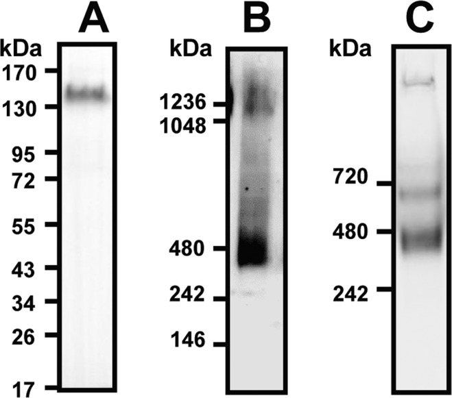
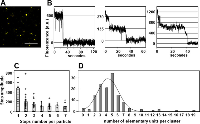
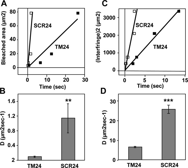
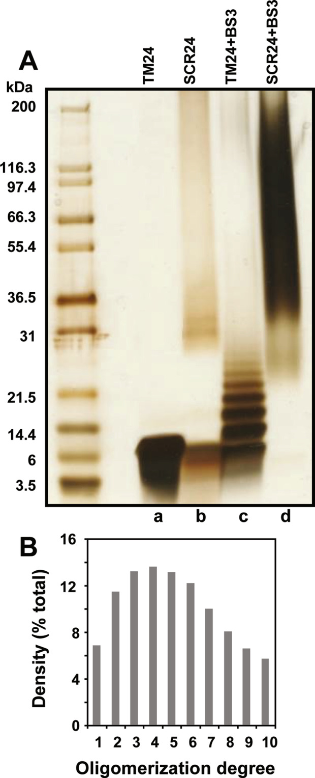
During inflammatory response, blood leukocytes adhere to the endothelium. This process involves numerous adhesion molecules, including a transmembrane chemokine, CX3CL1, which behaves as a molecular cluster. How this cluster assembles and whether this association has a functional role remain unknown. The analysis of CX3CL1 clusters using native electrophoresis and single molecule fluorescence kinetics shows that CX3CL1 is a homo-oligomer of 3 to 7 monomers. Fluorescence recovery after photobleaching assays reveal that the CX3CL1-transmembrane domain peptide self-associates in both cellular and acellular lipid environments, while its random counterpart (i.e. peptide with the same residues in a different order) does not. This strongly indicates that CX3CL1 oligomerization is driven by its intrinsic properties. According to the molecular modeling, CX3CL1 does not associate in compact bundles but rather with monomers linearly assembled side by side. Finally, the CX3CL1 transmembrane peptide inhibits both the CX3CL1 oligomerization and the adhesive function, while its random counterpart does not. This demonstrates that CX3CL1 oligomerization is mandatory for its adhesive potency. Our results provide a new direction to control CX3CL1-dependent cellular adherence in key immune processes.


Conflict of interest statement

The authors E.M.D. and A.J. are employees of CALIXAR that have patents applications that cover CALX173ACE described in this manuscript. Apart from that, the authors declare that they have no conflict of interest.

Figures

Figure 1
Figure 1
Electrophoresis of the CX3CL1-YFP fusion protein. (A) Cell lysate of LCX3CL1 cells stably transfected with CX3CL1-EYFP was analyzed by SDS-PAGE and Western blot (full length blot in Fig. S1A). (B) Cell lysate (40 µg of total proteins) of LCX3CL1 was analyzed by native electrophoresis using Nu-Page gel in the presence of 1% DDM (dodecylmaltoside) and Western blot. (C) The affinity-purified CX3CL1 from LCX3CL1 membranes was analyzed by native PAGE. CX3CL1 was solubilized using CALX173ACE and affinity purified (CX3CL1 antibody). Mini-Protean TGX gel electrophoresis (BioRad) was used for Native PAGE.
Figure 2
Figure 2
Single–particle fluorescence analysis of CX3CL1. (A) Fluorescence of the membrane preparation of LCX3CL1 diluted in DOPC analyzed by TIRF. Bar 10 µm. (B) Examples of the fluorescence of particles tracked during one or two minutes. (C) Distribution of the fluorescence step amplitude according to the step number per particle. The grey bars represent the mean of the step amplitude in each case. (D) Distribution of the number of elementary fluorescence units per particle calculated after analysis of the fluorescence kinetic of 126 different particles. The Gaussian curve fits the data with an amplitude of 27.5, a mean of 4.3 and a standard deviation of 1.8.
Figure 3
Figure 3
Diffusion rate of CX3CL1 transmembrane peptides analyzed by FRAP and FRAPP. (A) The fluorescence kinetics of Giant Unilamellar Vesicles containing either TM24-FITC (filled squares) or SC24-FITC (empty squares) were analyzed after bleaching by circles of various diameter (1, 2, 5 and 10 µm). The recovery constant time was reported versus the bleached area. Each measurement was the mean of triplicates. The slopes of the linear fit are 2.63 and 29.93 for TM24 and SCR24 respectively. (B)The diffusion rates were calculated based on the mean ± SEM of 12 measurements data used to give the (A). (C) The fluorescence kinetics of lipidic cubic “sponge” phase containing either TM24-FITC (filled squares) or SC24-FITC (empty squares) were analyzed after bleaching by interference pattern with various interfringe (20, 27, 46 and 58 µm). The recovery constant time was reported versus the interfringe distance. Each measurement is the mean of triplicates. The slopes of the linear fit are 252 ± 10 and 1093 ± 144 for TM24 and SCR24, respectively. (D) The diffusion rates were calculated based on the mean ± SEM of 12 measurements data used to give the (C). Note that the diffusion rate appeared dramatically higher in sponge phase since the fluorescent molecules could move in tridimensional milieu.
Figure 4
Figure 4
TM24 peptide polymerization analyzed by cross-linking and SDS-PAGE. (A) SDS-PAGE of the TM24 and SCR24 peptides dissolved in DPC with a ratio [DPC]/[peptide] = 10 and cross-linked or not with 1.9 mM SB3 crosslinker. The gel is then silver stained. The left lane contains markers of various molecular weights (full length gel in Fig. S5). (B) Densitogram of the “TM + SB3” lane in A.
Figure 5
Figure 5
Oligomer formation observed by coarse-grained molecular dynamics simulations of TM24 peptides in DOPC lipids. (A) Topology of the main oligomers observed in the three simulations, as seen from the top. The red arrow within each circle indicates an arbitrary axis in the middle of the TM24 helix enabling to indicate the rotational orientation of each monomer and allowing to distinguish between parallel, orthogonal and symmetrical dimers. (B) Helical wheels representing the most frequent mutual orientation of monomers in dimer (parallel), as seen from the top. Its monomer-monomer interface involves Ala7, Leu11, Ala18 and Leu5, Leu12, Gly16 from each monomer, respectively. Dotted arrows illustrate the angular fluctuation observed both for one dimer along the simulations and between the different dimers within this parallel arrangement.
Figure 6
Figure 6
Diffusion rate in cellular membrane of TM24 peptide and various proteins with a known number of TM domains. The lateral diffusion rate of the TM24-FITC peptide and of other proteins with known TM number were assayed by FRAP after transitory expression in COS-7 cell line. Each point is the mean of duplicates, except for CX3CR1 and TM24 (mean of pentaplicates±SEM).
Figure 7
Figure 7
Diffusion rate in cellular membrane of the CX3CL1 protein in the presence of TM24 and SCR24 peptides. The lateral diffusion rate of the CX3CL1-EYFP protein was assayed after transient expression in COS-7 cell line after 15 min preincubation in the presence or not of 3 or 10 µM of TM24 or SCR24 peptides. Each point is the mean of triplicates ± SD.
Figure 8
Figure 8
Specific inhibition of the CX3CL1-dependent cell adherence by the peptide TM24. (A) Real time adherence of CHOCX3CR1 cells to L929 (dashed traces) or LCX3CL1 cells (solid traces) as assayed by the LigandTracer technique, in the presence of 5 µM TM24 (red traces), 5 µM SCR24 (blue traces) or none (black traces). The data were normalized using the control trace with LCX3CL1 cells in the absence of peptide (100% after 60 minutes). The curves are the mean of three independent experiments. (B) Specific adherence of CHOCX3CR1 cells to LCX3CL1 cells using data of (A). The data obtained with LCX3CL1 cells were subtracted from data obtained with L929 and normalized at 100% at the 60 minutes time. The bars in the right show the specific adherence after 60 minutes (mean of triplicates ± SD) in the presence of 5 µM TM24 (red) and 5 µM SCR24 (blue). (C) Real time binding of 100 nM of fluorescent CX3CL1 to coated CHOCX3CR1 cells using the LigandTracer technique, in the presence of 5 µM TM24 (red trace), 1 µM unstained CX3CL1 (dashed trace) or none (black trace). The data were normalized using the control trace without peptide (100% after 60 minutes). (D) Specific adherence of CHOCX3CR1 cells to LCX3CL1 cells after 60 minutes in the presence of various TM24 peptide concentrations and in the presence (filled squares) or in absence (empty squares) of 1 µg/ml of anti-CX3CL1 antibody. The data were calculated and normalized as in (B). Experiments are performed in duplicate except for the 1 µM TM24 concentration done in triplicates (mean ± SD).

References

    1. Ingersoll MA, Platt AM, Potteaux S, Randolph GJ. Monocyte trafficking in acute and chronic inflammation. Trends Immunol. 2011;32:470–477. doi: 10.1016/j.it.2011.05.001. - DOI - PMC - PubMed
    1. Langer HF, Chavakis T. Leukocyte-endothelial interactions in inflammation. J Cell Mol Med. 2009;13:1211–1220. doi: 10.1111/j.1582-4934.2009.00811.x. - DOI - PMC - PubMed
    1. Luster, A. D., Alon, R. & von Andrian, U. H. Immune cell migration in inflammation: present and future therapeutic targets. Nat Immunol6, 1182–1190, doi:10.1038/ni1275 (2005). - PubMed
    1. Charo IF, Ransohoff RM. The many roles of chemokines and chemokine receptors in inflammation. N Engl J Med. 2006;354:610–621. doi: 10.1056/NEJMra052723. - DOI - PubMed
    1. Ortega-Gomez A, Perretti M, Soehnlein O. Resolution of inflammation: an integrated view. EMBO Mol Med. 2013;5:661–674. doi: 10.1002/emmm.201202382. - DOI - PMC - PubMed

Publication types