. 2020 Sep;585(7824):293-297.
doi: 10.1038/s41586-020-2374-x. Epub 2020 Jun 3.

The CDK inhibitor CR8 acts as a molecular glue degrader that depletes cyclin K

Affiliations

The CDK inhibitor CR8 acts as a molecular glue degrader that depletes cyclin K

Mikołaj Słabicki et al. Nature. 2020 Sep.

Abstract

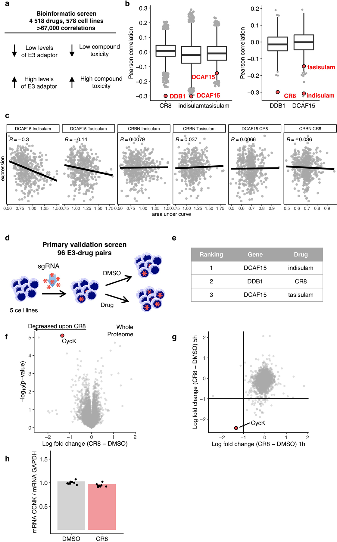
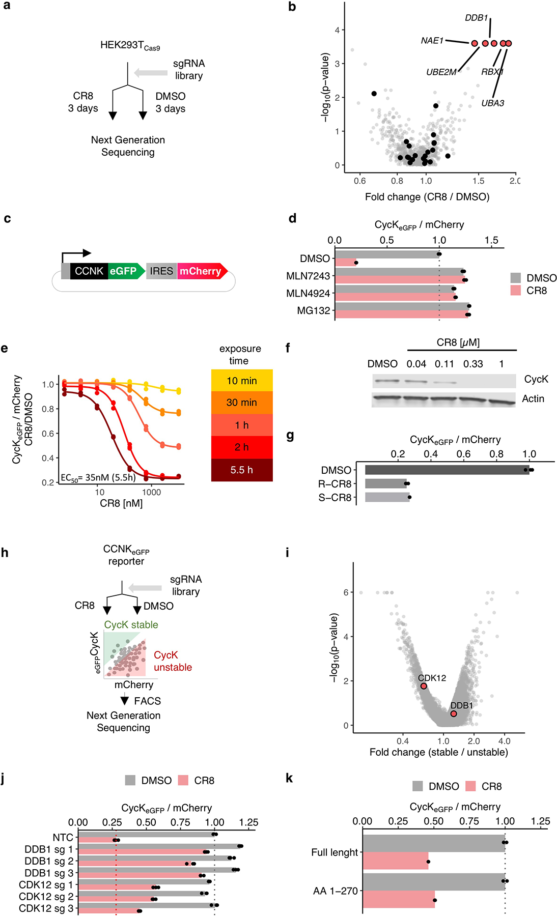
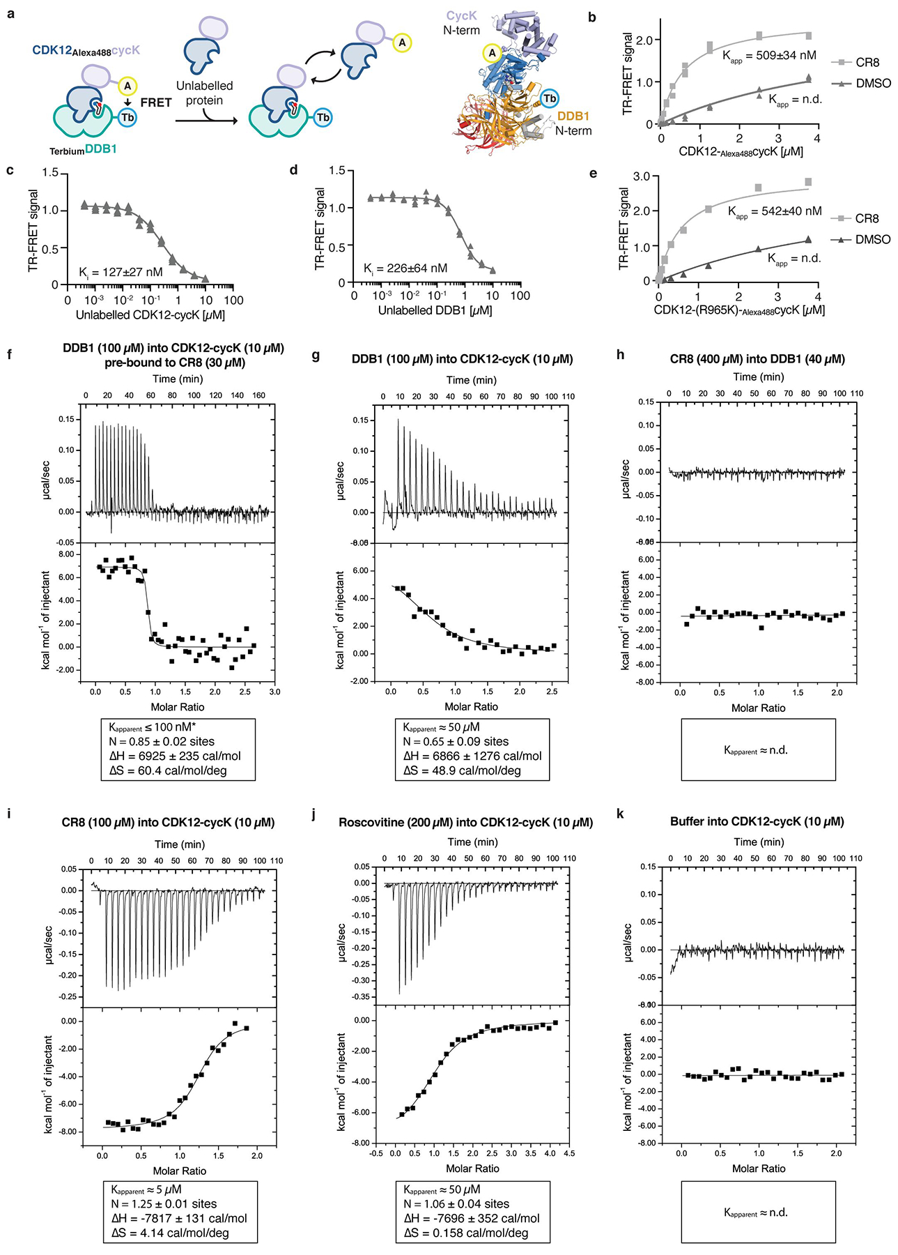
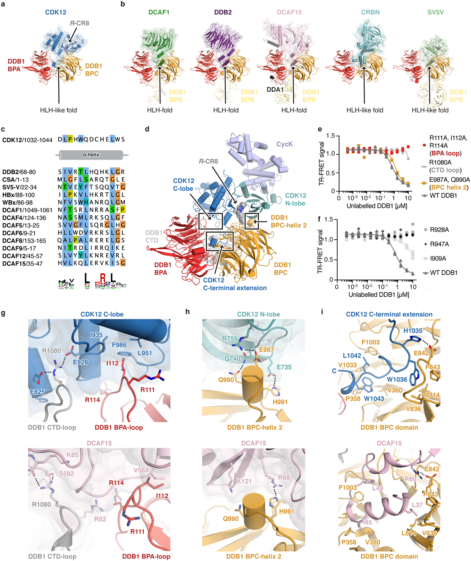
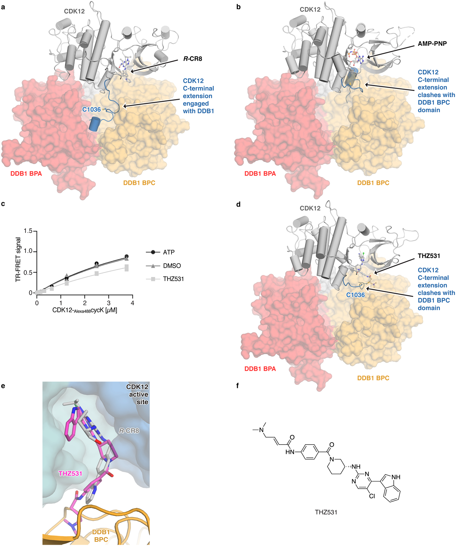
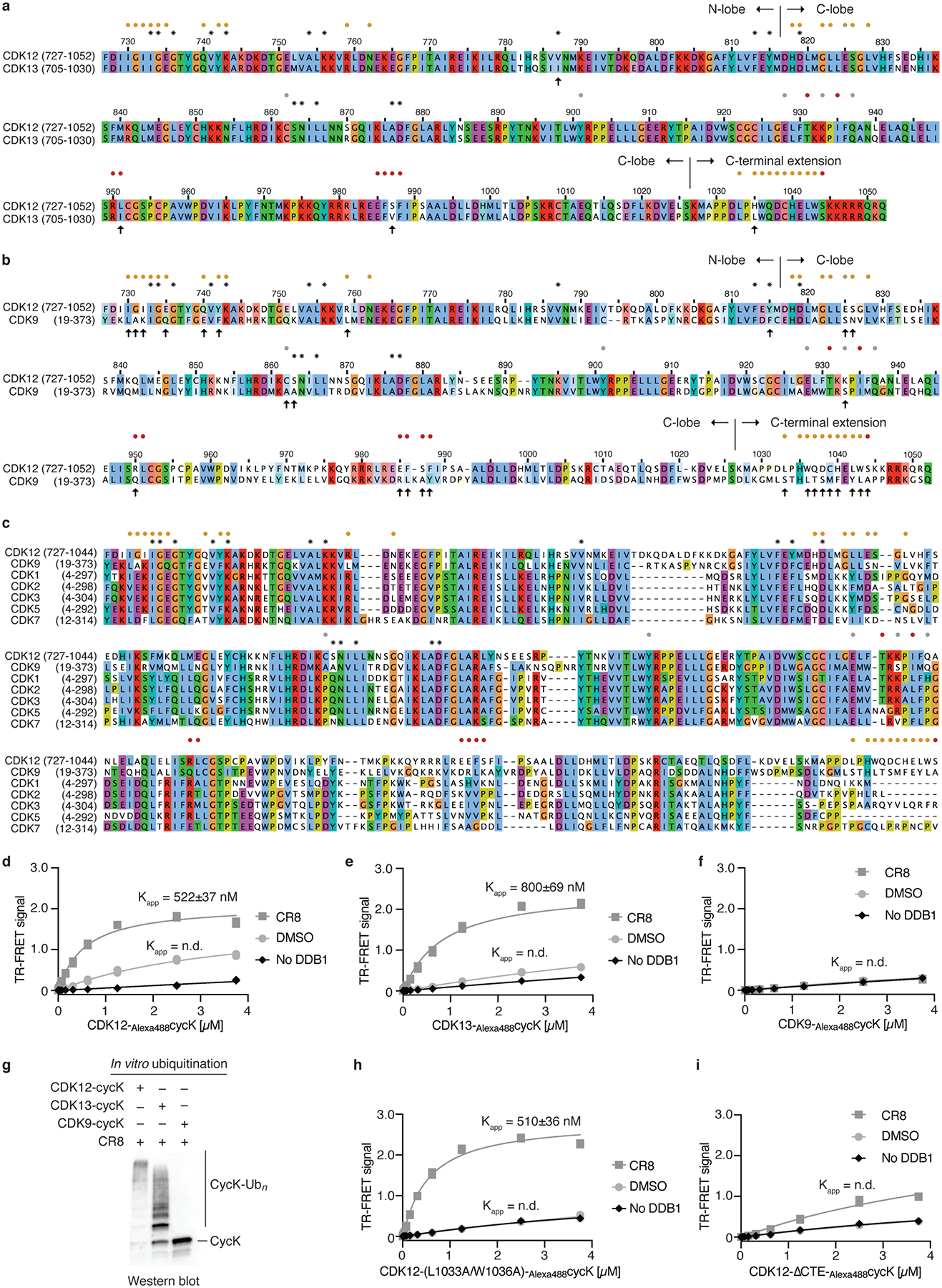
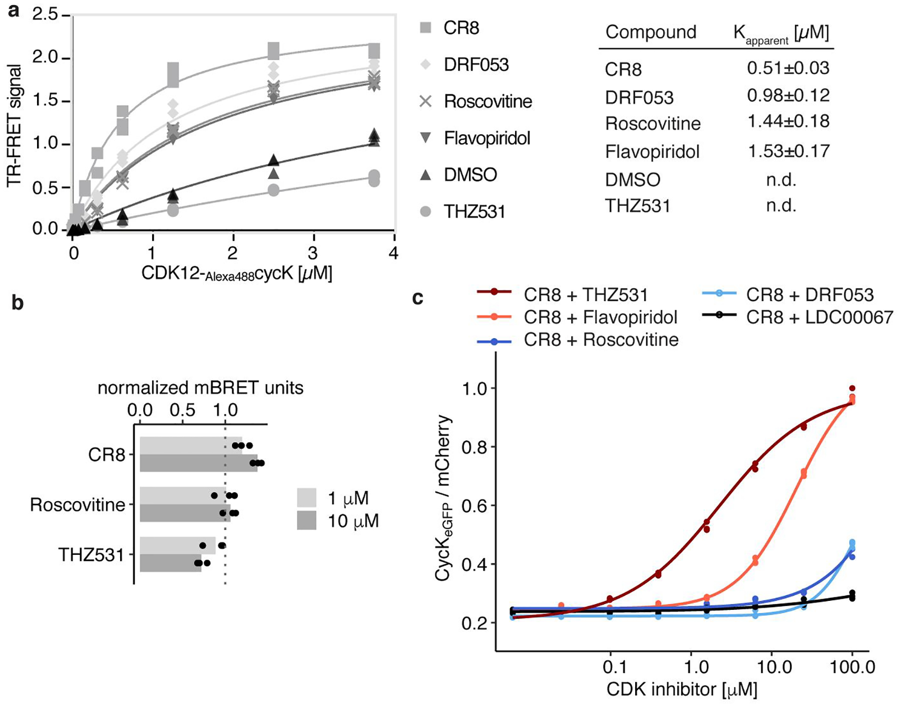
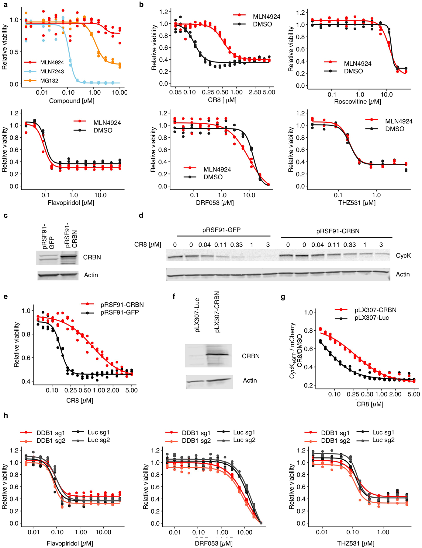
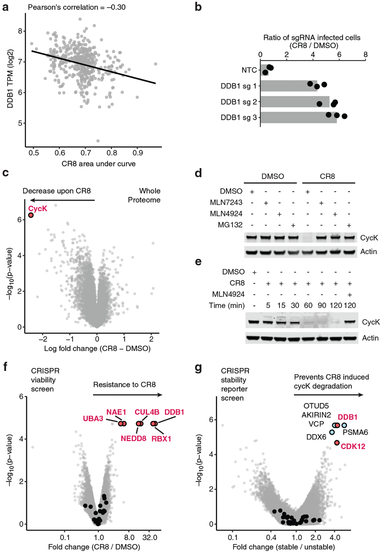
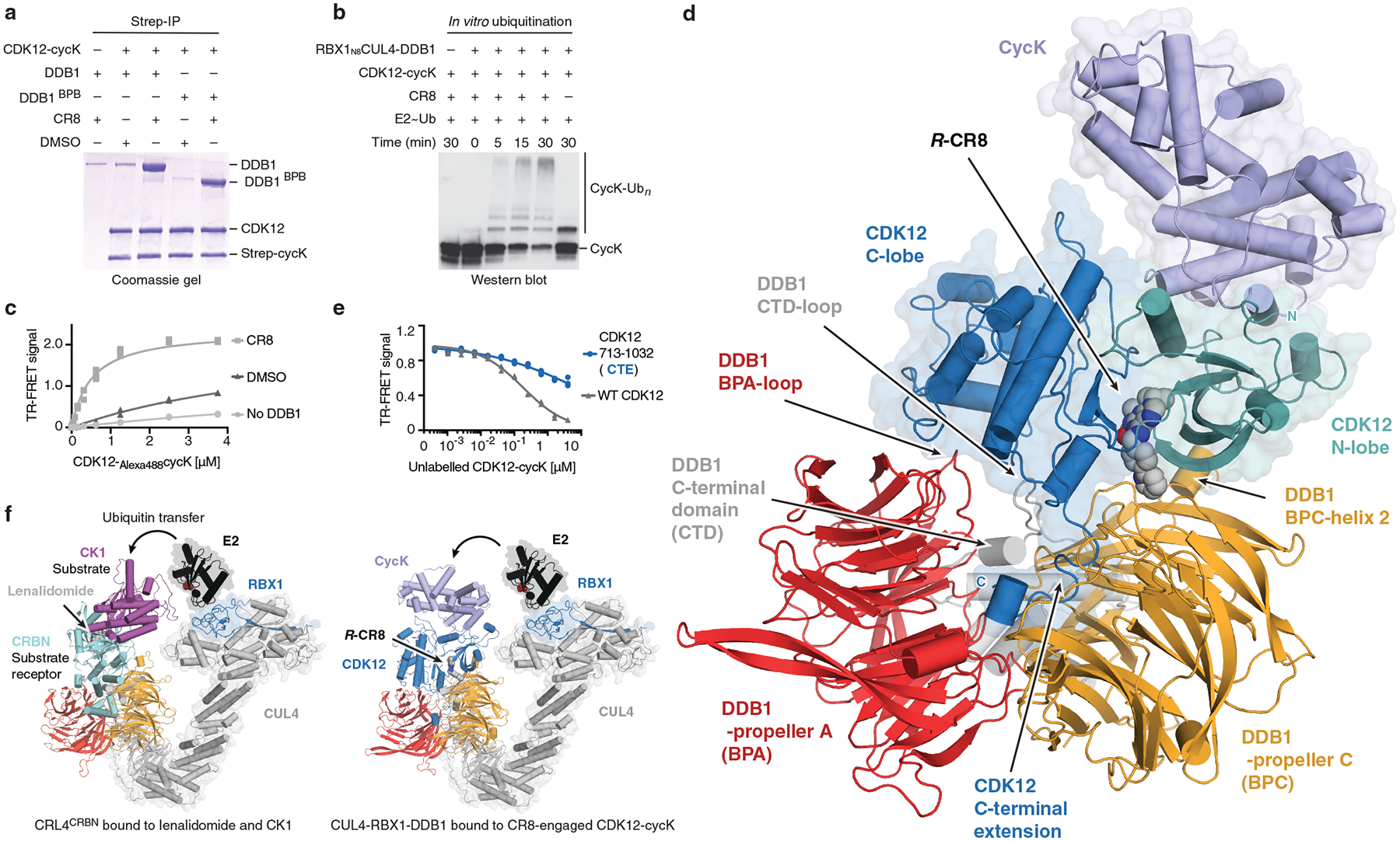
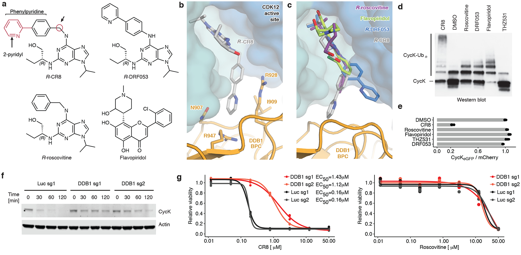
Molecular glue compounds induce protein-protein interactions that, in the context of a ubiquitin ligase, lead to protein degradation1. Unlike traditional enzyme inhibitors, these molecular glue degraders act substoichiometrically to catalyse the rapid depletion of previously inaccessible targets2. They are clinically effective and highly sought-after, but have thus far only been discovered serendipitously. Here, through systematically mining databases for correlations between the cytotoxicity of 4,518 clinical and preclinical small molecules and the expression levels of E3 ligase components across hundreds of human cancer cell lines3-5, we identify CR8-a cyclin-dependent kinase (CDK) inhibitor6-as a compound that acts as a molecular glue degrader. The CDK-bound form of CR8 has a solvent-exposed pyridyl moiety that induces the formation of a complex between CDK12-cyclin K and the CUL4 adaptor protein DDB1, bypassing the requirement for a substrate receptor and presenting cyclin K for ubiquitination and degradation. Our studies demonstrate that chemical alteration of surface-exposed moieties can confer gain-of-function glue properties to an inhibitor, and we propose this as a broader strategy through which target-binding molecules could be converted into molecular glues.


Conflict of interest statement

Competing interests

B.L.E. has received research funding from Celgene and Deerfield. He has received consulting fees from GRAIL, and he serves on the scientific advisory boards for and holds equity in Skyhawk Therapeutics and Exo Therapeutics. E.S.F. is a founder and/or member of the scientific advisory board (SAB), and equity holder of C4 Therapeutics and Civetta Therapeutics and a consultant to Novartis, AbbVie and Pfizer. N.H.T receives funding from the Novartis Research Foundation and is an SAB member of Monte Rosa Therapeutics. The Fischer lab receives or has received research funding from Novartis, Deerfield and Astellas. S.F. has had a consulting or advisory role, received honoraria, research funding, and/or travel/accommodation expenses funding from the following for-profit companies: Bayer, Roche, Amgen, Eli Lilly, PharmaMar, AstraZeneca, and Pfizer. R.B. is now an employee of Monte Rosa Therapeutics. S.M.C and T.R.G receive research funding from Bayer HealthCare. T.R.G. was formerly a consultant and equity holder in Foundation Medicine (acquired by Roche). T.R.G. also is a consultant to GlaxoSmithKline and is a founder of Sherlock Biosciences.

Figures

Extended Data Figure 1 |
Extended Data Figure 1 |. CR8-induced degradation of cycK correlates with DDB1 expression.
a, Schematic of bioinformatic screen for drug-E3 pairs. b, Box plot (centre, median; box, interquartile range (IQR); whiskers, 1.5 × IQR; outliers, points) for expression–sensitivity correlations (CR8 n=19110; indisulam, tasisulam n= 19109, DDB1, DCAF15 n=1618). c, Example Pearson correlation of selected drug-E3 pairs: positive controls (Indisulam-DCAF15; Tasisualm-DCAF15) and no correlation controls (others), (Indisulam n=452, Tasisulam n=418, CR8 n=471) d, Schematic of flow-based primary validation screen. e, Top three hits from the primary validation screen in 5 cell lines, performed according to the schematic in d. f, Whole proteome quantification of Molt-4 cells treated with 1 μM CR8 (n=1) or DMSO (n=3) for 1 hour (two-sided moderated t-test, n=3). g, The log2 fold changes in whole proteome quantification after 1 and 5 hours of exposure to CR8 plotted against each other. h, mRNA levels quantified by qPCR in HEK293TCas9 cells following 1 μM CR8 for 2 hours. Bars represent the mean (n=9).
Extended Data Figure 2 |
Extended Data Figure 2 |. CDK12 is required for CR8-induced cycK degradation.
a, Schematic of the genome-wide CRISPR-Cas9 resistance screen. b, `BISON` CRISPR/Cas9 viability screen for CR8 resistance. Guide counts were collapsed to gene-level (n = 4 guides/gene; two-sided empirical rank-sum test-statistics). c, Schematic of the cycK (CCNK) stability reporter. eGFP, enhanced green fluorescent protein, IRES, internal ribosome entry site. d, Flow analysis of CycKeGFP degradation in HEK293TCas9 cells pre-treated with 0.5 μM MLN7243, 1 μM MLN4924, or 10 μM MG132 for 4 hours followed by exposure to 1 μM CR8 for 2 hours (n=3). e, Flow analysis of CycKeGFP degradation in HEK293TCas9 cells treated with CR8 (n=3). f, Immunoblots of CycK degradation in HEK293TCas9 cells treated with CR8 for 2 hours (n=2). g, Flow analysis of CycKeGFP degradation in HEK293TCas9 cells treated with 1μM compound for 2 hours (n=3). h, Schematic of the genome-wide CRISPR-Cas9 cycK stability reporter screen. i, Genome-wide CRISPR/Cas9 reporter screen for cycKeGFP stability with DMSO treatment in HEK293TCas9 cells. Guide counts were collapsed to gene-level (n = 4 guides/gene; two-sided empirical rank-sum test-statistics). j, Flow analysis of CycKeGFP degradation in HEK293TCas9 cells following 1μM CR8 for 2 hours (n=3). k, Flow analysis of CycKeGFPFull Length or CycKeGFPAA1−270 in HEK293TCas9 following 1 μM CR8 for 2 hours (n=3). Bars represent the mean in d, g, j and k.
Extended Data Figure 3 |
Extended Data Figure 3 |. CR8-induced cycK degradation is not dependent on a canonical DCAF substrate receptor.
a, Drug sensitivity of K562Cas9, P31FUJCas9, THP1Cas9 and MM1SCas9 cells exposed to CR8 for 3 days (n=3). b, mRNA expression levels for genes in DCAF library. “|” represents mean (n=4). c, Flow analysis of K562Cas9, P31FUJCas9, THP1Cas9 and MM1SCas9 cells expressing sgRNAs and a BFP marker (blue fluorescent protein) after a 3-day treatment with 1 μM CR8. “|” represents mean (n>2).
Extended Data Figure 4 |
Extended Data Figure 4 |. Characterization of DDB1-CDK12-cycK complex formation.
a, Schematic of the TR-FRET setup. Positions of the FRET donor (terbium-streptavidin (Tb)) and acceptor (Alexa488SpyCatcher (A)) are indicated in the structural model. b, Titration of CDK12-Alexa488cycK (0–3.75 μM) to 50 nM terbiumDDB1 and 5 μM CR8 or DMSO (n=3). c, Counter-titration of unlabelled wild-type CDK12-cycK (0–10 μM) to 50 nM terbiumDDB1, 500 nM CDK12-Alexa488cycK and 12.5 μM CR8 (n=3). d, Counter-titration of unlabelled wild-type DDB1 (0–10 μM) to 50 nM terbiumDDB1, 500 nM CDK12-Alexa488cycK and 1 μM CR8 (n=3). e, Titration of CDK12(R965K)-Alexa488cycK (wild-type sequence of canonical isoform of CDK12; 0–3.75 μM) to 50 nM terbiumDDB1 and 5 μM CR8 or DMSO (n=3). The CDK12 K965R variant, which was used throughout our in vitro studies (see Methods), shows a binding affinity indistinguishable from that of wild-type CDK12 (residue distal from the interface with DDB1 and cycK). f, Isothermal titration calorimetry (ITC) experiment (n=2, additional replicates for this and following panels are provided in Supplementary ITC Data). Specifications of the titration are given in the panel. Asterisk marking the approximate Kapparent value denotes that the binding affinity was too high to allow confident fitting of the binding curve. g, ITC experiment as in f (n=2). h, ITC experiment as in f (n=1). i, ITC experiment as in f (n=3). j, ITC experiment as in f (n=3). k, ITC experiment as in f (n=1).
Extended Data Figure 5 |
Extended Data Figure 5 |. CDK12 contacts on DDB1 otherwise implicated in DCAF binding.
a, Structure of the CDK12-R-CR8-DDB1ΔBPB complex. The CDK12 C-terminal domain binds a cleft between the DDB1 BPA and BPC domains (arrow) and adopts an helix-loop-helix (HLH)-like fold. b, Diverse DCAFs bind DDB1 through HLH- or HLH-like folds. c, Sequence alignment of identically positioned helices of different HLH-domains. d, Overview of protein-protein interaction hotspots. e, Counter-titration of unlabelled wild-type or mutant DDB1 (0–10 μM) into pre-assembled terbiumDDB1-CR8-CDK12-Alexa488cycK complex (n=3). f, Counter titration of unlabelled wild-type or mutant DDB1 (0–10 μM) into pre-assembled terbiumDDB1-CR8-CDK12-Alexa488cycK complex (n=3). g-i, Close-up of DDB1 residues contacted by CDK12 (top) that are also otherwise involved in DCAF binding (bottom).
Extended Data Figure 6 |
Extended Data Figure 6 |. CDK12 C-terminal extension adopts different conformations.
a, Conformation of the C-terminal extension in the structure of the CDK12-CR8-DDB1ΔBPB complex. b, Structure of CDK12 bound to AMP-PNP (PDB entry 4CXA) superimposed onto CDK12 in the CDK12-CR8-DDB1ΔBPB complex. c, Titration of CDK12-Alexa488cycK (0–3.75 μM) to 50 nM terbiumDDB1 in the presence of 5 μM THZ531, ATP or DMSO (n=3). d, Structure of CDK12 bound to THZ531 (PDB entry 5ACB) superimposed onto CDK12 in the CDK12-CR8-DDB1ΔBPB complex. e, THZ531 binding pose in the active site of CDK12 as in d. f, Chemical structure of THZ531.
Extended Data Figure 7 |
Extended Data Figure 7 |. Differences between CDK12 and other CDKs highlight residues involved in CR8-induced DDB1 recruitment.
a, Sequence alignment of CDK12 and CDK13. In this and later panels asterisks denote contacts with CR8 and circles indicate contacts with DDB1 (coloured according to DDB1 domains, see Fig. 2). Arrows mark differences at the DDB1-CR8-CDK interface. b, Sequence alignment of CDK12 and CDK9. c, Multiple sequence alignment of different human CDKs. d, Titration of CDK12-Alexa488cycK (0–3.75 μM) to 50 nM terbiumDDB1 and 5 μM CR8 or DMSO (n=3). No DDB1 only contains streptavidin-terbium and shows concentration-dependent fluorophore effects. e, Titration of CDK13-Alexa488cycK (0–3.75 μM) to 50 nM terbiumDDB1 and 5 μM CR8 or DMSO (n=3). f, Titration of CDK9-Alexa488cycK (0–3.75 μM) to 50 nM terbiumDDB1 and 5 μM CR8 or DMSO (n=3). g, RBX1N8CUL4-DDB1 in vitro ubiquitination of cycK bound to CDK12, CDK13 or CDK9 (n=2). h, Titration of CDK12-Alexa488cycK (CDK12 mutant (L1033A, W1036A); 0–3.75 μM) to 50 nM terbiumDDB1 and 5 μM CR8 or DMSO (n=3). i, Titration of CDK12-Alexa488cycK (CDK12 tail truncation (713–1032); 0–3.75 μM) to 50 nM terbiumDDB1 and 5 μM CR8 or DMSO (n=3).
Extended Data Figure 8 |
Extended Data Figure 8 |. CDK inhibitors block CR8-induced cycK degradation.
a, Titration of CDK12-Alexa488cycK into terbiumDDB1 in the presence of 10 μM compound (n=3). b, NanoBRET of HEK293T cells transfected with NanoLucCDK12713−1052 and HaloTagDDB1ΔBPB constructs and treated with compound for 2 hours. Bars represent the mean (n=3). c, Flow analysis of CycKeGFP degradation in HEK293TCas9 cells treated with 1 μM CR8 and competitive CDK inhibitor (n=3).
Extended Data Figure 9 |
Extended Data Figure 9 |. Cytotoxicity of CR8 analogues does not depend on CRL4 components.
a, Drug sensitivity of HEK293TCas9 cells exposed to inhibitors for 3 days (n=3). b, Drug sensitivity of HEK293TCas9 cells exposed to 100 nM MLN4924 or DMSO in combination with the indicated compound for 3 days (n=3). c, Immunoblots of HEK293TCas9 cells transfected with control (pRSF91-GFP) or CRBN overexpression vectors (pRSF91-CRBN) (n=2). d, Immunoblots of HEK293TCas9 cells expressing pRSF91-GFP or pRSF91-CRBN and exposed to CR8 for 3 days (n=2). e, Drug sensitivity of HEK293TCas9 cells expressing pRSF91-GFP or pRSF91-CRBN and exposed to CR8 for 3 days (n=3). f, Immunoblots of HEK293TCas9 cells transfected with control (pLX307-Luc) or CRBN overexpression vectors (pLX307-CRBN) (n=2). g, Flow analysis of CycKeGFP degradation in HEK293TCas9 cells expressing pLX307-Luc or pLX307-CRBN and treated with CR8 for 2 hours (n=3). h, Drug sensitivity of HEK293TCas9 cells expressing sgRNAs targeting DDB1 or Luc and exposed to inhibitor for 3 days (n=3).
Figure 1 |
Figure 1 |. CR8-induced degradation of cycK depends on DDB1 and CDK12.
a, Pearson correlation between CR8 toxicity and mRNA expression of DDB1. Dots represent cancer cell lines. Smaller area under the curve (AUC) corresponds to higher drug toxicity. TPM, transcripts per million (n=471). b, Flow analysis of HEK293TCas9 cells expressing sgRNAs and a BFP marker (blue fluorescent protein) after a 3-day treatment with 1 μM CR8 (bars represent mean, n=3). c, Whole proteome quantification of Molt-4 cells treated with 1 μM CR8 (n=1) or DMSO (n=3) for 5 hours (two-sided moderated t-test, n=3). d, Immunoblots of CycK degradation in HEK293TCas9 cells pre-treated with 0.5 μM MLN7243, 1 μM MLN4924, or 10 μM MG132 for 4 hours followed by exposure to 1 μM CR8 for 2 hours (n=3). e, Immunoblots of CycK degradation time course in HEK293TCas9 cells treated with 1 μM CR8 (n=3). f, Genome-wide CRISPR/Cas9 viability screen for CR8 resistance in HEK293TCas9 cells. Guide counts were collapsed to gene-level (n=4 guides/gene; two-sided empirical rank-sum test-statistics). Black dots denote DCAF substrate receptors. g, Genome-wide CRISPR/Cas9 reporter screen for cycKeGFP stability upon 1μM CR8 treatment in HEK293TCas9 cells. Guide counts were collapsed to gene-level (n=4 guides/gene; two-sided empirical rank-sum test-statistics). Black dots denote DCAF substrate receptors.
Figure 2 |
Figure 2 |. CR8-bound CDK12 binds DDB1 in a DCAF-like manner.
a, Co-immunoprecipitation (IP) experiments with recombinant proteins (n=3). b, In vitro ubiquitination of cycK by the RBX1N8CUL4-DDB1 ubiquitin ligase core (n=2). c, TR-FRET signal for CDK12-Alexa488cycK titrated to TerbiumDDB1 in DMSO or 10 μM CR8 (n=3). No DDB1 only contains streptavidin-terbium and shows concentration-dependent fluorophore effects. d, Cartoon representation of the DDB1ΔBPB-R-CR8-CDK12-cycK crystal structure. e, TR-FRET counter titration of unlabelled wild-type or mutant CDK12-cycK (0–10 μM) into pre-assembled TerbiumDDB1-CR8-CDK12-Alexa488cycK complex (n=3). f, Structural models of CRL4CRBN bound to lenalidomide and CK1α and RBX1-CUL4-DDB1 (CRL4) bound to the R-CR8-CDK12-cycK complex (bottom). The E2 active site cysteine (red spheres) binds ubiquitin through a thioester bond.
Figure 3 |
Figure 3 |. A surface-exposed 2-pyridyl moiety of CR8 confers glue degrader activity.
a, Chemical structures of CDK inhibitors. Arrows indicate differences between R-CR8, R-DRF053 and R-roscovitine. b, Close-up of the CDK12-CR8-DDB1 interface. The phenylpyridine moiety of CR8 contacts DDB1 residues. c, R-roscovitine (PDB entry 2A4L), R-DRF053 and flavopiridol (3BLR) in the active site of CDK12 in the DDB1-CR8-CDK12-cycK complex through superposition of kinase domains or the purine moiety (for DRF053). d, In vitro ubiquitination of CDK12-cycK complex by RBX1N8CUL4-DDB1 in the absence (DMSO) or presence of 2 μM compound (n=2). e, Flow analysis of CycKeGFP degradation in HEK293TCas9 cells treated with 1 μM compound for 2 hours (n=3). f, Immunoblots of CycK in HEK293TCas9 cells transfected with the indicated sgRNAs and treated with 1 μM CR8 (n=2). g, Drug sensitivity of sgRNA-transfected HEK293TCas9 cells after a 3-day exposure to CR8 or roscovitine (n=3).

Comment in

  • Prospecting for molecular glues.
    den Besten W, Lipford JR. den Besten W, et al. Nat Chem Biol. 2020 Nov;16(11):1157-1158. doi: 10.1038/s41589-020-0620-z. Nat Chem Biol. 2020. PMID: 32747810 No abstract available.

References

    1. Stanton BZ, Chory EJ & Crabtree GR Chemically induced proximity in biology and medicine. Science 359, (2018). - PMC - PubMed
    1. Chopra R, Sadok A & Collins I A critical evaluation of the approaches to targeted protein degradation for drug discovery. Drug Discov. Today Technol 31, 5–13 (2019). - PMC - PubMed
    1. Yu C et al. High-throughput identification of genotype-specific cancer vulnerabilities in mixtures of barcoded tumor cell lines. Nat. Biotechnol 34, 419–423 (2016). - PMC - PubMed
    1. Corsello SM et al. Non-oncology drugs are a source of previously unappreciated anti-cancer activity. Biorxiv (2019). doi:10.1101/730119 - DOI
    1. Ghandi M et al. Next-generation characterization of the Cancer Cell Line Encyclopedia. Nature 569, 503–508 (2019). - PMC - PubMed

Publication types

MeSH terms