Review
. 2021 Jan;28(1):35-51.
doi: 10.1038/s41418-020-0565-5. Epub 2020 Jun 3.

Organoid systems to study the human female reproductive tract and pregnancy

Affiliations
Review

Organoid systems to study the human female reproductive tract and pregnancy

Lama Alzamil et al. Cell Death Differ. 2021 Jan.

Abstract

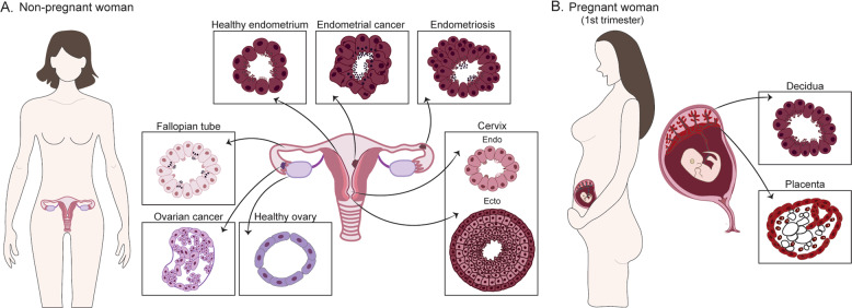
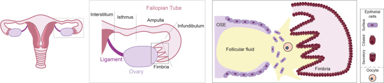
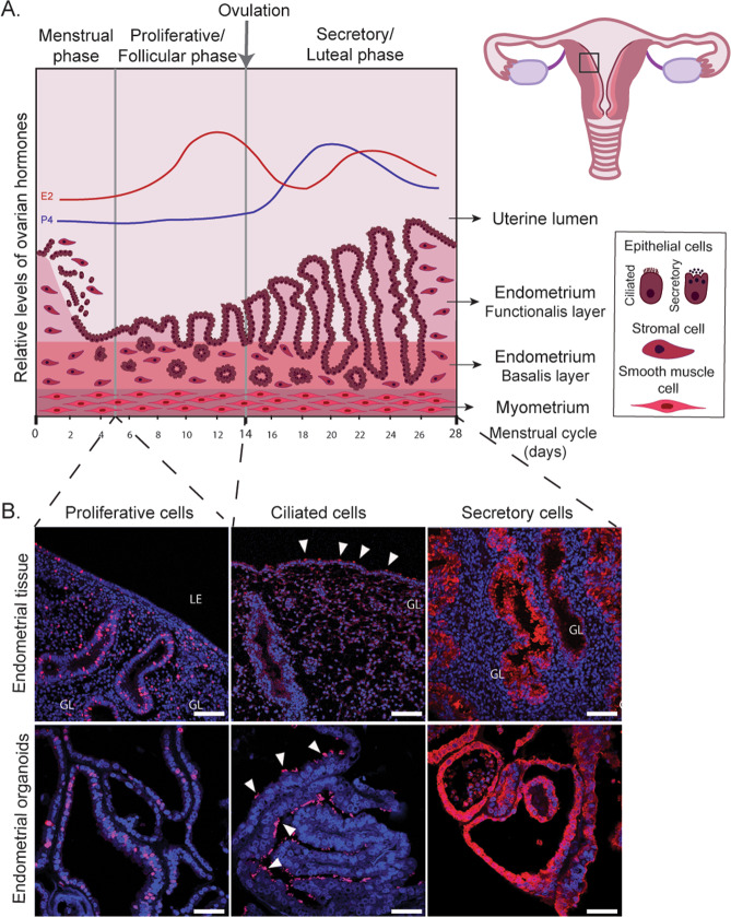
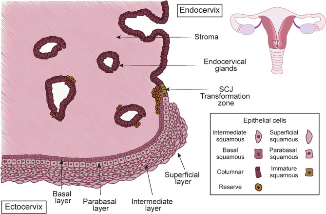
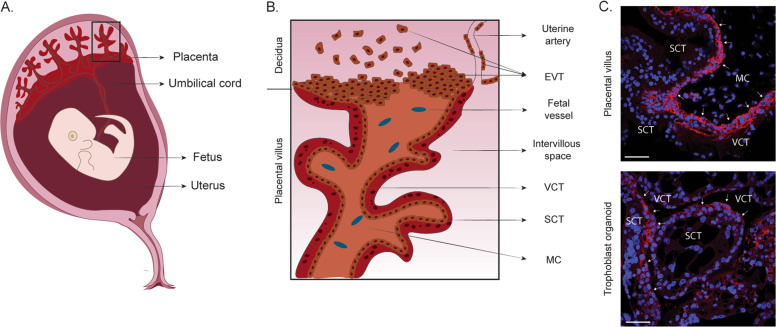
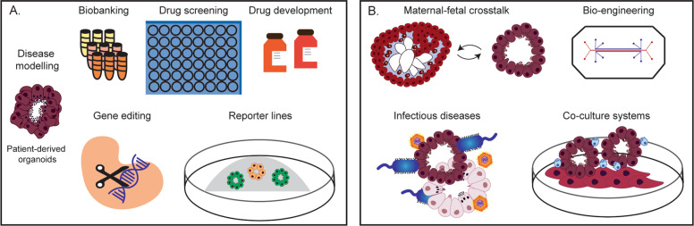
Both the proper functioning of the female reproductive tract (FRT) and normal placental development are essential for women's health, wellbeing, and pregnancy outcome. The study of the FRT in humans has been challenging due to limitations in the in vitro and in vivo tools available. Recent developments in 3D organoid technology that model the different regions of the FRT include organoids of the ovaries, fallopian tubes, endometrium and cervix, as well as placental trophoblast. These models are opening up new avenues to investigate the normal biology and pathology of the FRT. In this review, we discuss the advances, potential, and limitations of organoid cultures of the human FRT.


Conflict of interest statement

The authors declare that they have no conflict of interest.

Figures

Fig. 1
Fig. 1. Tissue-derived organoids of the human FRT and placenta.
a Types of organoids derived from the human FRT using tissue samples (normal and pathological) from non-pregnant women: ovaries, fallopian tubes, endometrium, and cervix. Organoids can also be derived from disorders of the FRT such as endometriosis and cancer. The different organoid systems show specific features that recapitulate the epithelial organization of their tissue of origin. b Organoid systems that have been derived from the pregnant endometrial lining (decidua) and the first-trimester placenta (fetal origin).
Fig. 2
Fig. 2. Anatomical relationship of the ovary and the fallopian tube at the time of ovulation.
The fallopian tubes consist of: the fimbriae, projections bordering the ovaries; the infundibulum, opening to which fimbriae are attached; the ampulla, the longer section of the tube where fertilization usually occurs; the isthmus, narrow section adjacent to the uterine cavity and the interstitium that extends from the uterine cavity through the uterine muscle (central box). The ovaries are encapsulated by the single-cell layered ovarian surface epithelium (OSE). The fimbria of the fallopian tube envelops the ovaries and consists of the columnar ciliated and secretory epithelium. During ovulation, the OSE ruptures resulting in the release of follicular fluid and the oocyte into the fimbria. The oocyte will travel down the fallopian tube guided by the movement of the cilia where it will be fertilized by a sperm cell (right box).
Fig. 3
Fig. 3. Endometrial organoids recapitulate essential features of proliferation and differentiation of human endometrium.
a The endometrium is organized into two layers, the functional layer adjacent to the uterine lumen and the basal layer adjacent to the myometrium. It undergoes cyclical growth, differentiation, and shedding under the influence of ovarian hormones, estrogen (E2) (red line), and progesterone (P4) (blue line). The menstrual phase is followed by an E2 dominated proliferative phase. Ovulation marks the start of the secretory phase during the decidualization of the endometrium. In the absence of implantation hormone levels drop, resulting in shedding of the functional layer. The different cell types are depicted in the box. b Immunofluorescence of endometrial tissue (in vivo) and organoids (in vitro) to visualize proliferative (Ki67 positive, red), ciliated (acetylated-a-tubulin positive, red), and secretory cells (PAEP positive, red). Nuclei are counterstained with Dapi (blue). White arrowheads indicate ciliated cells. Scale bars, 100 μm (in vivo), 50 μm (in vitro). LE luminal epithelium, GL glands.
Fig. 4
Fig. 4. The anatomy of the human cervix.
The cervix consists of two distinct epithelia; the columnar epithelium of the endocervix and stratified epithelium of the ectocervix, which merge in the squamocolumnar junction (SCJ). The reserve cells (in green) are localized under the columnar epithelium and are believed to regulate the process of metaplasia, which results in the formation of a new stratified epithelium, producing the transformation zone (TZ).
Fig. 5
Fig. 5. The pregnant uterus of the first trimester and the placenta.
a The placenta is firmly embedded into the pregnant lining of the uterus, the decidua, on one side and on the other, is connected to the fetus via the umbilical cord. b The placenta is made up of a system of branching villi that is covered by a bilayered epithelium—a multinucleated outer layer called the syncytium (SCT) and the mononuclear epithelium, the villous cytotrophoblast (VCT). The mesenchymal core (MC) of the villi contains stromal and endothelial cells and macrophages. At the tips of the villi, extravillous trophoblast (EVT) attach and invade into the stroma and the spiral arteries of the decidua. c Trophoblast organoids derived from human first-trimester placenta. Immunofluorescence of a placental villus (in vivo) and trophoblast organoids (in vitro) for VCT marker EPCAM (in red). VCT is shown with white arrows. Nuclei are stained with Dapi (blue). Scale bars, 50 μm.
Fig. 6
Fig. 6. Applications of tissue-derived organoids of the female reproductive tract and placenta.
a Organoid systems can be used for studying the physiology and pathologies of the FRT. They can be used as. tools for testing drug responses and drug development. Gene function can be assesed by CRISPR/Cas9 based genetic engineering of organoids. b The possibility to combine organoids with different cell types as well as pathogens will allow studies on their interactions. Bioengineering methods may allow the generation of more complex tissue-like models that include non-epithelial populations such as fibroblasts, immune and endothelial cells. The interactions between two different tissues can also be studied by co-culture of organoids, which is of particular relevance for maternal–fetal crosstalk using placental (trophoblast) and endometrial organoids.

References

    1. Cunha GR, Robboy SJ, Kurita T, Isaacson D, Shen J, Cao M, et al. Development of the human female reproductive tract. Differ Res Biol Divers. 2018;103:46–65. doi: 10.1016/j.diff.2018.09.001. - DOI - PMC - PubMed
    1. Hashimoto R. Development of the human Müllerian duct in the sexually undifferentiated stage. Anat Rec A Discov Mol Cell Evol Biol. 2003;272:514–9. doi: 10.1002/ar.a.10061. - DOI - PubMed
    1. Behringer RR, Cate RL, Froelick GJ, Palmiter RD, Brinster RL. Abnormal sexual development in transgenic mice chronically expressing müllerian inhibiting substance. Nature. 1990;345:167–70. doi: 10.1038/345167a0. - DOI - PubMed
    1. Berman DM. Pathology of the Female Reproductive Tract. Int J Gynecol Pathol. 2002;21:426. doi: 10.1097/00004347-200210000-00018. - DOI
    1. Onstad MA, Schmandt RE, Lu KH. Addressing the role of obesity in endometrial cancer risk, prevention, and treatment. J Clin Oncol. 2016;34:4225–30. doi: 10.1200/JCO.2016.69.4638. - DOI - PMC - PubMed

Publication types