Review
. 2017 May 15;8(5):2702-2716.
doi: 10.19102/icrm.2017.080502. eCollection 2017 May.

Strategies for Transvenous Lead Extraction Procedures

Affiliations
Review

Strategies for Transvenous Lead Extraction Procedures

Laurence M Epstein et al. J Innov Card Rhythm Manag. .

Abstract

Transvenous lead extraction (TLE) has undergone an explosive evolution since its inception as a rudimentary skill with limited technology and therapeutic options. Early techniques involved simple manual traction that frequently proved ineffective for chronically implanted leads, and carried a significant risk of myocardial avulsion, tamponade, and death. The morbidity and mortality associated with these early extraction techniques limited their application to use only in life-threatening situations, such as infection and sepsis. The past four decades, however, have witnessed significant advances in lead extraction technology, resulting in more efficacious techniques and tools, providing the skilled extractor with a well-equipped armamentarium. With the development of the discipline, we have witnessed a growth in the community of TLE experts coincident with a marked decline in the incidence of procedure-related morbidity and mortality, with recent registries at high-volume centers reporting high success rates with exceedingly low complication rates. Future developments in lead extraction are likely to focus on new tools that will allow for us to provide comprehensive device management, develop alternative systems for extraction training, and focus on the design of new leads conceived to facilitate future extraction.

Keywords: Defibrillator; lead extraction; lead management; pacemaker.


Conflict of interest statement

Dr. Epstein reports no relevant disclosures or financial arrangements. Dr. Maytin reports personal fees from Medtronic, Spectranetics, St. Jude Medical, and Biotronik, outside the scope of the submitted work.

Figures

Figure 1:
Figure 1:
Risk versus risk. The decision regarding lead extraction or abandonment requires comparison of the current risks of lead extraction with the future risks of both lead abandonment and potential lead extraction.
Figure 2:
Figure 2:
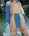
Patient preparation. We prepare our patients with a chlorhexidine solution and drape so as to allow for access for contralateral implant or for emergent pericardiocentesis, thoracentesis, thoracotomy, sternotomy, or cardiopulmonary bypass.
Figure 3:
Figure 3:
Locking stylets for transvenous lead extraction. A: The Liberator® Locking Stylet (Cook Medical, Bloomington, IN, USA) fits leads with lumen diameters of 0.016 to 0.032 inches. An undeployed Liberator® locking stylet is shown above a deployed Liberator® locking stylet. When deployed, the wound spring at the end of the stylet opens up, locking into place. Courtesy of Cook Medical, Inc. B: The Lead Locking Device (LLD®) EZ (Spectranetics, Colorado Springs, CO, USA) has a radiopaque tip and accommodates inner coil diameters of 0.015 to 0.026 inches (undeployed stylet, top). In contrast to the Liberator® locking stylet, the LLD® locking stylet has a braided mesh over the entire length of a solid lead that expands when deployed (bottom). Courtesy of The Spectranetics Corporation.
Figure 4:
Figure 4:
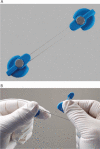
The Bulldog™ Lead Extender (Cook Medical, Bloomington, IN, USA). This useful tool is meant for leads that cannot receive a locking stylet, owing to either extensive damage or a solid-core design. The exposed end of the lead is passed through the loop of the Bulldog™ (arrow), and the metal sleeve (asterisk) is advanced over the loop grasping the lead. Courtesy of Cook Medical, Inc.
Figure 5:
Figure 5:
One-Tie® Compression Coil (Cook Medical, Bloomington, IN, USA). A: Undeployed tool. B: The One-Tie® Compression Coil (Cook Medical, Bloomington, IN, USA) is wound around the lead, compressing its components.
Figure 6:
Figure 6:
Telescoping non-powered countertraction sheaths. Telescoping sheaths are available in a range of sizes from 7F to 16F, and are made of different materials with varying properties, including A: stainless steel; B: Teflon™ (Chemours Co., Wilmington, DE, USA); and C: polypropylene. Courtesy of Cook Medical, Inc.
Figure 7:
Figure 7:
SLS® II Excimer Laser Sheaths (Spectranetics, Colorado Springs, CO, USA). A: The Excimer laser sheath utilizes ultraviolet laser energy to vaporize tissue in contact with the tip of the sheath, where the optical fibers terminate. The sheath is available in a range of sizes (12F, 14F, and 16F), displayed from top to bottom. B: End-on view of the laser sheath showing the distal end, where the optical fibers terminate. Both images courtesy of The Spectranetics Corporation.
Figure 8:
Figure 8:
Mechanism of photoablation. The laser sheath applies circumferential pulses of energy at its distal end. The ultraviolet energy disrupts molecular bonds to a depth of 50 mm, causing cells to rupture and fibrotic tissue to dissolve, forming a vapor bubble. The vapor bubble expands and implodes, clearing debris from the distal end of the sheath. Courtesy of The Spectranetics Corporation.
Figure 9:
Figure 9:
The TightRail™ Mechanical Dilator Sheaths (Spectranetics, Colorado Springs, CO, USA). The flexible shaft, shielded bidirectional rotating blade and static sheath are unique features that allow for the device to pass through dense and calcific scarring while requiring less traction force. The inset image is a magnified view of the sheath tip with the shielded metal blade. Courtesy of the The Spectranetics Corporation.
Figure 10:
Figure 10:
The Evolution® and Evolution® Shortie Mechanical Dilator Sheaths (Cook Medical, Bloomington, IN, USA). These “hand-powered” mechanical sheaths consist of a flexible, braided stainless steel sheath with a stainless steel spiral-cut dissection tip (inset). The sheath is attached to a trigger handle that rotates the sheath and allows for the threaded metal end to auger out the scar tissue. Courtesy of Cook Medical, Inc.
Figure 11:
Figure 11:
Transfemoral snaring of a lead. Transfemoral lead retrieval with the Byrd Workstation™ (Cook Medical, Bloomington, IN, USA) is a necessary skill for successful lead extraction, particularly in cases in which the lead is not accessible from the implant vein, as with a cut or fractured lead. Here, the lead has been snared and wound up by the Needle’s Eye® snare (Cook Medical, Bloomington, IN, USA) (arrow), allowing for successful removal of the lead.
Figure 12:
Figure 12:
The Bridge™ Occlusion Balloon (Spectranetics, Colorado Springs, CO, USA). A: The Bridge™ balloon is an 8-cm, low-pressure compliant balloon designed to conform to the anatomy of the superior vena cava (SVC) in the majority of individuals. The proximal radiopaque marker (circled) should be positioned at the SVC-right atrial junction to insure complete coverage of the SVC. B: The Bridge™ balloon ((Spectranetics, Colorado Springs, CO, USA) is easily compressed with minimal pressure. Both images courtesy of The Spectranetics Corporation.
Figure 13:
Figure 13:
Stepwise approach to transvenous lead extraction. We routinely employ a stepwise approach to lead extraction so as to achieve the highest rate of complete success while utilizing the fewest number of tools. ES: extraction sheath.
Figure 14:
Figure 14:
Schematic representation of the forces of counterpressure, traction and countertraction. Counterpressure is the force applied by the non-powered or powered sheath as it is advanced over the lead, interrupting areas of adherent scar tissue. Traction is the pulling force on the lead needed to provide a straight “rail” so as to allow for the sheath to follow the lead. Countertraction is the forward force applied by the sheath at the myocardium to limit the traction forces on an entrapped electrode to the circumference of the sheath at the lead tip-myocardium interface. Once the lead is released from the fibrous tissue, the myocardium falls away from the sheath, thereby reducing the risk of myocardial invagination or injury.

References

    1. Pakarinen S, Oikarinen L, Toivonen L. Short-term implantation-related complications of cardiac rhythm management device therapy: a retrospective single-centre 1-year survey. Europace. 2010;12(1):103–108. [CrossRef] [PubMed] - DOI - PubMed
    1. Sridhar AR, Lavu M, Yarlagadda V, et al. Cardiac implantable electronic device-related infection and extraction trends in the U.S. Pacing Clin Electrophysiol. 2017;40(3):286–293. [CrossRef] [PubMed] - DOI - PubMed
    1. Eckstein J, Koller MT, Zabel M, et al. Necessity for surgical revision of defibrillator leads implanted long-term: causes and management. Circulation. 2008;117(21):2727–2733. [CrossRef] [PubMed] - DOI - PubMed
    1. Voigt A, Shalaby A, Saba S. Continued rise in rates of cardiovascular implantable electronic device infections in the United States: temporal trends and causative insights. Pacing Clin Electrophysiol. 2010;33(4):414–419. [CrossRef] [PubMed] - DOI - PubMed
    1. Cabell CH, Heidenreich PA, Chu VH, et al. Increasing rates of cardiac device infections among Medicare beneficiaries: 1990–1999. Am Heart J. 2004;147(4):582–586. [CrossRef] [PubMed] - DOI - PubMed

LinkOut - more resources